39 monocot root diagram
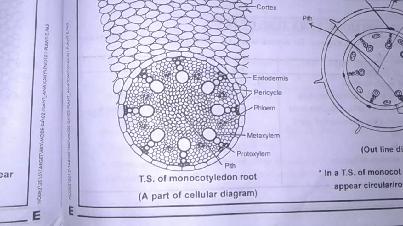
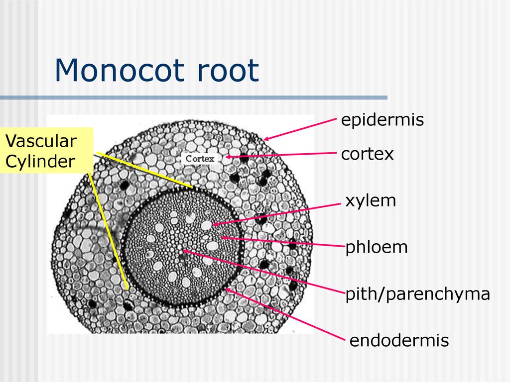
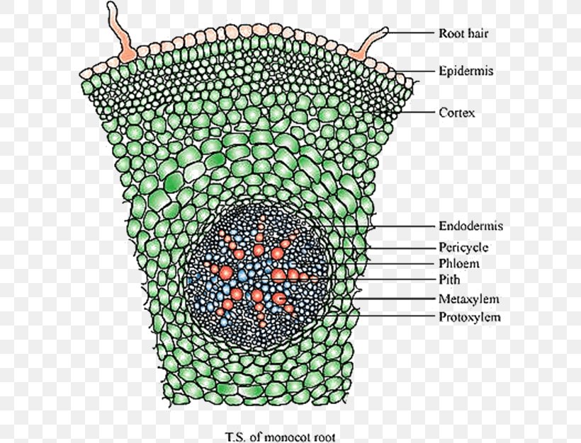
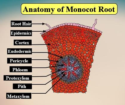
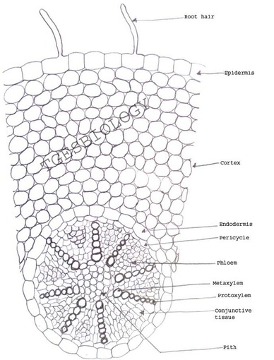
Anatomy of Monocot Root (Monocot Root Cross Section Under Microscope with Diagram) ... Ø The anatomical features of a monocot root can be studied through a cross ... Anatomy of Monocot Root. The transverse section (TS) of a typical monocotyledonous root shows the following structures: 1. Epiblema: It is the outermost layer consisting of a single row of thin-walled cells without any intercellular spaces. The structure and fate of this layer are more or less similar to that of dicot roots. 2.
Germination is usually the growth of a plant contained within a seed; it results in the formation of the seedling. It is also the process of reactivation of metabolic machinery of the seed resulting in the emergence of radicle and plumule. The seed of a vascular plant is a small package produced in a fruit or cone after the union of male and female reproductive cells.

Monocot root diagram
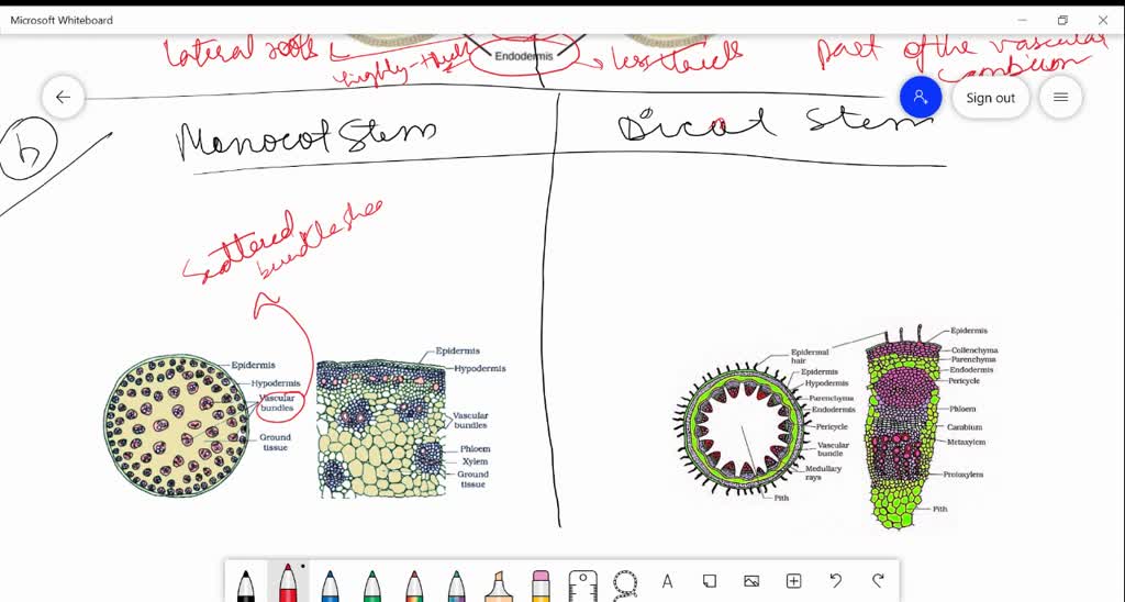
Read about: Difference between Dicot and Monocot Root. ... Besides this layer, there are other components as well like the Pericycle, the vascular bundles and pith. The below diagram of the Transverse section of Dicot stem will provide a better understanding of the location of all the layers of the cell. Aug 31, 2018 - Anatomy of a Typical Monocot Root Cross Section Structure (TS / CS) Under Microscope with Labelled Diagram, Description and PPT. T.S. of Monocot root. Answer: Question (C) Draw neat labelled diagrams of T.S. of dicot stem. Answer: Question 6. Write the information related to diagram given below. Answer: [Note: The labelled part can be considered as the 'region of maturation ' of root apical however, the region of maturation does not contain meristematic tissue ]
Monocot root diagram. Draw a neat labelled line-diagram of a monocot root cross section. (9) 1.2 Construct a table and give the anatomical differences between dicot and monocot roots. (4) 1.3 Differentiate between root hairs and lateral roots (. Place your order now for a similar paper and have exceptional work written by our team of experts to guarantee you A Results. Soln.(a) Differences between monocot root and dicot root are illustrated in the following figure and table. (b) Differences between monocot and dicot stems are illustrated in the following figure and table. ... Explain the structure of stomata with a labelled diagram. Soln.Stomata are structures present in the epidermis of leaves. Stomata ... This article provides a diagram of monocot root. Also learn about:- 1. Exodermis 2. Origin of Lateral Roots 3. Root-Stem Transition. The main characteristic feature to distinguish the dicot and monocot leaf is the type of venation a leaf have. One can easily observe either the veins are striking or parallel by seeing a leaf. Below is the diagram of dicot and monocot leaf, where we can see the venation pattern.
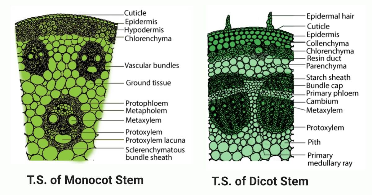
Monocot plants have a fibrous or adventitious root system. Dicot plants have a tap root system. Primary root. The development of the primary root stops during the postembryonic development of the roots. The primary root continues to grow throughout the life of the plant in the form of the taproot. Epidermal covering. A monocot is a plant with one seed leaf, referred to as cotyledon. Learn the more extensive definition of monocot through exploring examples of cotyledon and its function in the ecological system. Draw well labelled diagram of T.S. of monocot root. 645806160 000+ 300+ 1:39 Draw a well labelled diagram of T.S of dicot and monocot stem. 646580909 000+ 400+ Compare T.S. of monocot and dicot root with the help of well labelled diagrams only. ... (a) Dicot stem and monocot stem (b) Dicot stem and dicot root (c) Dicot root and monocot root (d) Dicot stem and monocot root. Ans. d. 29. Which one of the following is the correct sequence of tissues present in dicot stem during secondary growth? (a) Primary cortex, secondary cortex, phellogen, cork (b) Cork, primary cortex, secondary cortex ...
Diagram. T.S Of Dicot Stem. T.S Of Monocot Stem. Procedure Taking Sections. ... To study of the transverse section of monocot root, maize root. Theory. The transverse section of monocot root depicts the structures as listed below: Epiblema or Epidermis It is a single outermost layer with no cuticle; Aug 31, 2021 · Monocotyledon plants have just one cotyledon, which is an important part of the embryo of a seed, and the first thing to appear above ground as a plant begins its growth cycle. (a) Monocot root and dicot root (b) Monocot stem and dicot stem Solution: Question 5. Cut a transverse section of the young stem of a plant from your school garden and observe it under a microscope. How would you ascertain whether it is a monocot stem or a dicot stem? Give reasons. Solution: Monocot Root Cross Section Root Diagram Root Plant Science . Plant Cell Diagram Animal Cell Diagram Animal Cell Plant And Animal Cells Science Cells . An Ecosystem Is A Community Of Living Things And Their Non Living Environment And May Be As Large As A Desert Or Ecosystems Desert Ecosystem Biology Projects .

Plant Microscope Exercise Biology4friends
831 monocot root stock photos, vectors, and illustrations are available royalty-free. See monocot root stock video clips. of 9. plant cell typical cell root cross section monocot dicot xylem phloem plant pith corn cross section vascular bundles cambium root systems. Try these curated collections.
Draw A Well Labelled Diagram Of T S Of Monocot Root Sarthaks Econnect Largest Online Education Community
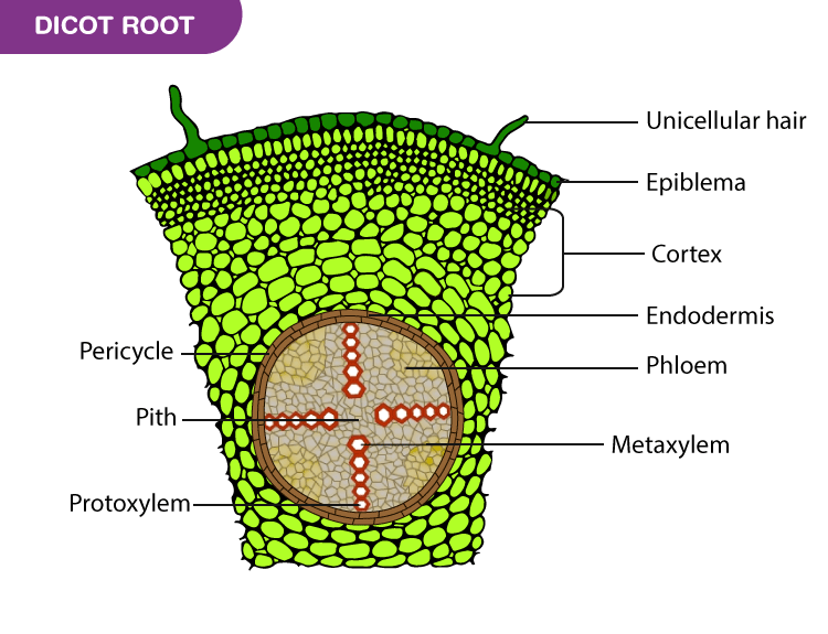
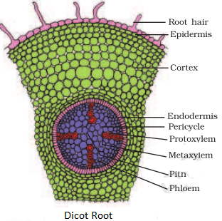
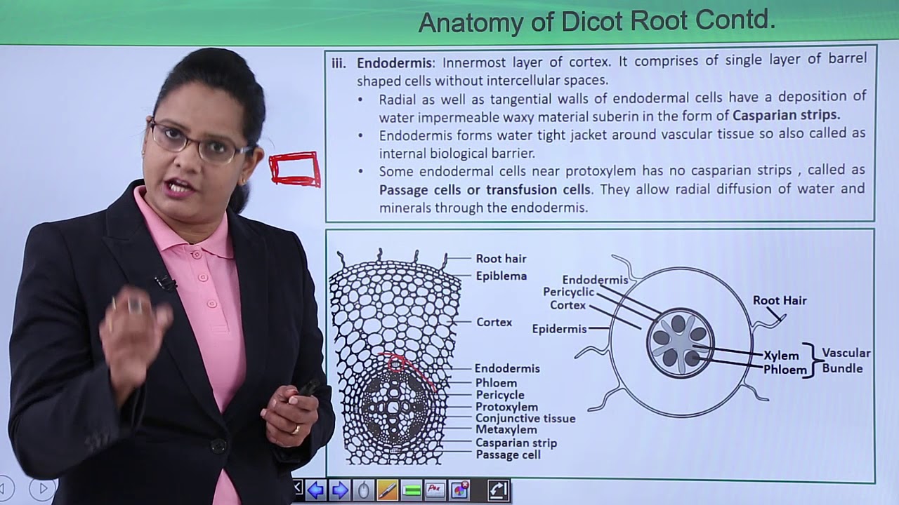
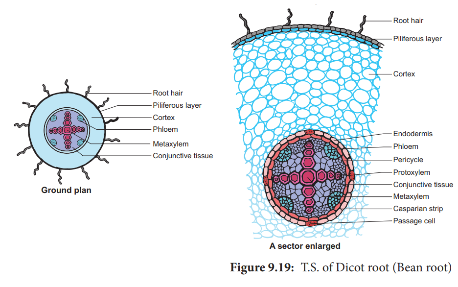
Following are the anatomical structure of the dicot root: Epiblema or epidermis or piliferous layer: It is outermost uni-layered with several unicellular root hairs. It consists of thin-walled, compactly arranged living parenchymatous cells. Epiblema of a root differs from the epidermis of the stem in being devoid of distinct cuticle and stomata.
Monocot Stem
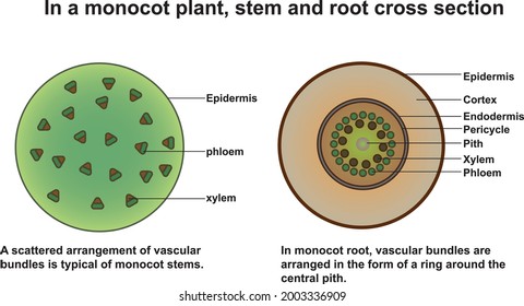
The secondary growth is absent in closed vascular bundles. These are mainly found in Monocots. Fascicular cambium is absent. Example: Asparagus stem. According to Mode of Occurrence of the Elements Radial. In the roots of monocots and dicots, xylem and phloem are arranged in an alternate manner on different radii. It is called radial arrangement.
Preparation And Study Of T S Of Dicot And Monocot Roots And Stems Primary
Also, read Anatomy of Monocot and Dicot Plants. Monocot Root. These plant roots have a comparatively wider, and fibrous root-like structure. Dicot Root. These plant roots have a comparatively narrow, and tap root-like structure. Normally, dicots and monocots differ in four aspects which include stems, flowers, leaves, and roots.
Solved Cross Section Of Monocot Root Plant Histology Study Card 1 Page 68 Figure 5 3 In Textbook Draw A Labeled Outline Diagram Of The Whole Cross Section 2 Points Also Draw A Well Labeled
Fully Labelled Diagram Of A Plant Cell As Seen Under A Light Microscope. angelo. November 1, 2021. Structure Of Animal Cell And Plant Cell Under Microscope Diagrams Cell Diagram Animal Cell Plant And Animal Cells. Monocot Root Cross Section Root Diagram Root Plant Science. Plant And Animal Cells Revised Plant And Animal Cells Plant Cell Drawing ...

Monocot Root Model Transverse Section Precise Hand Painted Details Eisco Labs
Monocot Stem vs Dicot Stem (22 Key Differences) Monocot stem is a circular-shaped hollow axial part of the plant which gives rise to nodes, internodes, leaves, branches, flowers with roots at the basal end. Dicot stem is the solid cylindrical axial part of a plant consisting of nodes and internodes giving rise to leaves, branches, and flowers.
Monocot Vs Dicot Stem Definition Structure 22 Differences Examples
Eudicots. As the plant embryo grows at germination, it sends out a shoot called a radicle that becomes the primary root, and then penetrates down into the soil.After emergence of the radicle, the hypocotyl emerges and lifts the growing tip (usually including the seed coat) above the ground, bearing the embryonic leaves (called cotyledons), and the plumule that gives rise to the first true leaves.
T S Of Monocot Root By Praatibaa Part 1 Youtube
Draw well labelled diagram of T.S. of monocot root. 645806160 000+ 300+ 1:39 Draw a well labelled diagram of T.S of dicot and monocot stem. 646580909 000+ 400+ With the help of a neat labelled diagram, describe the T.S. of a monocot root. ...
Monocot And Eudicot Dicot Roots Ppt Download
The root distribution in the soil is one of the elements that comprise the root system architecture (RSA). In monocots, RSA comprises radicle and crown roots, each of which can be basically represented by a single curve with lateral root branches or approximated using a polyline. Moreover, RSA vectorization (polyline conversion) is useful for RSA phenotyping.

Root Structure Monocot And Dicot Stems Cross Sections Of Plants Roots Vector Diagram For Educational Biological And Scientific Use Royalty Free Cliparts Vectors And Stock Illustration Image 128230639
Label the diagram below, paying particular attention to the events occurring when there is a change from n to 2n and from 2n to n. ... Label the different meristem types on the shoot and root longitudinal section diagrams. Note: Each label will be used twice. Ch. 4 ... True or False: In a monocot stem, xylem is located closer to the center of ...

Draw And Label The Parts Of Transverse Section Of Monocot Root Brainly In
A carrot is an example of a dicot root. Diagram illustrating the tissue layers and their organization within monocot and dicot roots. Is a buttercup a Dicot? Class. The class Dicotyledon means that buttercups produce two seed leaves when they germinate, instead of just one. In this category, buttercups diverge from monocotyledons such as magnolias.

Internal Structure Of Monocot Root Definition Examples Diagrams
Draw neat and labelled diagrams of dicot and monocot root and differentiate between their anatomical characters. Answer: The transverse section of a typical dicotyledonous root shows following anatomical features: 1. Epiblema: It is the outermost single layer of cells without cuticle. Some epidermal cells prolong to form unicellular root hairs. 2.
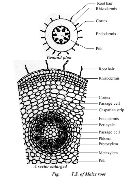
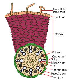
Primary Structure Of Monocotyledonous Root Maize Root
The roots of seed plants have three major functions: anchoring the plant to ... Dicots have a tap root system, while monocots have a fibrous root system.

Roots Stems And Leaves Diagrams
In this tutorial, we have explained the root anatomy along with the monocot root cross section and the dicot root cross section. Additionally, we did a difference between monocot vs dicot root. ROOT ANATOMY: DICOT ROOT CROSS SECTION Dicot Root Diagram Reveals Internal Structure of Dicot Root A thin transverse section of the young dicot root of Gram, Sunflower or Pea reveals the following ...
Dicotyledon Monocotyledon Anatomy Plant Stem Root Png 600x626px Watercolor Cartoon Flower Frame Heart Download Free
If you dig a monocot seed plant and have a look at its roots, you will find out that its roots are adventitious, which means the roots arise from parts of the plant other than just the roots, for example, the stem. The roots are smaller, thin and thread-like, that is why they are called fibrous roots.

Monocot Dicot Images Stock Photos Vectors Shutterstock
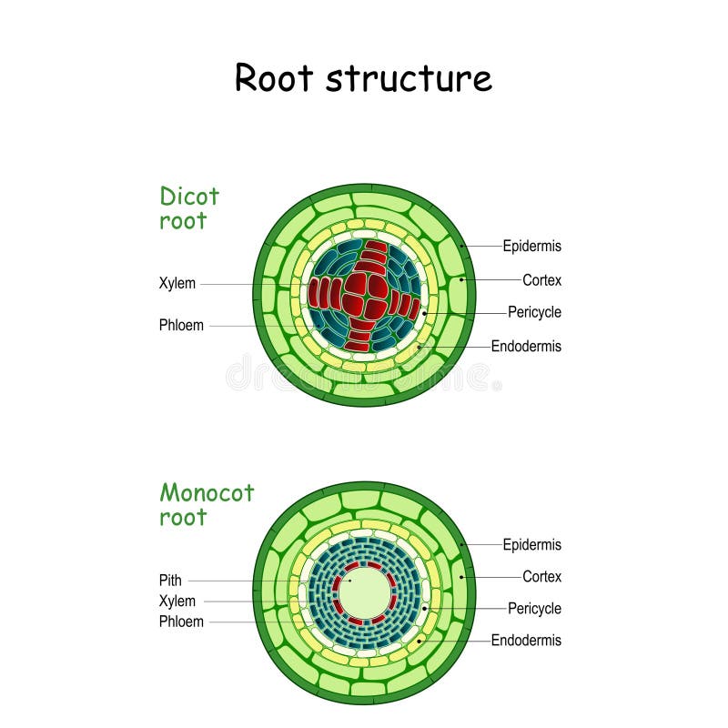
In monocot roots, the vascular bundles are arranged in a circular pattern. ... Monocots and dicots contain two main types of vascular tissue: the xylem and phloem ...
1
Labeled Hair Follicle Diagram Labeled Hair Follicle Diagram 133231 Layers Of The Skin Skin Anatomy Anatomy And Physiology Integumentary System Monocot Root Cross Section Root Diagram Root Plant Science Vascular Plant Biological Structure Labeled Diagram Vector Illustration In 2021 Vascular Plant Biology Plants Plants Shows A Root Hair Cell The Water Can Pass Through The […]

Latest Monocot Root Cross Section Structure With Ppt Easy Biology Class Latest Cross Section
A number of these differences are not unique to the monocots, and, while still useful, no one single feature will infallibly identify a plant as a monocot. For example, trimerous flowers and monosulcate pollen are also found in magnoliids, and exclusively adventitious roots are found in some of the Piperaceae. Similarly, at least one of these traits, parallel leaf veins, is far from universal ...

Biologypictures1108 Snail Monger Monocot Root 4x 40x 33 86x
Jun 06, 2007 · Monocot roots, interestingly, have their vascular bundles arranged in a ring. Dicot roots have their xylem in the center of the root and phloem outside the xylem. A carrot is an example of a dicot root. Diagram illustrating the tissue layers and their organization within monocot and dicot roots.
Monocot Stem Images Stock Photos Vectors Shutterstock
Jun 6, 2007 — Diagram illustrating the tissue layers and their organization within monocot and dicot roots. Image from Purves et al., Life: The Science of ...
Discuss The Internal Structure Of Monocot Roots Class 11 Biology Cbse
Roots, Stems and Leaves Diagrams. External Root Structure. Monocot Root. Dicot Root. External Structure of a Woody Stem. Monocot Stem. Woody Dicot Stem.
Root Structure Monocot And Dicot Stems Stock Vector Illustration Of Botanic Stem 167594771
Monocots, on the other hand, possess alternating xylem and phloem tissues. The xylem vessels are another factor that marks the difference. The vessels in the roots of dicots are angular or polygonal. Monocot roots, on the other hand, have vessels that are rounded or oval.

Cross Section Of A Monocot Root Diagram Quizlet
Feb 04, 2021 · Structure of Monocot and Dicot Seed. The structure of monocot and dicot seeds can be described based on the following parts; 1. Seed Coat. The seed coat is the outermost covering of the seed which in some cases might remain fused with the fruit wall.
1
What are Monocot Roots. Monocot roots are fibrous or adventitious roots or shoot-borne roots found in monocotyledonous plants, consisting of a vast network of thin roots and root fibers originating from the stem.The number of roots varies depending on the species and age of the plant.. However, most of the monocots are herbaceous with weak cambium that cannot hold woody tissues.
Anatomy Of Monocot Roots Zea Mays Sciencetopia
Aug 16, 2021 · Root System Growth. As plants grow above the surface, there is also growth that occurs within the soil. Roots need to grow in order to better support the …
Monocot Root 3d Warehouse
Monocot root *The cortex region is wide. *There is no formation of cork and only the epiblema is peeled off. *Older monocot roots show a covering of exodermis. *Endodermis cells are highly thick and the Casparian stripes are only visible in young roots. *Passage cells are present and they are thin-walled. *Pericycle only produces lateral roots.
Monocot And Dicot Stems
T.S. of Monocot root. Answer: Question (C) Draw neat labelled diagrams of T.S. of dicot stem. Answer: Question 6. Write the information related to diagram given below. Answer: [Note: The labelled part can be considered as the 'region of maturation ' of root apical however, the region of maturation does not contain meristematic tissue ]
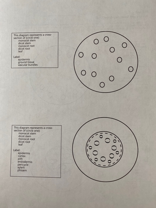
Solved This Diagram Represents A Cross Section Of Circle Chegg Com
Aug 31, 2018 - Anatomy of a Typical Monocot Root Cross Section Structure (TS / CS) Under Microscope with Labelled Diagram, Description and PPT.
Anatomy Of Dicotyledonous And Monocotyledonous Plants By Biology Experts Notes Medium
Read about: Difference between Dicot and Monocot Root. ... Besides this layer, there are other components as well like the Pericycle, the vascular bundles and pith. The below diagram of the Transverse section of Dicot stem will provide a better understanding of the location of all the layers of the cell.

Monocot Root Cross Section Corn Root Diagram Quizlet
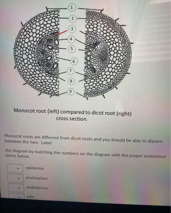
Solved Monocot Root Left Compared To Dicot Root Right Chegg Com
Anatomy Of Flowering Plant Dicot And Monocot Root Structure Youtube
Primary Structure Dicotyledonous Monocotyledonous Root Leaf Plantlet
Slide Preparation Biology4isc

Plant Structure Ii
Explain The Internal Structure Of Dicot Root With The Help Of Well Labelled Diagram And Also Differentiate Between Sarthaks Econnect Largest Online Education Community

T S Of Monocot Root Biology Notes Botany Microscopic Photography

5 2 Anatomy Of Dicotyledonous Plants Support And Transport Systems In Plants Siyavula

Monocot Root Nomenclature Book Montessori Print Shop Montessori Print Shop Usa


Comments
Post a Comment