42 labeling the parts of a cell on a diagram is an example of an exercise in
Labeling the parts of a cell on a diagram is an example of an exercise in anatomy positive feedback homeostasis physiology i don't know yet Categories Uncategorized Leave a Reply Cancel reply Pull down the digestive system diagram into each cell. Type the name of organs into the title boxes: Mouth, Esophagus, Stomach, Small Intestines, Large Intestines, and Anus. Identify the parts of the digestive system. Each cell should have one part of the diagram colored a different color than the rest, matching the title box.
Labeling the parts of a cell on a diagram is an example of an exercise in _____. physiology homeostasis anatomy positive stimuli

Labeling the parts of a cell on a diagram is an example of an exercise in
Label the diagram below with the following labels: Anaphase Interphase Mitosis . Cell division (M Phase) Interphase Prophase . Cytokinesis Interphase S-DNA replication . G1 - cell grows Metaphase Telophase . G2 - prepares for mitosis . Then on the diagram, lightly color the G1 phase . BLUE, the S phase YELLOW, Examples of circuit symbols used in drawing circuits. Examples of drawings of a simple circuit with a bulb, cell and the switch. Example of a simple circuit with the switch ON. Example of a simple circuit with the switch OFF. Example of a simple circuit with two bulbs with the switch ON. Look at each circuit diagram below. (ii) Fig. 3.2 also shows a diagram of part of the human alimentary canal and associated organs. On Fig. 3.2, draw label lines with letters to show: E where hydrochloric acid is made F where bile is made G where amylase is made H where egestion occurs. Fig. 3.2 [4] (c) (i) State where digested food is absorbed.
Labeling the parts of a cell on a diagram is an example of an exercise in. Animal cell diagram given the label on diagram identify the cell part. The cell membrane is the outer coating of the cell and contains the cytoplasm substances within it and the organelle. In both animals and plants cells generally become specialized to perform certain functions. Label the animal cell mitosis diagram. Match. Gravity. Labeling the parts of a cell on a diagram is an example of an exercise in _______. Click card to see definition 👆. Tap card to see definition 👆. anatomy. Click again to see term 👆. Tap again to see term 👆. At the cellular level, cells consist of _______. For each of the following printouts, first read the definitions, then label the diagram or map. Please let us know if there are any new Label Me! printouts that you need. Label Me! Printouts. Label the chicken egg cross-section diagram. Label the 72-hour old chicken embryo diagram. Label the 10-day old chicken embryo in egg diagram. If a cell undergoes mitosis but not cytokinesis, the product is __5__. The structure that acts as a scaffolding for chromosomal attachment and movement is called the __6__. __7__ is the period of cell life when the cell is not involved in division. Two cell populations in the body that do not routinely undergo cell division are __8__ and __9__.
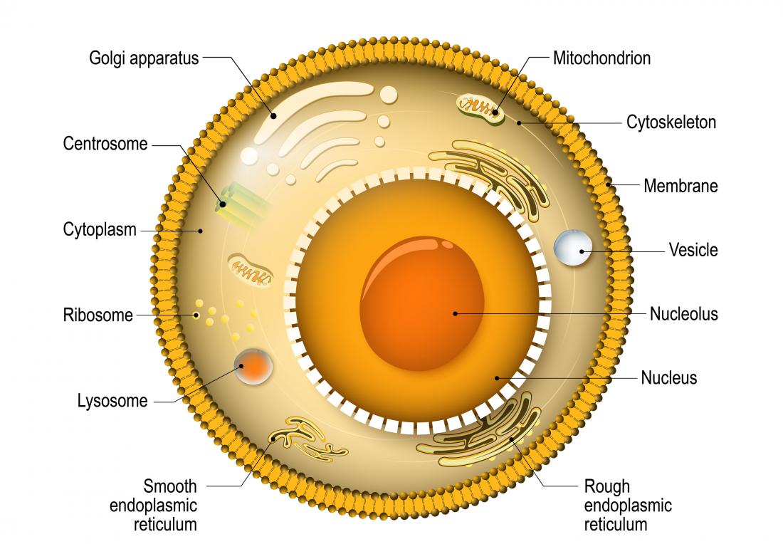
View BIO 210 Chapter 1 Reading Quiz.pdf from BIO 210 at Queens College, CUNY. 1. Labeling the parts of a cell on a diagram is an example of an exercise in _. Your Answer: anatomy 2. At the cellular Eukaryotic cells 2.3.1 Draw and label a diagram of the ultrastructure of a liver cell as an example of an animal cell.. Figure 2.3.1 - Annotated drawing of an animal cell. 2.3.2 Annotate the diagram from 2.3.1 with the functions of each named structure.. Ribosomes: Found either floating free in the cytoplasm or attached to the surface of the rough endoplasmic reticulum and in mitochondria and ... Eukaryotic cells 231 draw and label a diagram of the ultrastructure of a liver cell as an example. What differences are there between a prokaryotic and eukaryotic cell. Sorts modifies packages and distributes them to other parts of the cell or exit the cell. 233 identify structures from 231 in electron micrographs of liver cells. If there are some unlabeled parts left, check the diagram for key words. Then, scan the text for the right answer. Tips: Always underline new terms in the text - 99% that they will be used in the questions. The key words in the diagram may help you to complete it. Example: Label the parts of a leaf on the diagram below.
54 Step 1. Get a slide of the letter "e" from the tray on the side counter. This an example of a prepared slide, a slide that is already made for you and meant to be reused. (i.e., don't dispose of it, please return it to the tray when you are finished!Step 2. Use a piece of lens paper to clean any smudges (fingerprints, grease, etc.) off the slide. Biology Quiz : Animal Cell Diagram Given the LABEL (ON DIAGRAM), identify the CELL PART View Notes - A&P Skin & Body Membrane Quiz from A&P 2 at New York University. 1) Labeling the parts of a cell on a diagram is an example of an exercise in _. a. Positive stimuli b. Student Instructions. Label both a plant and animal cell on a poster layout. Include descriptions of what each part does. Click "Start Assignment". Find diagrams of a plant and an animal cell in the Science tab. Using arrows and Textables, label each part of the cell and describe its function. Color the text boxes to group them into organelles ...
Eukaryotic Cells Definition Parts Examples And Structure
Labeling the parts of a cell on a diagram is an example of an exercise in - Let's ques! Posted on. November 23, 2021 by hakan.
Easy Human
Label the Photosynthesis Diagram. Help students transit from passive listeners to active participants with this labelling the photosynthesis process worksheet pdf. Direct the 3rd grade and 4th grade students to use the words from the word box to label the diagram.

Plant Cell Definition Labeled Diagram Structure Parts Organelles
Figure-8 packaging of the cells Coordinated activity to act as a pump 7. Moves bones and the facial skin 8. Referred to as the muscular system 9. Voluntary 2. Identify the type of muscle in each of the illustrations in Figure 6—1. Color the diagrams as you wish. Intercalated discs S

Label The Skin Anatomy Diagram Tag Human Skin Diagram Label Human Anatomy Diagra Skin Anatomy Integumentary System Anatomy Organs
Answer. heart. 1. profile. Analisa1600. Labeling the parts of a cell on a diagram is an example of an excercise in ANATOMY. kattyahto8 and 1 more users found this answer helpful. heart outlined. Thanks 1.
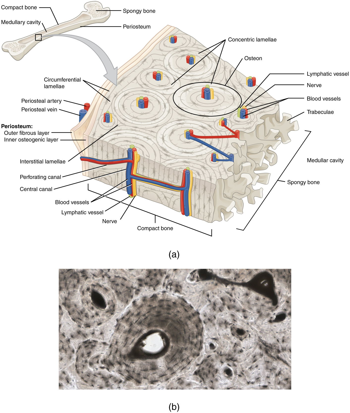
Bone Structure Anatomy And Physiology
Cells in the tail of a tadpole are digested by lysosomal enzymes during the gradual transition into a frog. Peroxisome: A membrane-bound organelle that contains specific enzymes imported from the cytoplasm (cytosol). For example, certain peroxisomes contain the enzyme catalase which rapidly breaks down toxic hydrogen peroxide into water and oxygen.

34 Human Cell Diagram To Label Labels Database 2020
he cells of the human body do not operate independently of one another. Instead, related cells live and work together in cell communities called tissues. A tissue is defined as a group of cells of similar structure that perform a common function. However, tissues do not consist entirely of cells: Between the cells is a nonliving
/profile-of-man-s-head-with-brain-anatomy-labeled-on-white-background-1093597090-f6a5470b98a4453997931b1cb72fb47d.jpg)
What Is The Cerebellum
Part 2: Cytoplasmic streaming in Elodea. Elodea is an freshwater, aquatic plant native to the Americas commonly used in aquariums. With leaves only two cells thick. Elodea is a good model to study living plant cells in action.In this experiment, you will see chloroplasts moving in the Elodea cells as they begin to photosynthesize. This movement is known as cytoplasmic streaming, which ...

2 3 Cell Structure And Function Cells The Basic Units Of Life Siyavula
Human Cell Diagram, Parts, Pictures, Structure and Functions The cell is the basic functional in a human meaning that it is a self-contained and fully operational living entity. Humans are multicellular organisms with various different types of cells that work together to sustain life.

Body Structures And Functions Chapter 3 Cell Labeling Diagram Quizlet
This vibrant worksheet contains the cross-section of an animal cell, vividly displaying the organelles. Examine the animal cell diagram and recognize parts like the centrioles, lysosomes, Golgi bodies, ribosomes and more indicated clearly. Label the Parts of an Animal Cell. Labels are important features of any scientific diagram.

Biology Ch 6 Hw Flashcards Quizlet
3. Go over the different parts of the two cells using the supplied "Teacher's Answer Guide" diagrams. As you discuss each part, have a student or yourself read out loud to the class the description and function of each part from the "Cell Parts" worksheet. Have the students complete the worksheets by filling in part as discussed.

Http Sciencevideos Wordpress Com Draw The Core 16
cells are also relatively large, but are long and spindle shaped. Red blood cells and sperm are both exam-ples of small cells. Red blood cells appear round, while sperm cells are streamlined with long flagella. Cell shape is often directly related to function. Epithelial cells fit tightly together and cover large areas.
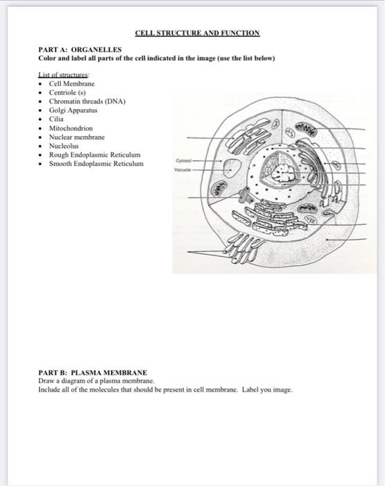
Solved Cell Structure And Function Part A Organelles Color Chegg Com
(ii) Fig. 3.2 also shows a diagram of part of the human alimentary canal and associated organs. On Fig. 3.2, draw label lines with letters to show: E where hydrochloric acid is made F where bile is made G where amylase is made H where egestion occurs. Fig. 3.2 [4] (c) (i) State where digested food is absorbed.
1
Examples of circuit symbols used in drawing circuits. Examples of drawings of a simple circuit with a bulb, cell and the switch. Example of a simple circuit with the switch ON. Example of a simple circuit with the switch OFF. Example of a simple circuit with two bulbs with the switch ON. Look at each circuit diagram below.

Skeletal System Definition Function And Parts Biology Dictionary
Label the diagram below with the following labels: Anaphase Interphase Mitosis . Cell division (M Phase) Interphase Prophase . Cytokinesis Interphase S-DNA replication . G1 - cell grows Metaphase Telophase . G2 - prepares for mitosis . Then on the diagram, lightly color the G1 phase . BLUE, the S phase YELLOW,
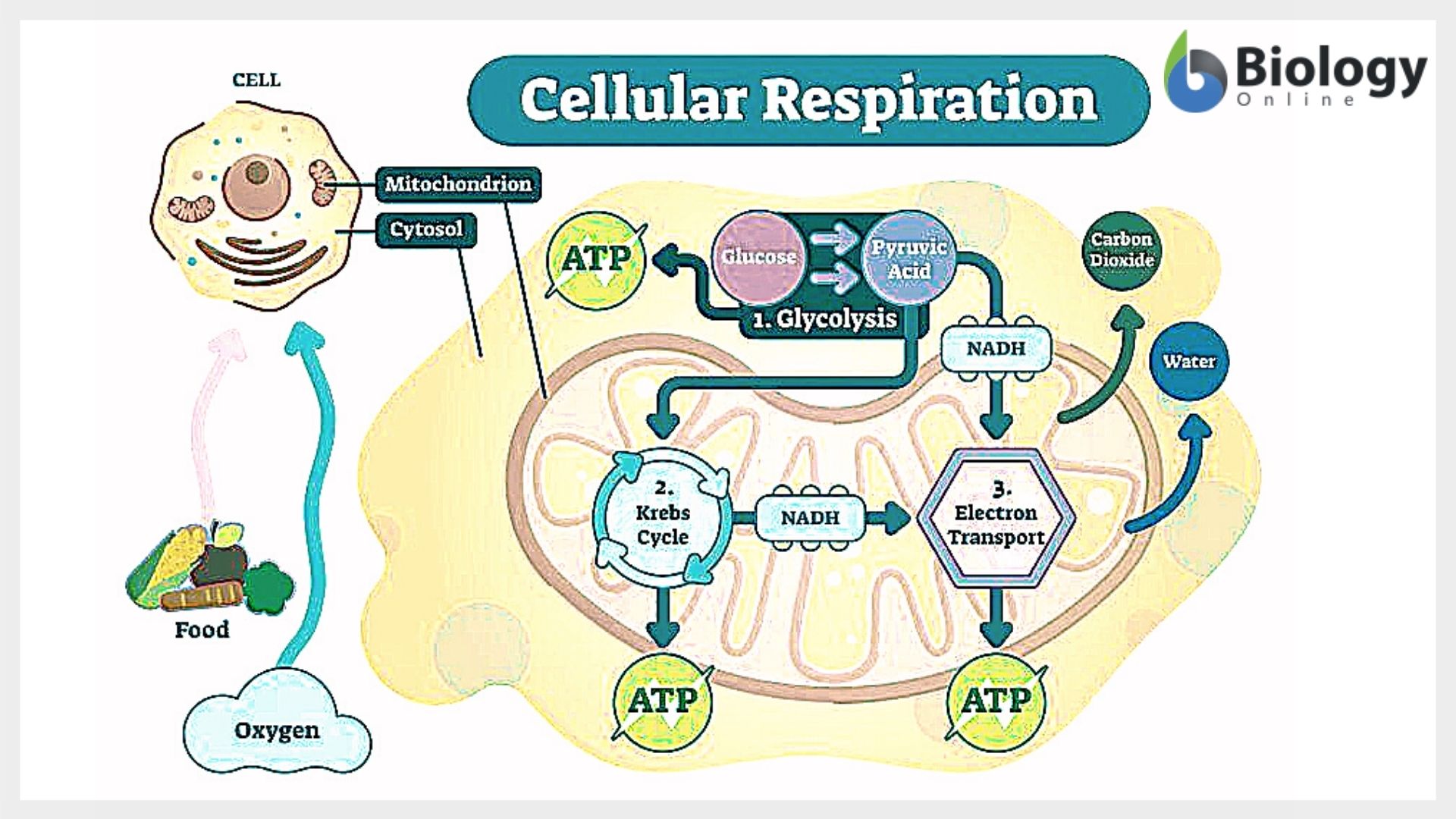
Cellular Respiration Definition And Examples Biology Online Dictionary

Cells Worksheets And Online Exercises
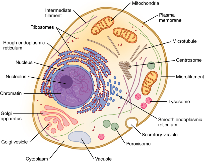
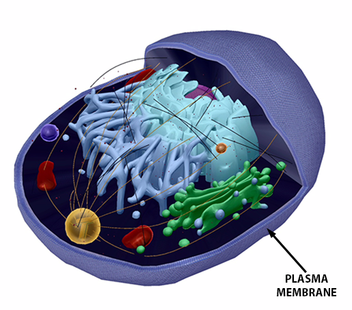
The Cytoplasm And Cellular Organelles Anatomy And Physiology

Labeling The Parts Of A Cell On A Diagram Is An Example Of An Exercise In Brainly Com

Lab Manual Exercise 1a

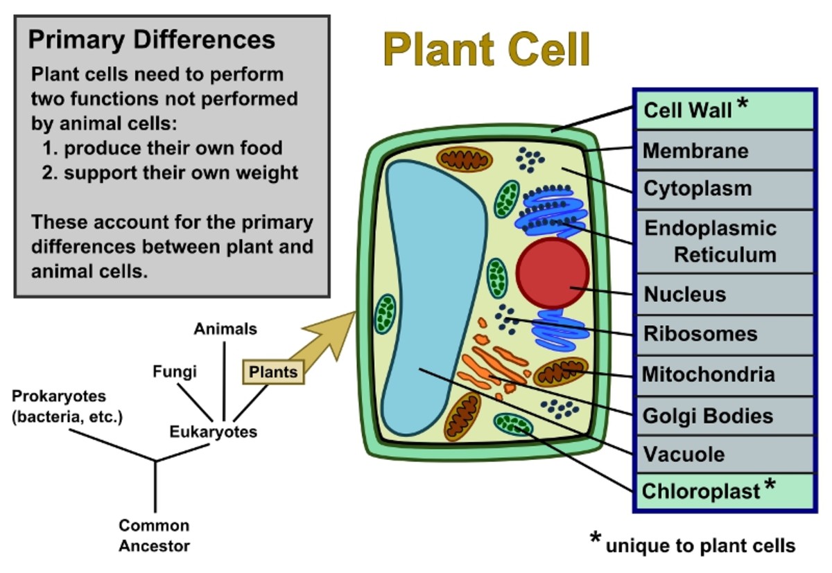
1 2 Difference Between Plant And Animal Cells Cells As The Basic Units Of Life Siyavula
/understanding-white-blood-cells-and-counts-2249217_final-0587074073ce4709b41e42a08db0f596.gif)
Types And Function Of White Blood Cells Wbcs
Cell Diagram To Label Pensandpieces
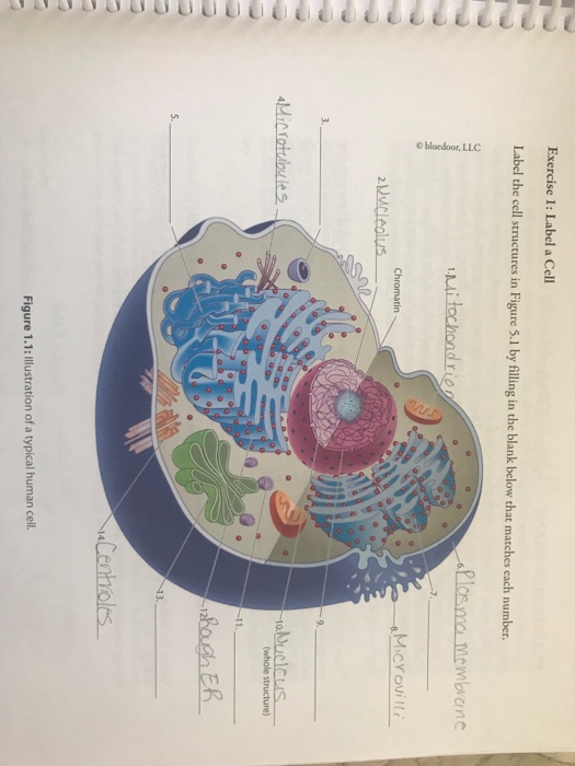
Solved Exercise 1 Label A Cell Label The Cell Structures In Chegg Com
2

31 Label Cell Parts Quiz Labels Database 2020
Types Of Plant Cell Definition Structure Functions Diagrams

Plant Cell Definition And Examples Biology Online Dictionary
Nervous System Structure Function And Diagram Kenhub

Eukaryotic Cells Definition Parts Examples And Structure

Lysosome Cell Organelle Vector Illustration Labeled Cross Section Diagram Stock Illustration Download Image Now Istock
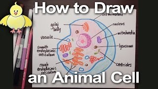
How To Draw An Animal Cell Diagram Homework Help Doodledrawart Youtube

Lab Manual Exercise 1a

3 5 The Cell Exercises Biology Libretexts
Anatomy And Physiology Parts Of A Human Cell
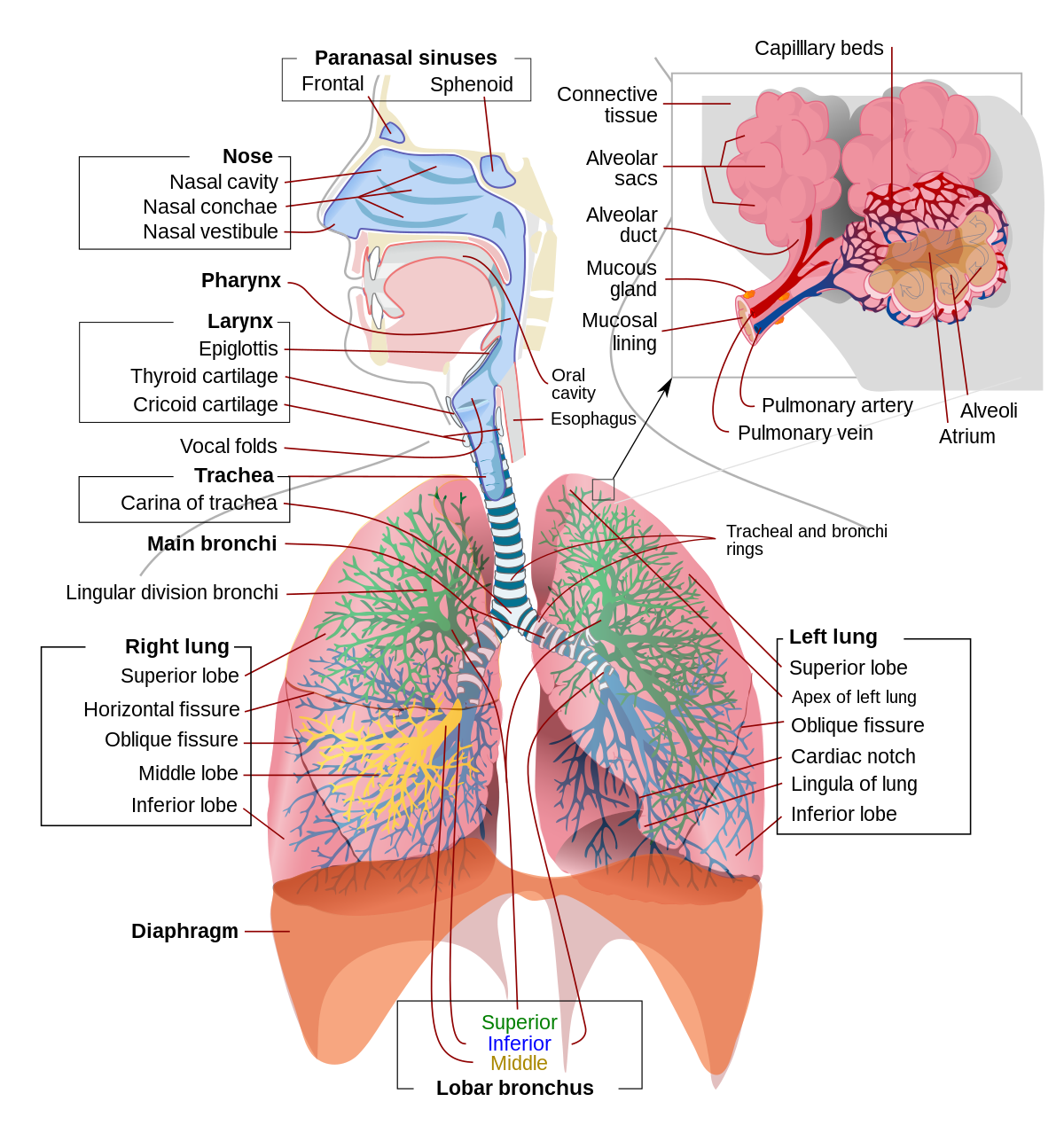
Respiratory System Wikipedia

Question 1 Labeling The Parts Of A Cell On A Diagram Is An Example Of An Exercise In Course Hero
Plant Cells Vs Animal Cells With Diagrams Owlcation

Cell Organelles And Function With Labels Flashcards Quizlet

Cell Structure Organisation Cie Igcse Biology Revision Notes

Question 1 Labeling The Parts Of A Cell On A Diagram Is An Example Of An Exercise In Course Hero
The Cell Types Functions And Organelles

Label Parts Of The Cell Brainly Com


Comments
Post a Comment