42 stingray anatomy diagram
Learn the ventricles of the brain along with their definition, function, location, anatomy, and cerebrospinal fluid (CSF) flow using labeled diagrams. The ventricular system contains the lateral, third, and fourth ventricles whose function is to produce cerebrospinal fluid. Learn where CSF is found, Florida Museum of Natural History Gainesville, FL 32611 352-392-1721 (Research) or 352-846-2000 (Exhibits)
Internal anatomy. The internal skeleton of skates and rays (or endoskeleton) - as with sharks - lacks true bone, and is instead made entirely of cartilage. Cartilage is a strong and durable material that is lighter and more flexible than bone, enabling elasmobranchs (which lack a swim bladder) to stay afloat and turn in a tighter radius than ...

Stingray anatomy diagram
Part 4: Digestive Anatomy A. Place your shark ventral side up on the dissection tray. • Using scissor make a mid-ventral incision just anterior to the cloacal opening. Cut in an anterior direction slightly to the right of the mid-ventral line. Cut all the way to the pectoral girdle. The inside of the large cavity will be exposed. After cleaning the fabric, allow it to air dry completely and then apply 303 in a thin, even coat. After allowing the first coat of 303 to air dry, apply a second thin, even coating of 303. Two light coatings are more effective in restoring fabric water resistance than a single heavy coating. Internal Anatomy: The internal organs of the fish perform the basic function of the body such as respiration, digestion, and sensory function. The brain, stomach, liver, and kidneys are same as in man for the fish and perform the same function. Some organs are different; man has lungs to breathe whereas fish has gills for the same purpose.
Stingray anatomy diagram. PDF | On Jun 1, 2017, Erin Culpepper and others published Stingray Anatomy and Ultrasound: Supplement to Chapter 30 Diagnostic Imaging | Find, read and cite all the research you need on ResearchGate Diagram Of A Whiptail Stingray By Andy Murch Stingray Anatomy Baby Stingray . A stingray can live between 15-25 years in the wild Fact 2. Life cycle of a stingray. The life cycle of a stingray is similar to the life cycles of most other living organisms. It is born usually as part of a litter ranging from five to ten. Download scientific diagram | The collected species of freshwater stingrays from the Potamotrygonidae family: Paratrygon aiereba [A], Potamotrygon orbignyi [B], P. scobina [C], P. motoro [D], P ... Stingray and Human Interaction. Stingrays very rarely attack humans, and the vast majority of injuries occur as self-defense. Especially with bottom-dwelling species, most stings occur when the fish is stepped on. In areas with high populations of stingrays it is advised to shuffle your feet while wading in the water to avoid stepping on the ...
Mar 20, 2012 - Diagram of a Whiptail Stingray by Andy Murch. Manta ray. Manta rays are large eagle rays belonging to the genus Manta. The animals called vertebrates get their name from vertebrae, the series of bones that make up the backbone. One lineage of vertebrates colonized land 365 million years ago. There are about 52,000 species of vertebrates, including the largest organisms ever to live on the Earth. Vertebrates have great disparity, a wide range of differences ... Stingray, any of a number of flat-bodied rays noted for the long, sharp spines on their tails. Stingrays are disk-shaped and have flexible, tapering tails armed, in most species, with one or more saw-edged, venomous spines. They inhabit warm temperate and tropical waters, sometimes in great abundance. Anatomy - Stingrays. Stingrays have an anatomy composed of a flattened body and on their body consists of pectoral fins joined to their head and trunk with their tail behind. A stingrays eyes are on the dorsal side of the body ( top/upper side). On the underside of the stingray lies the mouth, nostrils, and gill slits.
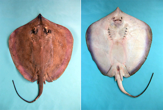
Internal Anatomy . Heart. This organ pumps blood throughout the body delivering oxygen and digested nutrients to the cells of various organs. It transports waste products from the cells to the kidneys and liver for elimination. In fish, the circulatory system is a single circuit, with a 2-chambered heart, unlike Stingrays are commonly found in the shallow coastal waters of temperate seas. They spend the majority of their time inactive, partially buried in sand, often moving only with the sway of the tide. The Digestive System: Anatomy Review 1. List two main divisions of the digestive system. a. Gastrointestinal tract b. Accessory digestive 2. The four main layers of the digestive tract wall are a. Mucosa b. Submucosa c. Muscularis Externa d. Serosa 3. Label the diagram below with the four main layers you listed in question 6. 4. External anatomy of a male stingray. Stingray jaw and teeth. The teeth are modified placoid scales. The stingray breathes though spiracles just behind the eyes when it hunts in seafloor sediment. Jaw and teeth. The mouth of the stingray is located on the ventral side of the vertebrate.
Structure And Mechanical Implications Of The Pectoral Fin Skeleton In The Longnose Skate Chondrichthyes Batoidea Sciencedirect
On the sides of the body the pectoral, large and triangular shaped fins are located. When the manta ray swims it often flaps its wings up-down rather than swelling them like rays living on the ocean floor, this makes them seem as if they were flying.

Skates Rays Of The Jersey Shore Save Coastal Wildlife
Diagram Of Male Groin Area / Low Back Pain - Disc Herniation ,Sciatica - Everything You - First, a lesson in the male pelvic anatomy.. The groin is the area in the body where the upper thighs meet the lowest part of the abdomen. The pelvis forms the base of the spine as well as the socket of the hip joint.
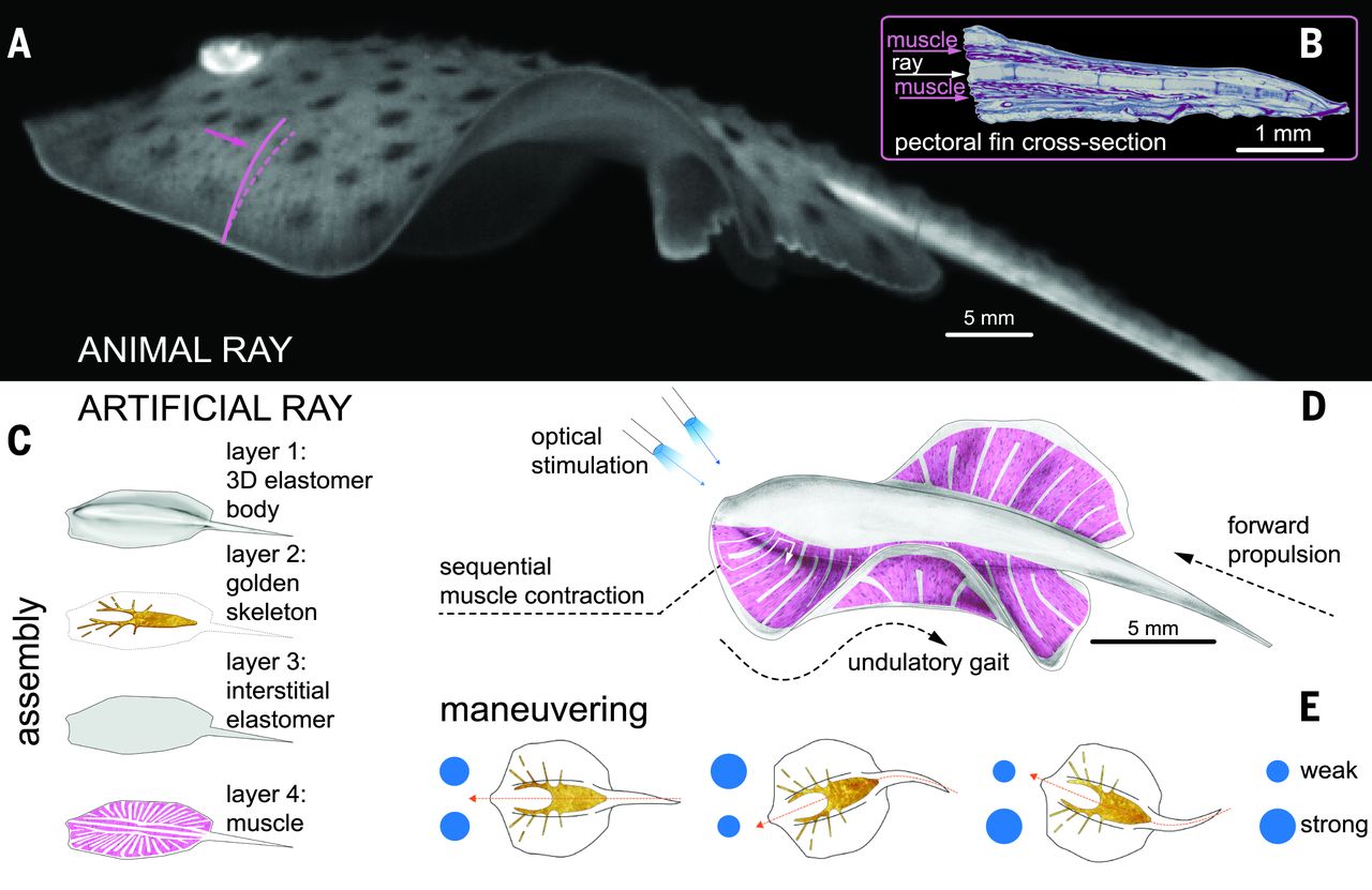
This Artificial Stingray Has A Gold Skeleton And Light Activated Rat Muscles Techcrunch
Fish are boney, and ornamental pond fish are no exception, as you'll see in the pond fish anatomy diagram. Unlike the bones of sharks and stingrays, their bones are truly bones and not cartilage. The bones of a fish are not meant for bearing weight because, in water, the fish is pretty much weightless.
Stingray Facts And Beyond Biology Dictionary
Stingrays were collected year round to encompass the range of gestational stages, though the two main breeding seasons occur roughly from late February to early August and late August through January (Fahy et al., 2007). The stingrays were temporarily maintained in a holding pen located in the Nova Southeastern University Oceanographic Center ...

Njdep Division Of Fish Wildlife Skates And Stingrays Of New Jersey
Stingrays, with their wide, flat bodies, may not look like fish, but they are. They are related to sharks, and like their shark cousins, they do not have bones. Instead, their bodies are supported by cartilage—the same material that you feel inside the tip of your nose. Stingrays have broad fins that run the full length of their bodies, giving them a flat, roundish shape. To swim, some ...

Taeniura Lymma Blue Spotted Fantail Ray Taxo4254 Wiki Nus
Manta rays (Manta birostris) are the largest rays and are closely related to sharks. These harmless rays have a short tail and no stinging spine. They are very acrobatic; they can even leap from the water.

Njdep Division Of Fish Wildlife Skates And Stingrays Of New Jersey
Stingray print Fish art print Vintage sea art print old prints Natural History Ocean Decor Antique prints Nature print Victorian art This gorgeous illustration is from a series of hand colored lithographs. It comes from the Belgium book published in the 1800's. This print is digitally enhanced with some odd blemishes left to enhance it's ...
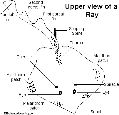
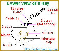
Rays Enchanted Learning Software
The stingrays are part of a unique group of fishes known as "batoids" and are closely related to sharks. A stingray's body is made of cartilage like a shark's body so sometimes they are called "flat sharks"! For more information check out the Chondrichthyan Tree of Life. Most batoids spend their time resting on the seafloor with ...

Diagram Of A Whiptail Stingray By Andy Murch Stingray Anatomy Lapbook
108 Stingray Internal Anatomy clipart free images in AI, SVG, EPS or CDR. Save 15% on iStock using the promo code. CLIPARTLOGO15 apply promocode. Beautiful Go Media Freebies Pack. Vitruvian man vector. Brain. Science Diagram Female Human Cartoon Diagrams Body Medicine Artfavor Medical Organs Internal Location Anatomy Organ Labeled. Anatomy of Eye.
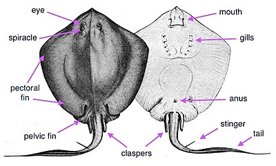
External Anatomy
a hard, pointed piece coming from a plant or an animal's body. not safe. stingray. a large venomous ray with large barbed spines near the base of…. unusual. not common, rare; different from others. 15 terms. AmandaQuerry. Stingray.
2
Ray's Anatomy. Ray's Anatomy. Stingrays are members of the group of fish that also includes sharks and skates. Sharks and rays have skeletons made of flexible cartilage instead of bone. What makes rays unusual is that their wing-like fins stretch out flat from their bodies, making them look like a disc with a tail. ...

Anatomy Of A Stingray
Internal Anatomy: The internal organs of the fish perform the basic function of the body such as respiration, digestion, and sensory function. The brain, stomach, liver, and kidneys are same as in man for the fish and perform the same function. Some organs are different; man has lungs to breathe whereas fish has gills for the same purpose.
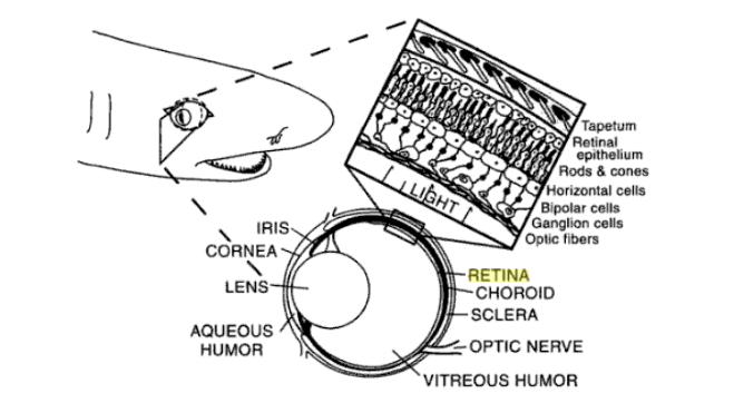
Sensory Systems Vision
After cleaning the fabric, allow it to air dry completely and then apply 303 in a thin, even coat. After allowing the first coat of 303 to air dry, apply a second thin, even coating of 303. Two light coatings are more effective in restoring fabric water resistance than a single heavy coating.
Stingray Wikipedia
Part 4: Digestive Anatomy A. Place your shark ventral side up on the dissection tray. • Using scissor make a mid-ventral incision just anterior to the cloacal opening. Cut in an anterior direction slightly to the right of the mid-ventral line. Cut all the way to the pectoral girdle. The inside of the large cavity will be exposed.
2

Manta Rays Of Penida

Shark And Ray Anatomy Sharklab Malta
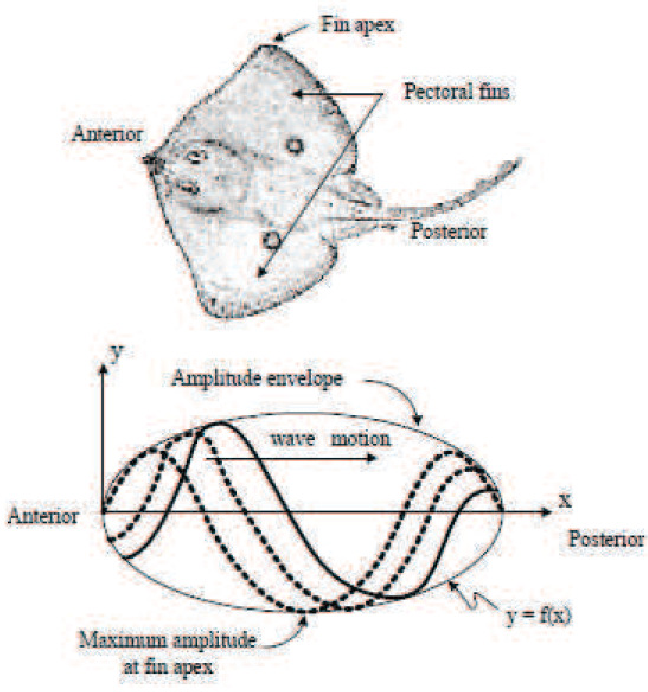
Figure 1 From Design Of An Angular Radial Robotic Stingray Semantic Scholar

Stingray Internal Anatomy Clipart Free Download
Stingray Wikipedia

Shark And Relatives Week S The Library Part Ii Free For All

The Anatomy Of Manta Rays Manta Ray Advocates Hawaii

Materials
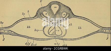
Quain S Elements Of Anatomy T Inflected Into Theouter Wall Of The Heart Alih Imi Inner Or Endovascular Lining Of The Heart Jpa Pericardialcavity Nvis Mesoblast Beyond The Rudiments
1
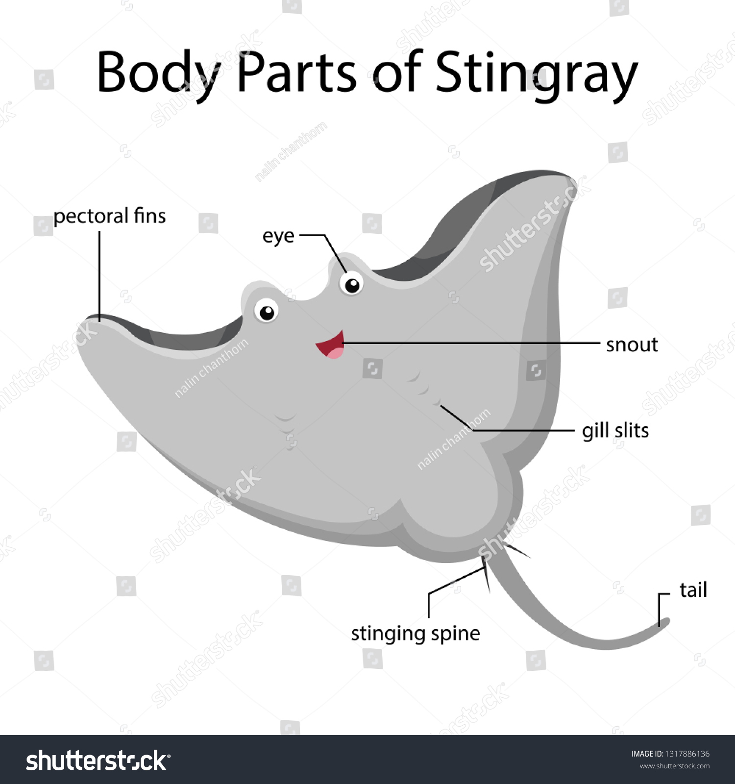
Illustrator Body Parts Stingray Stock Vector Royalty Free 1317886136

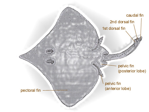
2 Basic Internal Anatomy Of The Brown Stingray Dasyatis Lata Showing Download Scientific Diagram
Shutterstock Puzzlepix
Dasyatis Margarita Discover Fishes

2 Basic Internal Anatomy Of The Brown Stingray Dasyatis Lata Showing Download Scientific Diagram

Antique Illustration Of Human Body Anatomy Bones Skull Zygomatic Bone Cheekbone Malar Bone Stock Illustration Download Image Now Istock
External Anatomy

External Anatomy

Chondrichthyes Electric Ray Torpedo Eod Electric Organ Discharge
2
Rays Enchanted Learning Software

Behavioral Responses Of Batoid Elasmobranchs To Prey Simulating Electric Fields Are Correlated To Peripheral Sensory Morphology And Ecology Sciencedirect

Taeniura Lymma Blue Spotted Fantail Ray Taxo4254 Wiki Nus
2

Skate Fish Wikipedia

2 Basic Internal Anatomy Of The Brown Stingray Dasyatis Lata Showing Download Scientific Diagram

Reproductive Biology Of The Cownose Ray In The Charlotte Harbor Estuarine System Florida


Comments
Post a Comment