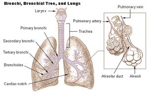
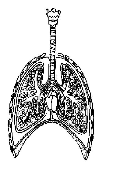
43 lungs diagram with labels
In the diagram to the left, provide the labels for the structures involved in the reflex act when a person steps on a tack and jerks their leg away. Brain Anatomy Provide the labels for the diagram on the left below and provide descriptions of the functions of each structure on the blank lines. The diagram of heart is beneficial for Class 10 and 12 and is frequently asked in the examinations. A detailed explanation of the heart along with a well-labelled diagram is given for reference. Well-Labelled Diagram of Heart. The heart is made up of two chambers: The upper two chambers of the heart are called auricles.
Advantages. We can combine values in the cells with the diagram to add labels for the dimensions. Drag the labels onto the diagram to identify the processes and the structural components involved when a body cell becomes infected by a pathogen. Drag the labels onto the diagram to identify the stages of the cell cycle. Superior vena cava 6.

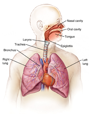
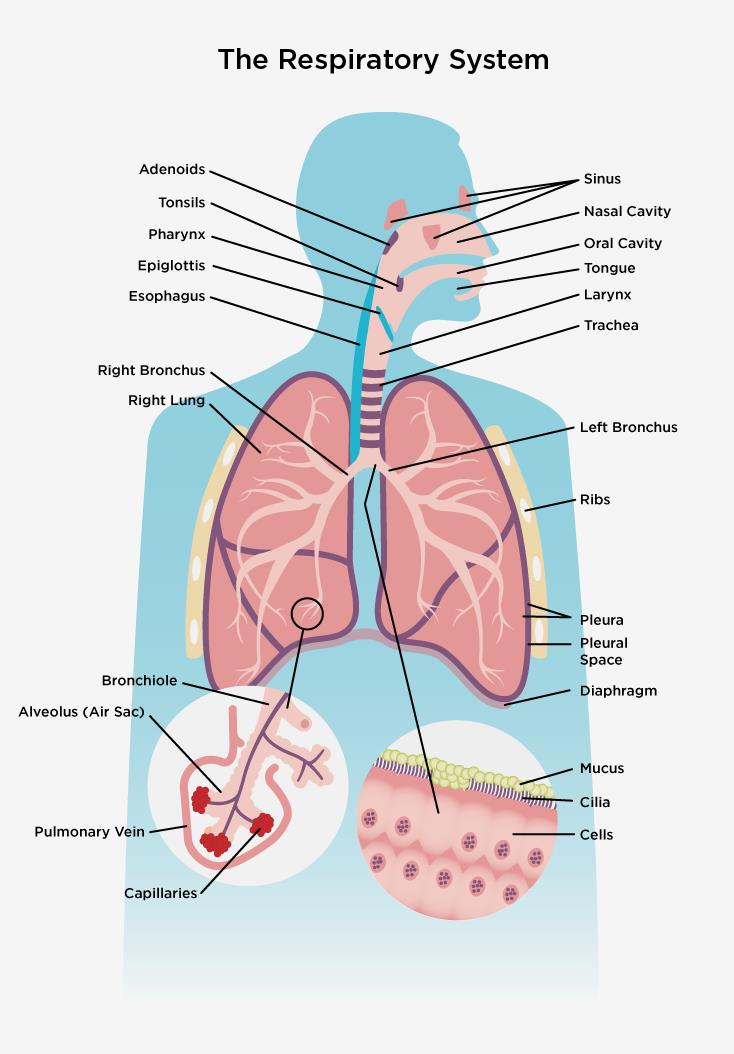
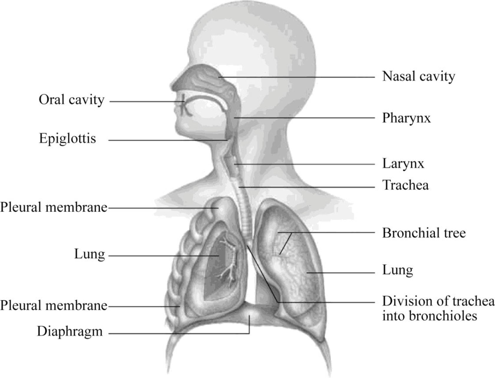
Lungs diagram with labels
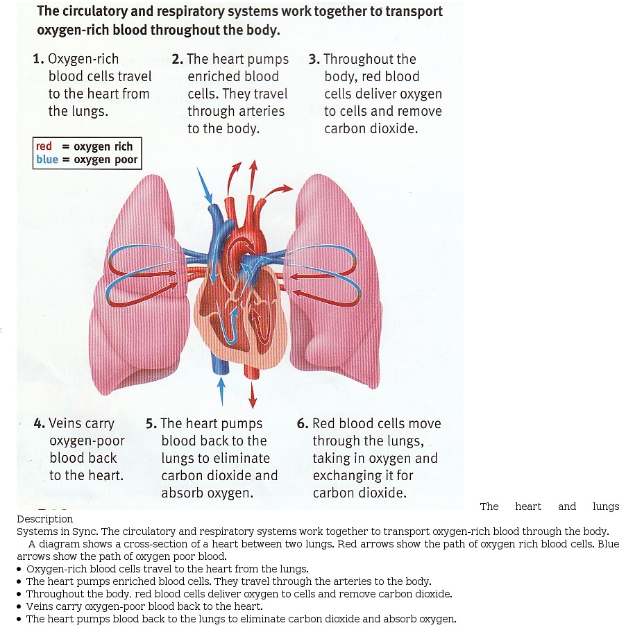
Jun 16, 2017 · Labels. Description. Vena cava. Carries deoxygenated blood from the body to the heart. Semilunar valve. Flaps that prevent backflow of blood. Left atrium. Receives oxygenated blood from the lungs. Left ventricle. Region of the heart that pumps oxygenated blood to the body. Pulmonary artery. Carries deoxygenated blood to the lungs. Right ventricle Lungs Diagram Diagram - Lungs Diagram Chart - Human anatomy diagrams and charts explained. This diagram depicts Lungs Diagram with parts and labels. Posted in Diagrams | Tagged human lungs , lungs , lungs chart , lungs diagram , lungs explained The heart is in the middle of the chest. It fits snugly between the two lungs. It is held in place by the blood vessels that carry the blood to and from its chambers. The heart is tipped somewhat so that there is a little more of it on the left side than on the right. The pointed tip at the bottom of the heart touches the front wall of the chest.
Lungs diagram with labels. They may come with or without labels. Common circulatory system diagrams show pulmonary circulation, coronary circulation, systematic circulation, veins, arteries, or a combination. The systemic circulation system is the most commonly illustrated of the systems that make up the circulatory system as it is the largest. The heart is in the middle of the chest. It fits snugly between the two lungs. It is held in place by the blood vessels that carry the blood to and from its chambers. The heart is tipped somewhat so that there is a little more of it on the left side than on the right. The pointed tip at the bottom of the heart touches the front wall of the chest. Lungs Diagram Diagram - Lungs Diagram Chart - Human anatomy diagrams and charts explained. This diagram depicts Lungs Diagram with parts and labels. Posted in Diagrams | Tagged human lungs , lungs , lungs chart , lungs diagram , lungs explained Jun 16, 2017 · Labels. Description. Vena cava. Carries deoxygenated blood from the body to the heart. Semilunar valve. Flaps that prevent backflow of blood. Left atrium. Receives oxygenated blood from the lungs. Left ventricle. Region of the heart that pumps oxygenated blood to the body. Pulmonary artery. Carries deoxygenated blood to the lungs. Right ventricle
Organs And Structures Of The Respiratory System Anatomy And Physiology Ii

Best Of Lungs Diagram With Labels Free Watch Download Todaypk

Label The Lungs 2 Diagram Quizlet

A Schematic Drawing Of The Lungs And Airway Tree In Which Several Download Scientific Diagram

Task 10 Label The Chest Lungs Yr7 Diagram Quizlet

Labeled Diagram Of The Human Lungs Human Lungs Asthma Cure Human Muscle Anatomy

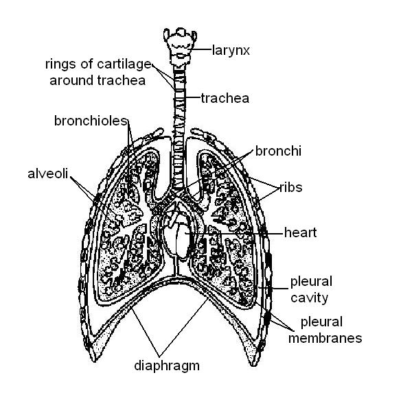
The Respiratory System Label Diagram

The Respiratory System Diagram Structure Function
Anatomy Of The Respiratory System

How To Draw Lungs Diagram Science Drawing Biology Drawing Biology Diagrams
Respiratory System The Lung Association
Sample 1 Heart And Lung Diagram Accessible Image Sample Book
3

Lesson Worksheet Introduction To The Respiratory System Nagwa
Lungs Label Teaching Resources

Diagram Lungs Stock Illustrations 2 192 Diagram Lungs Stock Illustrations Vectors Clipart Dreamstime

How To Easily Draw Respiratory System With Labeling For Students Youtube
1

Labelled Respiratory System Images Stock Photos Vectors Shutterstock
To Label The Parts Of The Respiratory System And The Structure That Surrounds The Parts Of Respiratory System Introduction The Process Of Gas Exchange That Occurs Between An Organism And Its Environment
How The Lungs And Respiratory System Work With Diagrams Macmillan Cancer Support
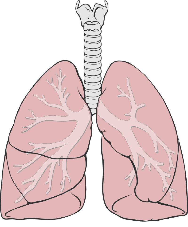
File Lungs Diagram Simple Svg Wikimedia Commons
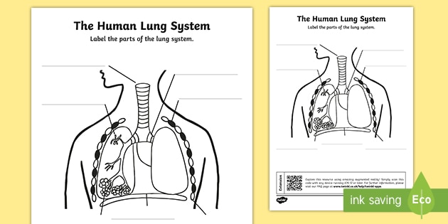
Lung Diagram Labelling Activity Primary Resources Twinkl
Lungs Labeled Diagram Stock Vector Image Art Alamy

Respiratory System Labeling Worksheet
The Anatomy And Physiology Of Animals Respiratory System Worksheet Worksheet Answers Wikieducator
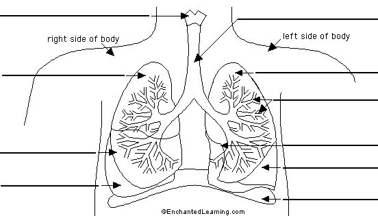
Label Lungs Diagram Printout Enchantedlearning Com
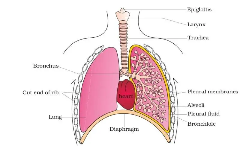
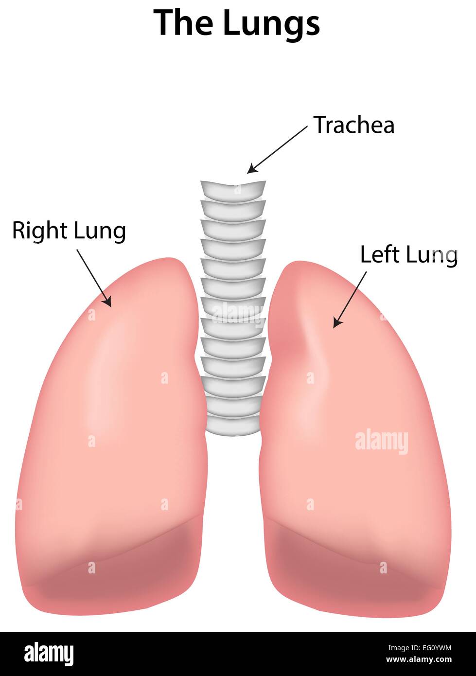
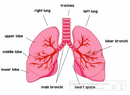
Diagram Of The Lungs
How The Lungs And Respiratory System Work With Diagrams Macmillan Cancer Support
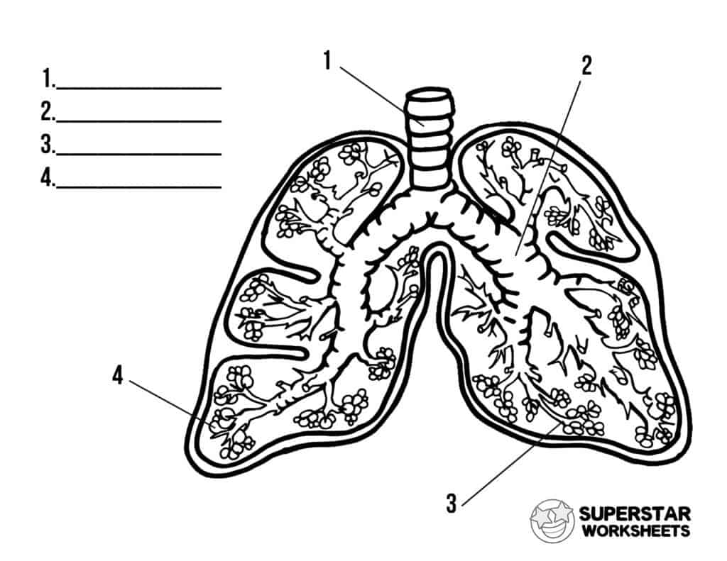
Human Lungs Worksheets Superstar Worksheets

Lungs Diagram Anatomy System Human Body Anatomy Diagram And Chart Images

The Lungs Anatomy And Physiology Ii
Lung Definition Function Facts Britannica

Lung Structure Bioninja
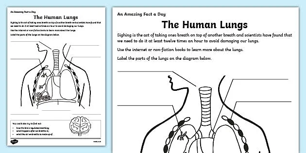
The Human Lungs Ks2 Worksheet Primary Resource
Anatomy Clipart Anatomy Lungs Labeled Clipart Classroom Clipart

The Respiratory System Lung Function And Chest Anatomy Patient
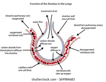
Fully Labelled Diagram Alveolus Lungs Showing Stock Vector Royalty Free 369984683

Overview Of The Respiratory System Lung And Airway Disorders Merck Manuals Consumer Version
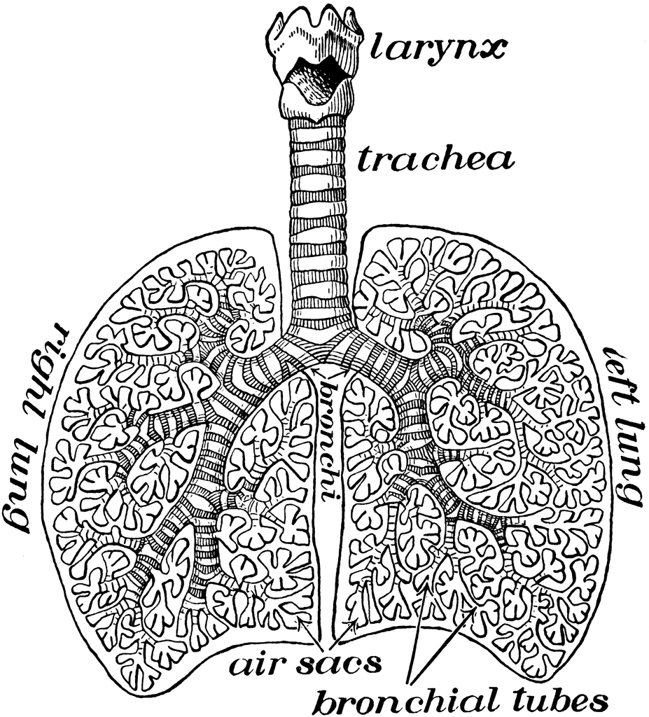
Diagram Of Air Tubes In The Lungs Clipart Etc

Human Respiratory System Description Parts Function Facts Britannica
Lung Anatomy Physiopedia
The Anatomy And Physiology Of Animals Respiratory System Worksheet Wikieducator


Comments
Post a Comment