38 onion cell diagram
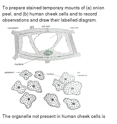
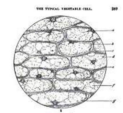
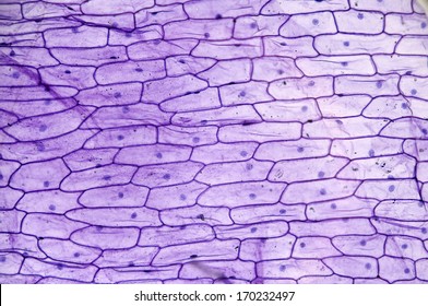
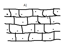
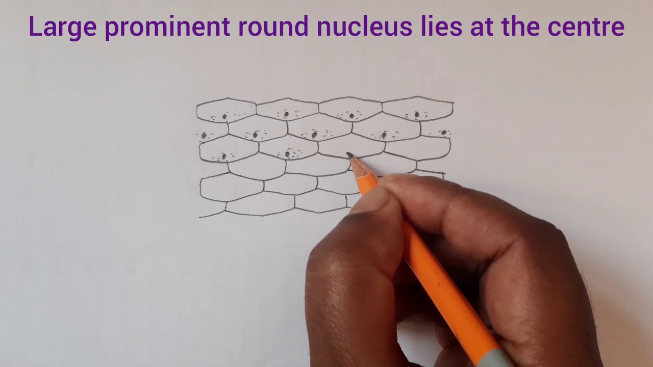
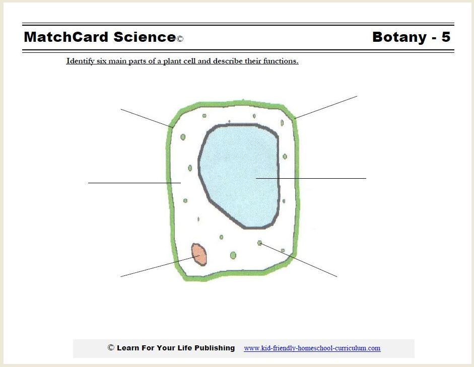
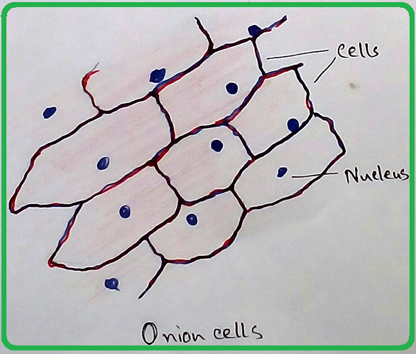
Diagram below shows the onion cell when viewed under a microscope. 3. Fig 1.1.3.jpg (353 KB) Fig 1.1.3: Onion epidermal cells stained with iodine seen under ... An onion is a multicellular (consisting of many cells) plant organism.As in all plant cells, the cell of an onion peel consists of a cell wall, cell membrane, ...
Observing onion cells under the microscope. For this microscope experiment, the thin membrane will be used to observe the cells.

Onion cell diagram
5 steps1.Peel the brown skin away from the outside of the onion. Take one layer of the onion flesh and carefully cut out a piece.2.On the inside of this piece is a thin sheet of the membrane. Use tweezers or dissection needle to peel the membrane away.3.Place the specimen in a small dish of stain (Eosin Y) and leave it for about 3 minutes. Find the perfect onion cells stock photo. Huge collection, amazing choice, 100+ million high quality, affordable RF and RM images. No need to register, ... Onion peel is an example of a plant cell whereas a human cheek cell is an example of an animal cell. Complete answer: The plant cell usually consists of an ...
Onion cell diagram. Create your own Onion Cells Drawing Diagram Biology Beyond 1 themed poster, display banner, bunting, display lettering, labels, Tolsby frame, story board, ... Find onion cell stock images in HD and millions of other royalty-free stock photos, illustrations and vectors in the Shutterstock collection. Onion peel is an example of a plant cell whereas a human cheek cell is an example of an animal cell. Complete answer: The plant cell usually consists of an ... Find the perfect onion cells stock photo. Huge collection, amazing choice, 100+ million high quality, affordable RF and RM images. No need to register, ...
5 steps1.Peel the brown skin away from the outside of the onion. Take one layer of the onion flesh and carefully cut out a piece.2.On the inside of this piece is a thin sheet of the membrane. Use tweezers or dissection needle to peel the membrane away.3.Place the specimen in a small dish of stain (Eosin Y) and leave it for about 3 minutes.
Microscopy How A Microscope Works Magnification Calculations How To Use A Microscope Slide Preparation Investigations Resolution Resolving Power Measuring Size Of Cell Electron Microscope Micrograph Light Micrograph Igcse O Level Gcse 9 1 Biology Revision

Onion Cell Diagram Diagram Quizlet
A Scientist Is Observing Onion Cells And Human Cheek Cells Under
Mic Uk

Onion Cell Labelling Diagram Quizlet

Artificial Muscles Created From Gold Plated Onion Cells
Onion Cell Images Stock Photos Vectors Shutterstock
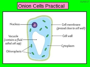
Grade 6 Looking At Onion Cells Practical By Active Science Tpt
What Are The Similarities Between The Cheek And Onion Cells Quora
1
Cells Part 2 Structure And Function Moss Cells Blood Cell Cheek Cells Onion Cells Ppt Download
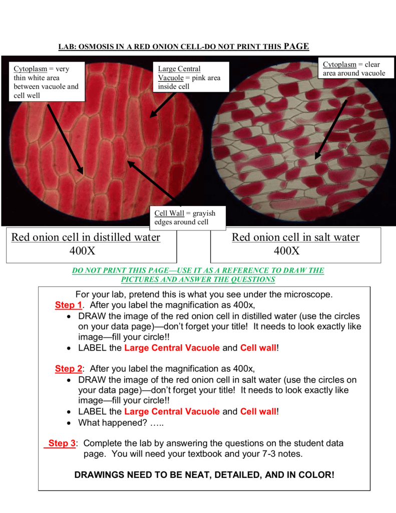
Red Onion Cell In Distilled Water 400x Red Onion Cell In Salt Water

Science 8 Onion Lab The Blog Site Of Carl Sommerfeld
1
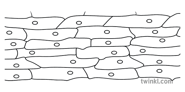
Onion Cells Drawing Diagram Biology Beyond 1 Illustration Twinkl
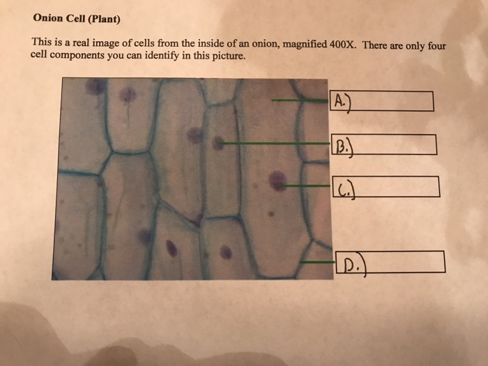
Solved Onion Cell Plant This Is A Real Image Of Cells From Chegg Com

Thethinkinglab Experiment Onion And Cheek Cells
The Following Diagram Shows Cells Of Onion Peel Label Class 11 Biology Cbse

Ruzivo Digital Learning

Stages Of Onion Mitotic Cell Division A Interphase B Prophase C Download Scientific Diagram
How To Draw Onion Cell Easy Way Step By Step Youtube
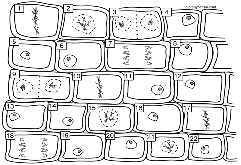
Onion Mitosis
Solved Assignment 1 Diagram Three Adjacent Epidermal Cells As Observed On Your Slide And Label The Parts You See This Question Was Created From L Course Hero
1
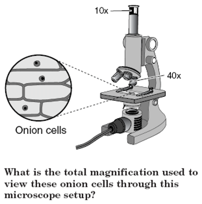
Ppt What Is The Total Magnification Used To View These Onion Cells Through This Microscope Setup Powerpoint Presentation Free To Download Id 557e41 Ztzhn

Lab Observing Cells Michael Eng Michael S Blog

Onion Epidermal Cell Wikipedia

Gcse Biology Microscope Drawing And Measuring Cell Size Edexcel 9 1 Youtube

Learning By Questions
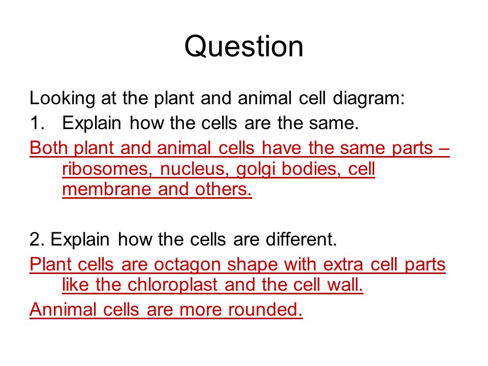
Tabulate Differences Between Plant And Animal Cell With Diagram Of Onion Peel Cells And Cheek Cells Sarthaks Econnect Largest Online Education Community

Microscope And The Cell

Onion Blood Cells Lab Ppt Video Online Download
Plant Cell Diagram
Human Cheek Cells Under The Microscope Haematoxylin Cell Membrane

Cellular Localization Of Scork11 A Epidermal Onion Cells Expressing Download Scientific Diagram

Biology Help Online Learning On Onion Cells

Cell Cycle Stages In An Onion Root Tip Diagram Quizlet
What Is The Shape Of An Onion Cell Why Quora


Comments
Post a Comment