40 relaxed sarcomere diagram
About Press Copyright Contact us Creators Advertise Developers Terms Privacy Policy & Safety How YouTube works Test new features Press Copyright Contact us Creators ... Skeletal muscle is the muscle type that initiates all of our voluntary movement. Label the parts of the sarcomere. Draw and label a diagram to show the structure of a sarcomere including z lines actin filaments myosin filaments with heads and the resultant light and dark bands. Draw your own diagram of two sarcomeres.
towards the centre of the sarcomere, like the stroking of a boat oar. This is called a POWER STROKE. 6. This power stroke pulls the thin filament inward only a small distance. 7. Once the head tilts, this allows release of ADP & phosphate ions. 8. At the site of release of ADP, a new ATP binds. This binding causes the detachment of the myosin head
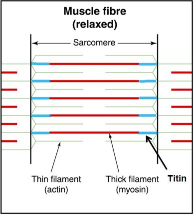
Relaxed sarcomere diagram
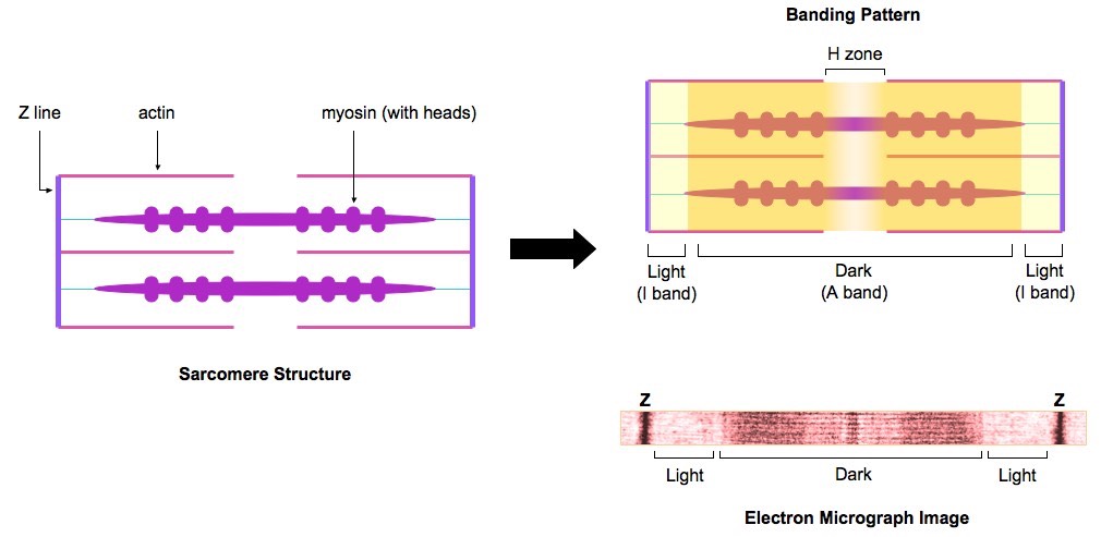
Sarcomere diagram to label. This is a quiz called label the sarcomere and was created by member deanne1480. Biology computers geography history languages math. Sarcomere of skeletal muscle mammalian. ... The first should be of a relaxed muscle. Draw your own diagram of two sarcomeres. Longitudinal tubules give rise to terminal cisternae closely ... Sarcomere H zone Thin (actin) filament Thick (myosin) filament Z disc Z disc M line (c) Small part of one myofibril enlarged to show the myofilaments responsible for the banding pattern. Each sarcomere extends from one Z disc to the next. Draw and label a diagram to show the structure of a sarcomere, including Z lines, actin filaments, myosin filaments with heads, and the resultant light and dark bands. 7. Explain how skeletal muscle contracts, including the release of calcium ions from the sacroplasmic reticulum, the formation of cross-bridges, the sliding of actin and myosin ...
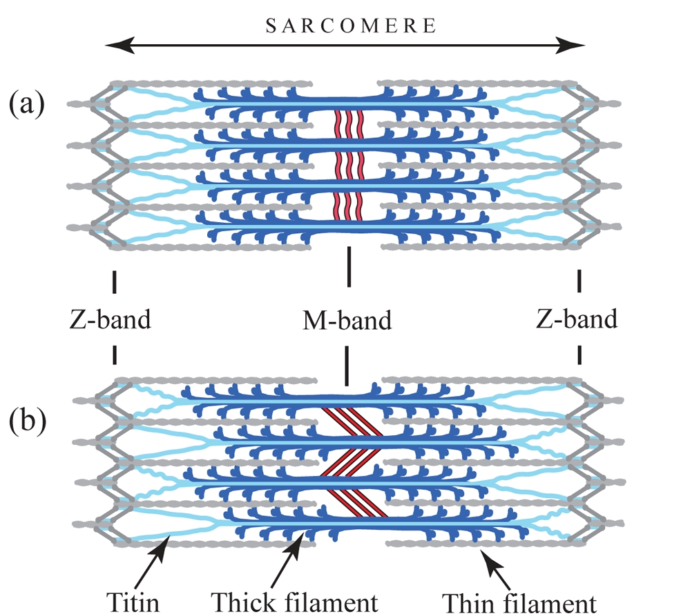
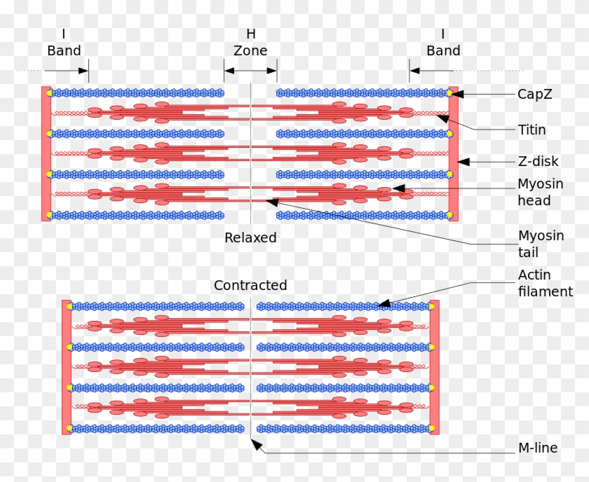
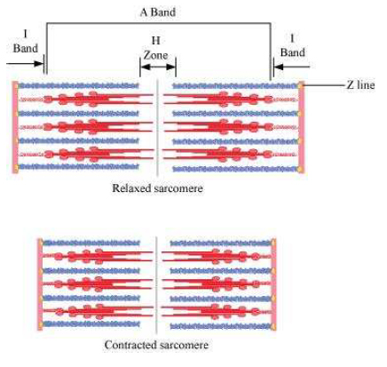
Relaxed sarcomere diagram. Each sarcomere divides into different lines, bands, and zone: "I" and "A" bands, "M" and "Z" lines, and the "H" zone. - Z-lines define the boundaries of each sarcomere. - The M-line runs down the center of the sarcomere, through the middle of the myosin filaments. - The I-band is the region containing only thin filaments. locations within a sarcomere. QUESTIONS: 10. Label the thick and thin filaments in Figs. A, B, and C above. 11. There are three sarcomeres shown in the diagram below. Sarcomere 1 Sarcomere 2 Sarcomere 3 a) In Sarcomere 1, identify the location within the sarcomere of the cross section indicated by Figure A in Model 3. The thin filament in a sarcomere is composed of actin, troponin, and tropomyosin. Troponin and tropomyosin are attached to one another, both overlaying actin. When a muscle is relaxed, tropomyosin blocks actin's myosin-binding sites. Calcium binds to troponin, initiating a shape change that removes the blocking action of tropomyosin. (b) Schematic diagram of a cardiac sarcomere. The sarcomere is the fundamental unit of contraction and is defined as the region between two Z-lines. Each sarcomere consists of a central A-band (thick filaments) and two halves of the I-band (thin filaments). The I-band from two adjacent sarcomeres meets at the Z-line.
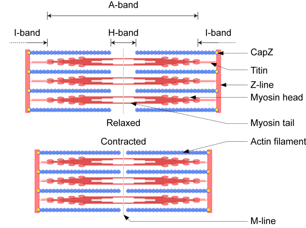
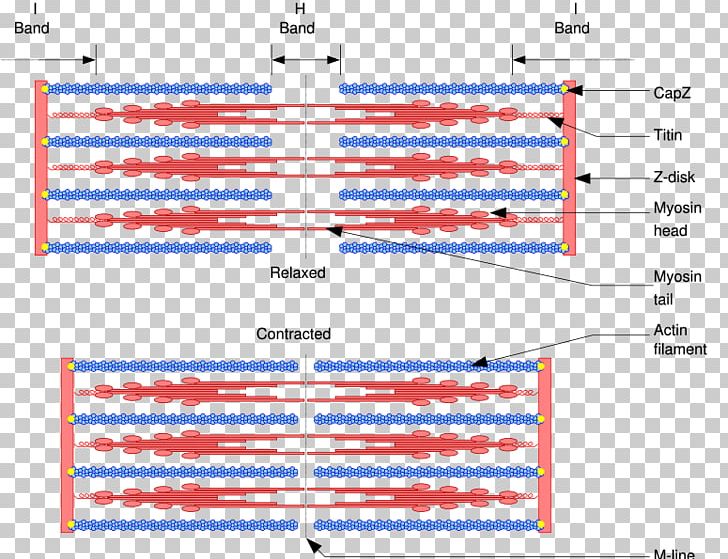
Sarcomere Structure. To understand the sliding filament model requires an understanding of sarcomere structure. A sarcomere is defined as the segment between two neighbouring, parallel Z-lines. Z lines are composed of a mixture of actin myofilaments and molecules of the highly elastic protein titin crosslinked by alpha-actinin. (b) Schematic diagram of a cardiac sarcomere. The sarcomere is the fundamental unit of contraction and is defined as the region between two Z-lines. Each sarcomere consists of a central A-band (thick filaments) and two halves of the I-band (thin filaments). The I-band from two adjacent sarcomeres meets at the Z-line. Start studying Sarcomere. Learn vocabulary, terms, and more with flashcards, games, and other study tools. Sarcomere Diagram. Sarcomere Anatomy: Anatomical is said to be the term of microanatomy. The sarcomere is the basic unit function with muscle fiber cells. ... First, in this theory muscle is relaxed. The actin has to be brought close together for the contraction of the muscle. The myosin has cross bridges to get actin close, which pull them ...
A sarcomere is the functional unit of striated muscle. This means it is the most basic unit that makes up our skeletal muscle. Skeletal muscle is the muscle type that initiates all of our voluntary movement. Herein lies the sarcomere's main purpose. Sarcomeres are able to initiate large, sweeping movement by contracting in unison. *32AB21104* *32AB21104* 10778 2 The diagrams below represent one sarcomere in both relaxed and contracted states. (Scale bars are included.) 2.2 µm Relaxed Contracted (a)State the name of the inorganic ion required for muscle contraction and describe its role. Ion _____ Role _____ Sarcomere coloring worksheet answers key. hinge joint d. 3 - Explain how skeletal muscle contracts by the sliding filament theory. b- is the distance between two Z membranes. Label an A band, an I band, and a sarcomere. com Sarcomere Coloring Key The sarcoplasmic reticulum (E) is a network of tubes that run parallel to the myofilaments. Start studying Relaxed and Fully Contracted Sarcomere. Learn vocabulary, terms, and more with flashcards, games, and other study tools.
Superhelical Architecture Of Myomesin
Sarcomere Diagram Labeled. 16.08.2018 16.08.2018 5 Comments on Sarcomere Diagram Labeled. ... The first should be of a relaxed muscle. The second should be of a contracted muscle. Label the Z line, M line. Start studying UNIT 5: Label the parts of the Sarcomere. Learn vocabulary, terms, and more with flashcards, games, and other study tools.

Sarcomere An Overview Sciencedirect Topics
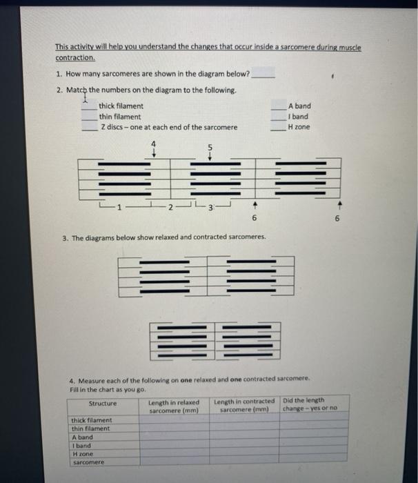
3. Observe the diagram in Model 2 and describe possible reasons why there is a limit to the amount of shortening that can occur in a sarcomere during muscle contraction. Model 3 - Cross Sections Through a Sarcomere The diagrams in Model 3 are cross sections of a sarcomere that show the filaments at various locations within a sarcomere. 1.

Lesson Explainer Muscle Contraction Nagwa
1) Sarcomere is the repeating units between 2 Z lines.In this diagram shows 4 z lines.So this diagram shows 3 sarcomeres. 2) 1)A band 2)I band 3)H zone 4)Thin filament 5)Thick filament 6)Z disc-one at each end of sarcomere 3)First diagram is relaxed… View the full answer
Shutterstock Puzzlepix
The diagrams show a sarcomere in different states of contraction. a. Name the parts labelled P, Q and R. b. Explain why there are no actin-myosin cross-bridges visible in diagram A. c. Muscle fibres are able to contract with more force in some states of contraction than others. Suggest which of the diagrams shows the state that can develop ...

Muscle Contraction And Relaxation Interdigitation Of Thick And Thin Filaments Allows Sarcomere Basic Anatomy And Physiology Physiology Anatomy And Physiology
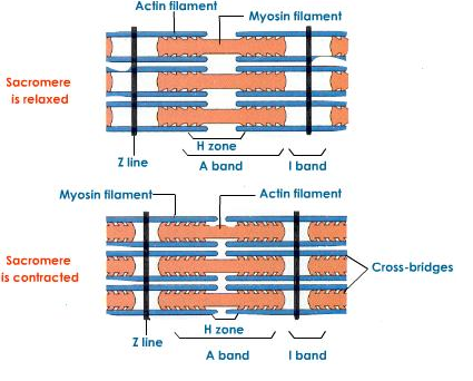
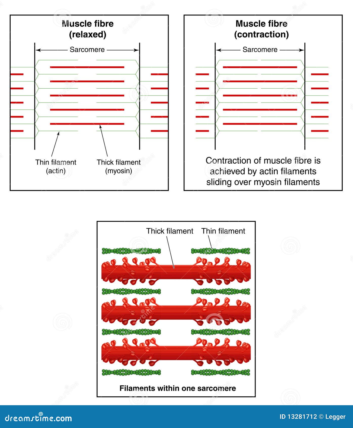
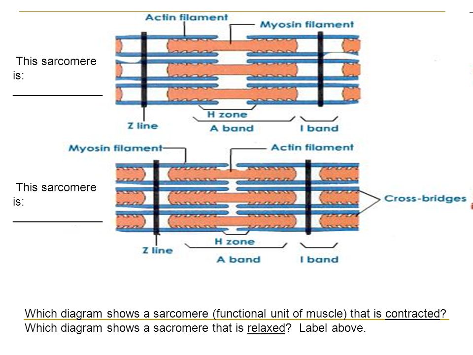
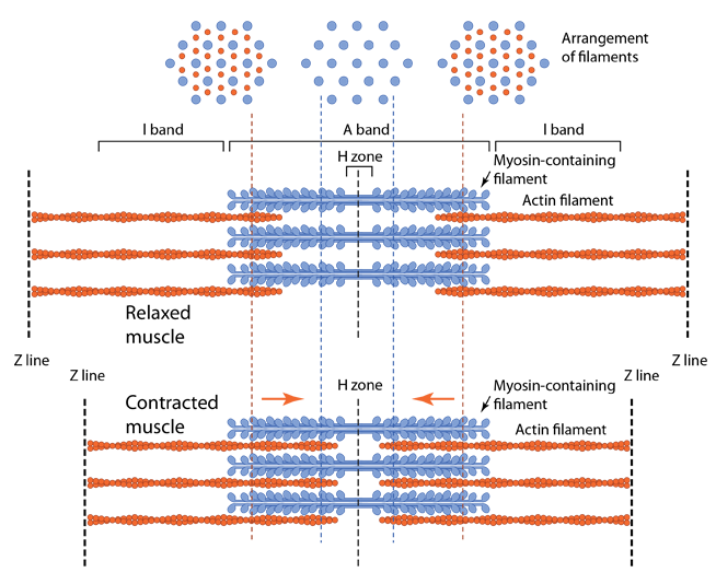
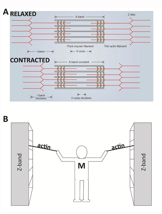
In this diagram, we can see two of the protein filaments that are present in a sarcomere: thick myosin filaments and thinner actin filaments. The diagram shows a sarcomere in a relaxed muscle at the top and in a contracted muscle below.

The Sarcomere And Sliding Filaments In Muscular Contraction Definition And Structures Video Lesson Transcript Study Com
Groups of thick and thin filaments that alternately overlap and move apart are called sarcomeres. The areas between the thick and thin filaments during a relaxed state are called I bands, H zones and A bands. During contraction and relaxation, the length of the filaments remains the same. Myosin heads grasp the thin filaments and either push or ...

Relaxed And Fully Contracted Sarcomere Diagram Quizlet
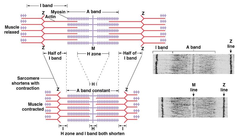
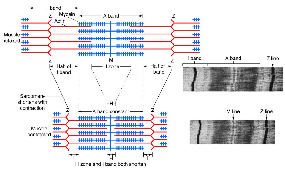
Figure \(\PageIndex{5}\): The top diagram shows a relaxed sarcomere, and the bottom diagram shows a contracted sarcomere. Please note the z discs, h zone, and M line. In a contracted sarcomere the H zone reduces as compared to relaxed sarcomere because actin fibers (greenish-yellow double helix) move towards the M line.
34 Sarcomere Diagram To Label Labels For Your Ideas
Download scientific diagram | 8. Schematic representation of a sarcomere in the relaxed ( a ) and contracted ( b ) state (according to Gault , 1992). 1 I band, 2 A band, 3 Z line, 4 M line, 5 thin ...
Locomotion And Movement Ncert Cbse Resources
Diagram and micrograph of a sarcomere The I band is that part of the sarcomere that contains thin filaments, while the A band contains an area of overlap between the thin and the thick filaments. Mass Haul Diagram Explained. Whirlpool Duet Dryer Parts Diagram. Minecraft Circle Diagram. Standing Rigging Diagram. 3 Position Switch Wiring Diagram.

Structure Skeletal Muscle Myofibril With Stock Illustration 71047270 Pixta
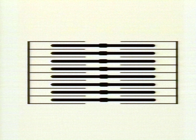
Diagram of the filament lattice of vertebrate striated muscle in the overlap region of the sarcomeres . Thick filaments are represented by solid circles; thin filaments are represented by smaller open circles. ... helps the students learn the relative longitudinal arrangement of the myofilaments when the sarcomere is in the relaxed or ...

0714 Arrangement Of Myofilaments In The Sarcomere Medical Images For Powerpoint Presentation Powerpoint Images Example Of Ppt Presentation Ppt Slide Layouts
(a) Electron micrograph showing a whole sarcomere from fish muscle in relaxing conditions (Z to Z distance about 2.3 µm). (b) Schematic diagram showing the sarcomere with titin molecules, green and blue, with the N-terminus of each titin molecule located at the Z-band and the C-terminus at the M-band.
Human Physiology Chapter 12 Flashcards Easy Notecards
The diagram above shows part a myofibril called a sarcomere. The diagram above shows a partially contracted muscle where there is more overlapping of the. Draw your own diagram of two sarcomeres. The first should be of a relaxed muscle. The second should be of a contracted muscle.
The Structure Of A Sarcomere The Basic Morphological Contracted And Relaxed Sarcomere Hd Png Download 800x619 4109504 Pngfind
A sarcomere is the basic unit of striated muscle tissue. It is the repeating unit between two Z lines. Skeletal muscles are composed of tubular muscle cells which. Sarcomeres are composed of thick filaments and thin filaments. The thin filaments Look at the diagram above and realize what happens as a muscle contracts.
Sarcomere Stock Illustrations 40 Sarcomere Stock Illustrations Vectors Clipart Dreamstime
Draw your own diagram of two sarcomeres. The first should be of a relaxed muscle. The second should be of a contracted muscle. Label the Z line, M line. Their observations led to the discovery of sarcomere zones. Sarcomere The figure depicts the structure of a Sarcomere. (Each zone is labeled). They first.

0714 Arrangement Of Myofilaments In The Sarcomere Medical Images For Powerpoint Presentation Powerpoint Images Example Of Ppt Presentation Ppt Slide Layouts
Download scientific diagram | Detection of the myosin spacing in relaxed sarcomeres by AFM. (a) 3D height-image of psoas half-sarcomere in relaxing buffer at slack SL (tapping mode); pixel size, 5 ...
Solved This Activity Will Help You Understand The Changes Chegg Com
The reaction, created from the arrival of an impulse stimulates the 'heads' on the myosin filament to reach forward, attach to the actin filament and pull actin towards the centre of the sarcomere. This process is carried out simultaneously in all sarcomere and the end process is the shortening of all sarcomeres.
Chapter 20 Locomotion And Movement
Draw and label a diagram to show the structure of a sarcomere, including Z lines, actin filaments, myosin filaments with heads, and the resultant light and dark bands. 7. Explain how skeletal muscle contracts, including the release of calcium ions from the sacroplasmic reticulum, the formation of cross-bridges, the sliding of actin and myosin ...

Depiction Of Muscle Sarcomere Left Micrograph Of A Sarcomere Right Download Scientific Diagram
Sarcomere H zone Thin (actin) filament Thick (myosin) filament Z disc Z disc M line (c) Small part of one myofibril enlarged to show the myofilaments responsible for the banding pattern. Each sarcomere extends from one Z disc to the next.

Sarcomere An Overview Sciencedirect Topics
Sarcomere diagram to label. This is a quiz called label the sarcomere and was created by member deanne1480. Biology computers geography history languages math. Sarcomere of skeletal muscle mammalian. ... The first should be of a relaxed muscle. Draw your own diagram of two sarcomeres. Longitudinal tubules give rise to terminal cisternae closely ...
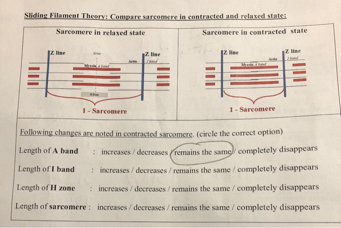
Solved Sliding Filament Theory Compare Sarcomere In Chegg Com
How Muscles Actually Work Jules Mitchell Yoga

Ch 7 2 Muscle Contraction Sarcomeres Diagram Quizlet

10 2 Skeletal Muscle Anatomy Physiology
Muscles Muscle Tissue Contains Many Mitochondria To Power Contractions Muscles Are Longer Than They Are Wide Muscles Are Divided Into Fibers Muscle Fibers Ppt Download
What Is The Zone That Disappears When Actin Filaments Slide Socratic

Muscular System Contraction Advanced Ck 12 Foundation

Muscle Fiber Contraction And Relaxation Anatomy And Physiology I

38 4c Sliding Filament Model Of Contraction Biology Libretexts
Mccc Edu
11 2 Muscles And Movement Bioninja

Muscle Physiology Chapter 13 Fundamentals Of Anaesthesia
Myosin Actin Sliding Filament Theory Myofilament Sarcomere Png Clipart Actin Angle Area Cell Diagram Free Png

Muscle Tissue Knowledge Amboss
Skeletal Muscle Physiology

Skeletal Muscle Tissue Anatomy And Structure
The Muscular System
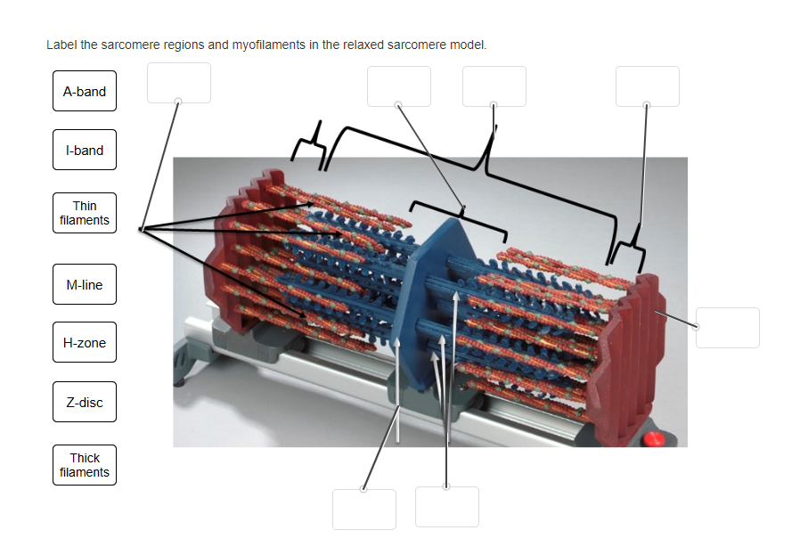
Solved Label The Sarcomere Regions And Myofilaments In The Chegg Com
What Is A Sarcomere Quora
Comparison Of A Relaxed And Contracted Sarcomere Learn Science At Scitable
00248733 Peir Digital Library


Comments
Post a Comment