42 blank skin diagram
This is an online quiz called Skin Labeling. There is a printable worksheet available for download here so you can take the quiz with pen and paper. From the quiz author. Epidermis, Dermis, Hypodermis Your Skills & Rank. Total Points. 0. Get started! Today's Rank--0. Today 's Points. One of us! 9+ Free Body Diagram - Free Printable Download. Studying the structure of a human body without visual aid is quite complicated. So much complicated, in fact, you won't understand anything. But the reason why many tutors don't use diagrams for visual demonstration is these venn diagrams are often complex to create. Luckily, though, you can ...
Clean, isolated white background. Biology illustration vector Skin anatomy, diagram. Basic human skin layer. Cubic cross section. Organ structure parts dermis, epidermis, subcutis, hypodermis. Clean, isolated white background. Biology illustration vector blank anatomy diagrams stock illustrations

Blank skin diagram
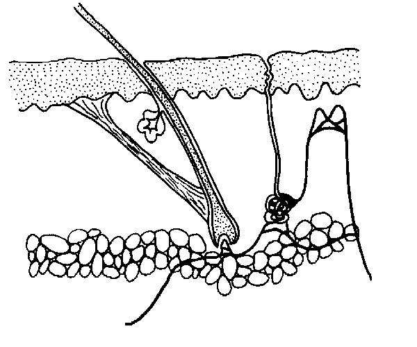
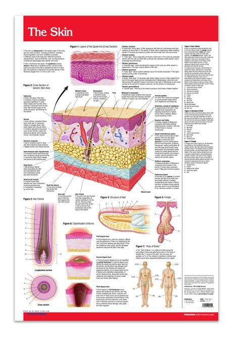
INTEGUMENTARY SYSTEM PART III: ACCESSORY STRUCTURES Integumentary Accessory Structures • Hair, hair follicles, sebaceous glands, sweat glands, and nails: - are made of epithelial tissue (part of epidermis) - are located in dermis - project through the skin surface The Hair Follicle • Is located deep in dermis - (made of epithleial tissue) Anatomy and Physiology of the Skin Paul A.J. Kolarsick, BS, Maria Ann Kolarsick, MSN, ARNP-C, and Carolyn Goodwin, APRN-BC, FNP CHAPTER 1 Introduction The skin is the largest organ of the body, accounting for about 15% of the total adult body weight. It performs many vital functions, including protection against external physical, SKIN MAPS. FREE PRINTABLE SKIN MAPS FOR DOGS AND CATS. Here are dog and cat skin maps to use at home or in your clinic to keep track of lumps and bumps. Use this with your calipers to note where the mass is and the size. No one can look at a mass or feel a mass and know what it is. Remember, my See Something, Do Something.
Blank skin diagram. make a note of any patches of skin that appear abnormal and how they look and feel, like the example provided (right). Each time you check your skin after that, find the spot that corresponds to those on your body map and record the new date, noting any changes in size, shape and colour. Also check your skin crocodile skin leather jacket. crusty skin allergies in dogs. custom skin epic fortnite skins. d&d 5e skin kite. dark skin jaundice. deep skin avulsion. default skin fortnite chapter 2. default skin funny fortnite. dog skin fungal infection treatment. The skin has a surface area of between 16.1-21.5 sq ft. for an adult human. The thickness of the skin differs over all parts of the body, and between men and women and the young and the old. For example, the skin on the forearm which is on average 1.3 mm in the human male and 1.26 mm in the human female. Beginning students may work with a partner or use their notes to help them label the diagram. Adaptations for Advanced Students Advanced students may research some additional parts of the skin (such as melanocyte, melanin, or corpuscles) or a related topic (such as sunburn, skin cancer, or moles and freckles) to explain to the class.
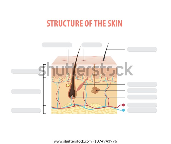
This skin diagram clearly shows all the layers of skin. We will now go over the skins layers in more detail. Epidermis layer. As can be seen in the skin diagram, the outermost layer of the skin is called the epidermis layer. There are no blood vessels in the epidermis but its deepest layer is supplied with lymph fluid. A Venn diagram is a design/illustration of the relationship between and among sets or group of objects that have something in common. Normally, Venn diagrams are used to highlight set intersections usually denoted by an upside-down letter U. the diagram is usually used in engineering and scientific presentations, in computer applications, theoretical mathematics and in statistics. Blank Skin Diagram. Lisa Potts. 102 followers. Skin Structure. Structure And Function. Human Integumentary System. Skin Anatomy. Sensory Nerves ... View a complete skin diagram. Full color diagram of skin anatomy. Information about the layers and parts of the skin. Tips and advice on skin care and skin problems. See all the layers of skin ex Blank Pale Skin Template. krissyroll. 1. 0. Blank santa hat. ElijahRock. 1. 0-Winter- ~PFPCE~ WolfieTWP42. 4. 0. Blank Bunny (for editing) mochicato. 0. 0 [BLANK] Shkon County Merch Long Sleeve (Christmas) Tactical123747. 0. 0 [BLANK] Shkon County Merch Short Sleeve (Chrismas) Tactical123747. 0. 0
The Skin Blank Diagram Pdf from imgv2-2-f.scribdassets.com Dead as it may be, your skin cells still need proper upkeep. This poster clearly labels the:. Browse skin layers resources on teachers pay teachers,. Neatniks take heart—even dead skin needs a . The epidermis also contains other cell structures. Read the definitions, then label the skin anatomy diagram below. blood vessels - Tubes that carry blood as it circulates. Arteries bring oxygenated blood from the heart and lungs; veins return oxygen-depleted blood back to the heart and lungs. dermis - (also called the cutis) the layer of the skin just beneath the epidermis. A professional-looking human skin layers science diagram template is here for you. With Edraw, you can hit the rich list of essential elements about human being bodies. Just type in your keywords for more related searches in the Edraw preset library. Lab Apparatus List. 64704. 211. Plant Cell Diagram. 19550. 173. Heart Diagram. Find free pictures, photos, diagrams, images and information related to the human body right here at Science Kids. Photo name: Skin Diagram Picture category: Human Body Image size: 64 KB Dimensions: 396 x 407 Photo description: This skin diagram lists all the important parts of human skin, including the dermis, epidermis, hypodermis, sweat pore, hair shaft, pigment layer, nerve fiber, dermal ...

301 Moved Permanently
Download the AAD's body mole map for information on how to check your skin for the signs of skin cancer. Keep track of the spots on your skin and make note of any changes from year-to-year. If you notice a mole that is different from others, or that changes, itches or bleeds, you should make an appointment to see a dermatologist.

Lab 6 Integumentary System
Skin Diagram Though skin may seem like nothing more than the source of things like zits and oil to your pre-teen, skin actually has many important jobs to do. Learn more about the skin (and the science behind pimples -- ew!) in this printable life science diagram.
Skin Diagram. The largest organ in the human body is the skin, covering a total area of about 1.8 square meters. The skin is tasked with protecting our body from the external elements as well as microbes. The skin is also responsible for maintaining our body temperature - this was apparent in victims who were subjected to the medival torture ...

Integumentary System Diagram to Label Luxury Quia Class ...
Skin Diagram Labeling . 1. Label the diagram with the . letters. below according to the structure/area they describe. You may label with a line or put the label directly onto the area described. Be as precise as possible. If you are worried about the precision of your label add a word after to explain exactly where your label should be.
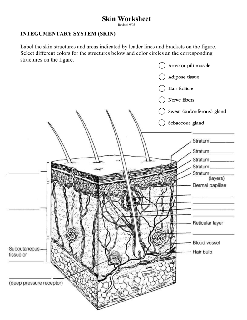
Skin Worksheet
Use this step-by-step guideline to fill out the Skin Monitoring Comprehensive CNA Shower Review form promptly and with ideal accuracy. How you can complete the Skin Monitoring Comprehensive CNA Shower Review form on the internet: To start the blank, utilize the Fill & Sign Online button or tick the preview image of the blank.
The Anatomy and Physiology of Animals/Skin Worksheet ...
28. $2.00. PDF. Students will read the definitions and label the skin anatomy diagram. Answer key included. This diagram had been modified from Enchanted Learning. I have used this worksheet as an in-class assignment as well as a homework assignment. You can also view a preview of this diagram :) ************.
Answers to some Questions
Skin Worksheet. 1. The outermost layer of the skin is: the dermis / the epidermis / fat layer. 2. Which is the thickest layer: the dermis / the epidermis? 3. Add the following labels to the diagram of the skin shown below.
It's time to label the diagram for yourself! Click below to download a free unlabeled version of the diagram above. Download PDF Worksheet (blank) Download PDF Worksheet (labeled) Skin anatomy. What if you want to test your knowledge of the skin only? No problem! With multiple layers and sublayers, there's plenty to learn about skin anatomy.
Diagrams are graphic representations of information. Diagrams can be simple lines or marks and can be complex drawings like architectural drawings and engineering plans. Charts, graphs, and schematics are types of diagrams. Chart-like diagrams include tree diagram, Venn diagram, existential graph, network diagram and flow chart.
Quantitative structure-skin permeability relationships - ScienceDirect
Skin is the largest organ in the body and covers the body's entire external surface. It is made up of three layers, the epidermis, dermis, and the hypodermis, all three of which vary significantly in their anatomy and function. The skin's structure is made up of an intricate network which serves as the body's initial barrier against pathogens, UV light, and chemicals, and mechanical injury.

Linee guida regionali per la gestione delle infezioni ...
SKIN MAPS. FREE PRINTABLE SKIN MAPS FOR DOGS AND CATS. Here are dog and cat skin maps to use at home or in your clinic to keep track of lumps and bumps. Use this with your calipers to note where the mass is and the size. No one can look at a mass or feel a mass and know what it is. Remember, my See Something, Do Something.
Mrs. Stewart's Science Page / Class Files/Links
Anatomy and Physiology of the Skin Paul A.J. Kolarsick, BS, Maria Ann Kolarsick, MSN, ARNP-C, and Carolyn Goodwin, APRN-BC, FNP CHAPTER 1 Introduction The skin is the largest organ of the body, accounting for about 15% of the total adult body weight. It performs many vital functions, including protection against external physical,

Quia - Skin Diagram
INTEGUMENTARY SYSTEM PART III: ACCESSORY STRUCTURES Integumentary Accessory Structures • Hair, hair follicles, sebaceous glands, sweat glands, and nails: - are made of epithelial tissue (part of epidermis) - are located in dermis - project through the skin surface The Hair Follicle • Is located deep in dermis - (made of epithleial tissue)

skin diagram quiz : Biological Science Picture Directory ...
Skin Diagram by Sandra Gibbs | Teachers Pay Teachers

Integumentary System Diagrams

Skin Structure Vector Illustration Stock Vector ...

Image from page 272 of "Diseases of the ovaries : their diagnosis and treatment" (1872)
Pop-up Human Body Skin

A Diagram of the Layers of the Skin | ClipArt ETC

Skin Labeling-detailed

Blank Layers Of Skin Diagram - Diagram Media
33 Blank Skin Diagram - Wiring Diagram Niche

DRAW IT NEAT : Resources
Solved Integumentary System 10 points Label the diagram | Chegg.com
How Does Diagram Microwave Oven Showing Stock Vector ...

What is menstrual cycle?
Skin Layers Diagram Blank - Diagram Media
Quiz - Skin - ABPI - Resources for Schools

The Life Of Brian
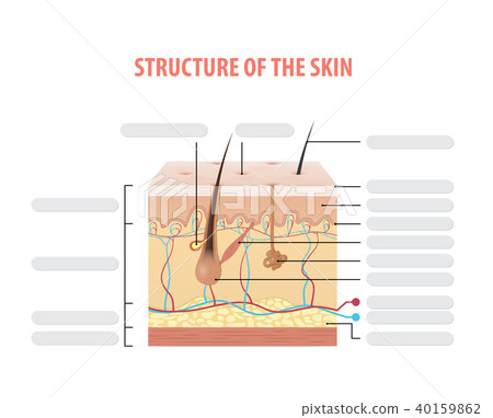
Vol.2 Structure of the skin info blank vector - Stock ...

Image from page 196 of "Annual report of the Bureau of Ethnology to the Secretary of the Smithsonian Institution" (1880)
Skin Poster - 24" x 36" Laminated Quick Reference
Skin - Teaching resources

Hair Unlabeled Example - SmartDraw | Teaching biology ...
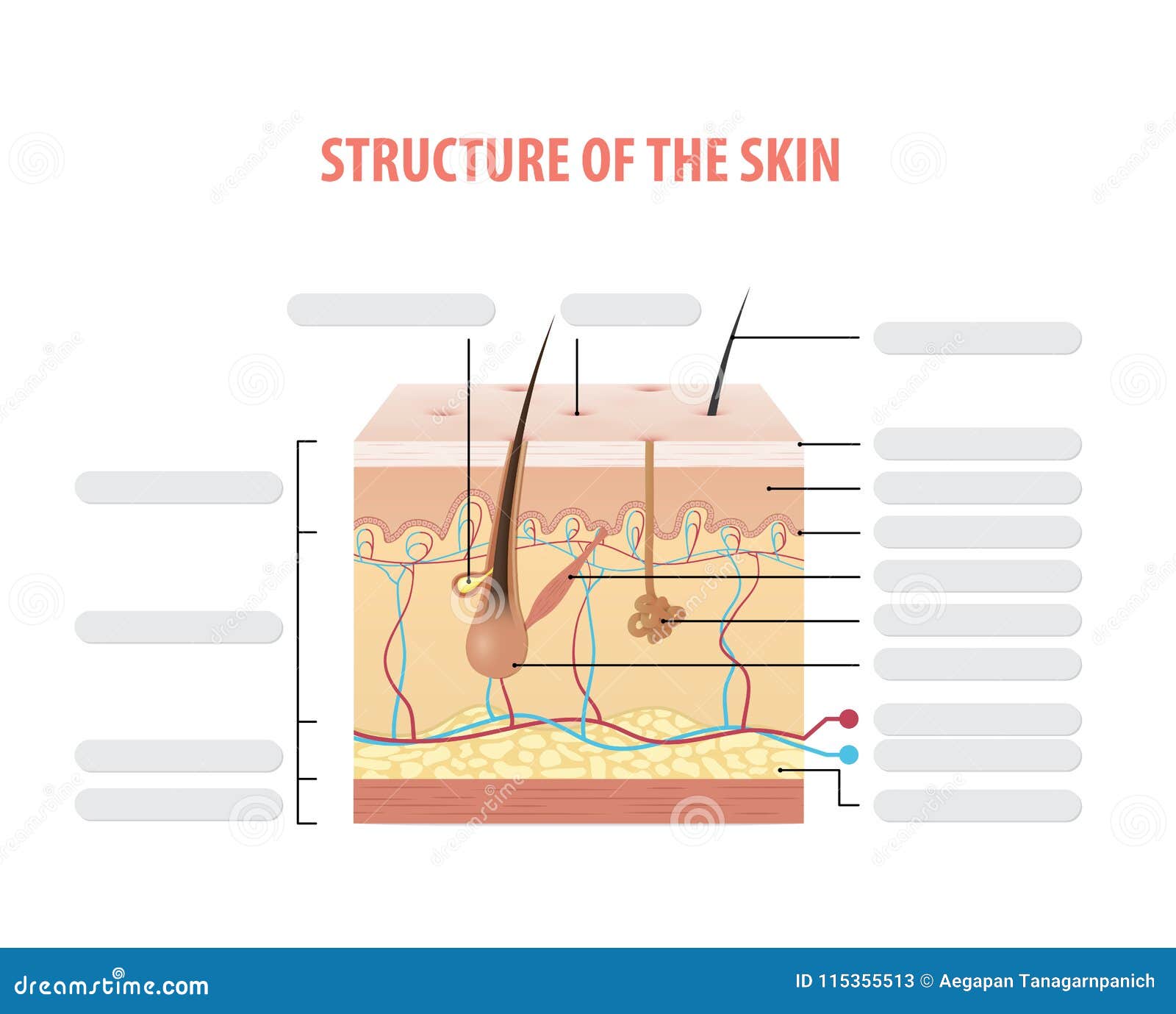
Vol.2 Structure Of The Skin Info Blank Illustration Vector ...
Animal Skin Diagram Quiz

Skin Layers Diagram Blank - Diagram Media

Blank Skin Diagram | Cakes | Anatomy, physiology, Anatomy ...

11 best images about Biology Diagrams on Pinterest | Dna ...


Comments
Post a Comment