42 transitional epithelium diagram
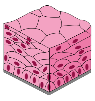
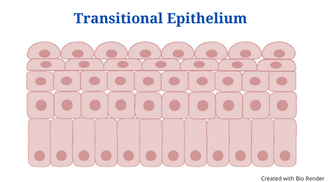
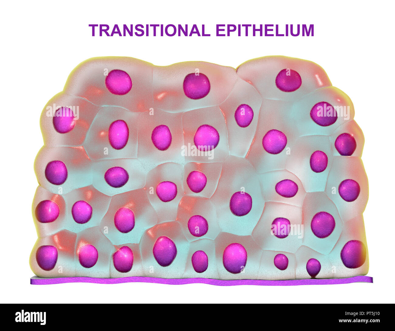
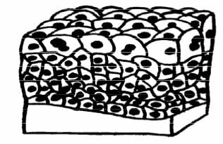
Information. Transitional epithelium is a stratified tissue made of multiple cell layers, where the cells constituting the tissue can change shape depending on the distention in the organ. When the organ is filled with fluid, cells on the topmost layer of this epithelium can stretch and appear flattened. SAMANTHA CURTIS. Transitional Epithelium Definition. Transitional epithelium is a stratified tissue made of multiple cell layers, where the cells constituting the tissue can change shape depending on the distention in the organ.When the organ is filled with fluid, cells on the topmost layer of this epithelium can stretch and appear flattened.
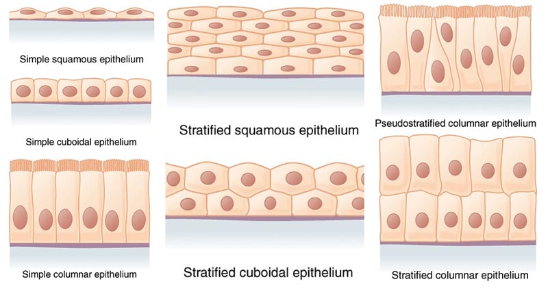
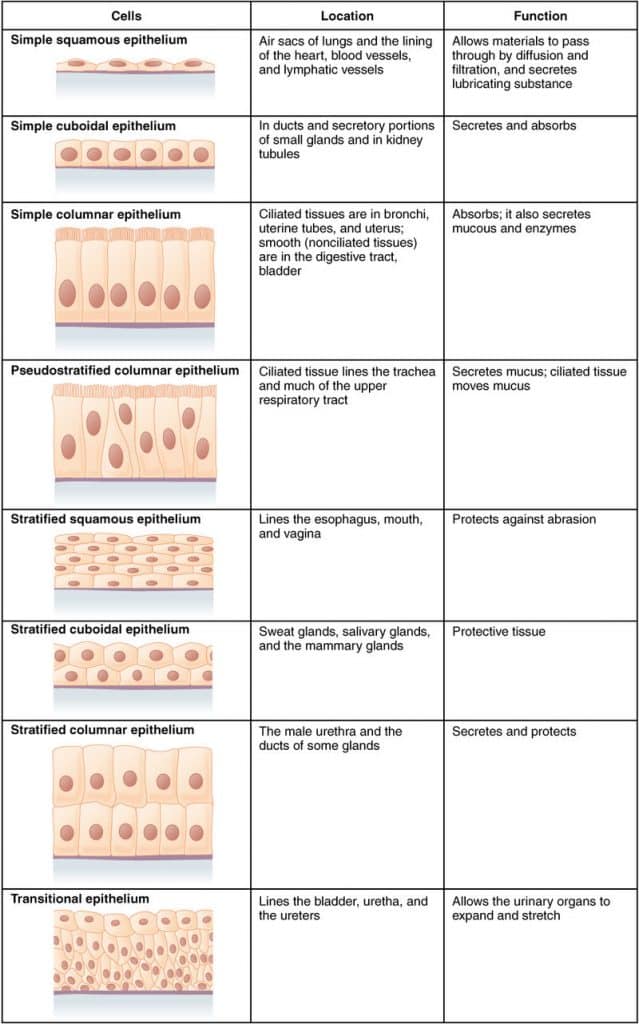
Form the Outer Covering of the skin and some internal organs. Form the Inner Lining of blood vessels, ducts and body cavities, and the interior of the respiratory, digestive, urinary and reproductive systems. Glandular epithelia. Constitute the secretory portion of glands. Simple squamous epithelium. Most delicate epithelium.

Transitional epithelium diagram
Transitional epithelium definition. Transitional epithelium is a type of stratified epithelium consisting of multiple layers of cells where the shape of the cell changes according to the function of the organ. The epithelium has a varying appearance as they appear cubical or round when in a relaxed state, except the apical layer which seems to be flattened when stretched. Examples of Transitional Epithelium: The transitional epithelium was commonly present in urinary and in the male reproductive tract in humans.These are the areas where volume and osmolarity of the organ can be changed fastly.. In the urinary system, the volume and concentration of solutes in urine are depending on a number of factors. The prostatic urethra in the male reproductive system is ... Diagram 4.4: Columnar epithelium with cilia. Columnar epithelium with cilia on the free surface (also known as the apical side of the cell) lines the respiratory tract, fallopian tubes and uterus (see diagram 4.4). The cilia beat rhythmically to transport particles. Diagram 4.5: Transitional epithelium
Transitional epithelium diagram. In keratinized epithelia, the most apical layers (exterior) of cells are dead and and contain a tough, resistant protein called keratin. An example of this is found in mammalian skin that makes the epithelium waterproof. Transitional epithelia are found in tissues such as the urinary bladder ... Transitional Epithelium Functions, Location and Diagram. Transitional epithelium is also called Uroepithelium or Urothelium (because it lines the urinary system), and it is a type of stratified epithelial tissue in which the surface cells change shape from being rounded to squamous in nature. Transitional epithelium is located in the urinary ... Transitional epithelium is a stratified tissue in which the cells are all have a fairly round shape when the organ it lines is not distended (stretched out). The image shows the wall of the urinary bladder in the relaxed state (not distended). When the tissue is stretched, the cells, especially those on the surface, become flat. A. Simple columnar epithelium. Slide 29 (small intestine) View Virtual Slide Slide 176 40x (colon, H&E) View Virtual Slide Remember that epithelia line or cover surfaces. In slide 29 and slide 176, this type of epithelium lines the luminal (mucosal) surface of the small and large intestines, respectively. Refer to the diagram at the end of this chapter for the tissue orientation and consult ...
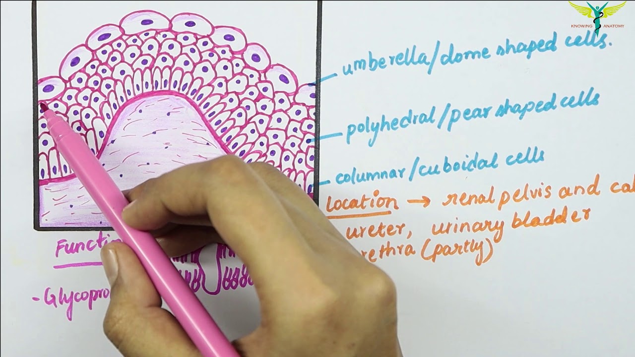
Transitional epithelium tissue is the somewhat self-explanatory name of a group of cells that can undergo a change in shape and composition. Discover the common functions of transitional ... Transitional epithelium Location and characteristics. Transitional epithelium (TE), also called urothelium, is a special type of stratified epithelium. It lines the urinary tract, specifically the major and minor calyces in the kidney, renal pelvis, ureters, bladder, the proximal part of the urethra, and the prostate gland in males. As it ... Transitional epithelium also known as urothelium is a type of stratified epithelium.[1] Transitional epithelium is a type of tissue that changes shape in response to stretching . The transitional epithelium usually appear cuboidal when relaxed and squamous when stretched.[1] This tissue consists ... The epithelium is found in various parts of the body. The simple squamous epithelium location specifically exists in the lining of the blood vessels like the arteries, veins, and capillaries. It ...
June 17, 2021 - It was thought that transitional epithelium was an intermediate type of epithelium, a “transition”, between the stratified squamous epithelium and the stratified columnar epithelium. That is why the name transitional. But it is not. The transitional epithelium is a stratified epithelium ... Transitional describes a form of specialized stratified epithelium in which the shape of the cells, and the number of layers present, can vary depending on the degree of stretch within a tissue. Figure 4.2.2 – Cells of Epithelial Tissue: Simple epithelial tissue is organized as a single layer of cells and stratified epithelial tissue is ... Transitional epithelium also known as urothelium is a type of stratified epithelium. Transitional epithelium is a type of tissue that changes shape in response to stretching (stretchable epithelium). The transitional epithelium usually appear cuboidal when relaxed and squamous when stretched. This tissue consists of multiple layers of epithelial cells which can contract and expand in order to ... Transitional Epithelium Definition Transitional epithelium is a sort of stratified epithelium made up of numerous layers of cells whose shape changes depending on the organ's function.When the epithelium is relaxed, it appears cubical or round, with the exception of the apical layer, which appears flattened when stretched.
Sox2 Specifies Primitive Transitional Epithelium Into Squamous Download Scientific Diagram
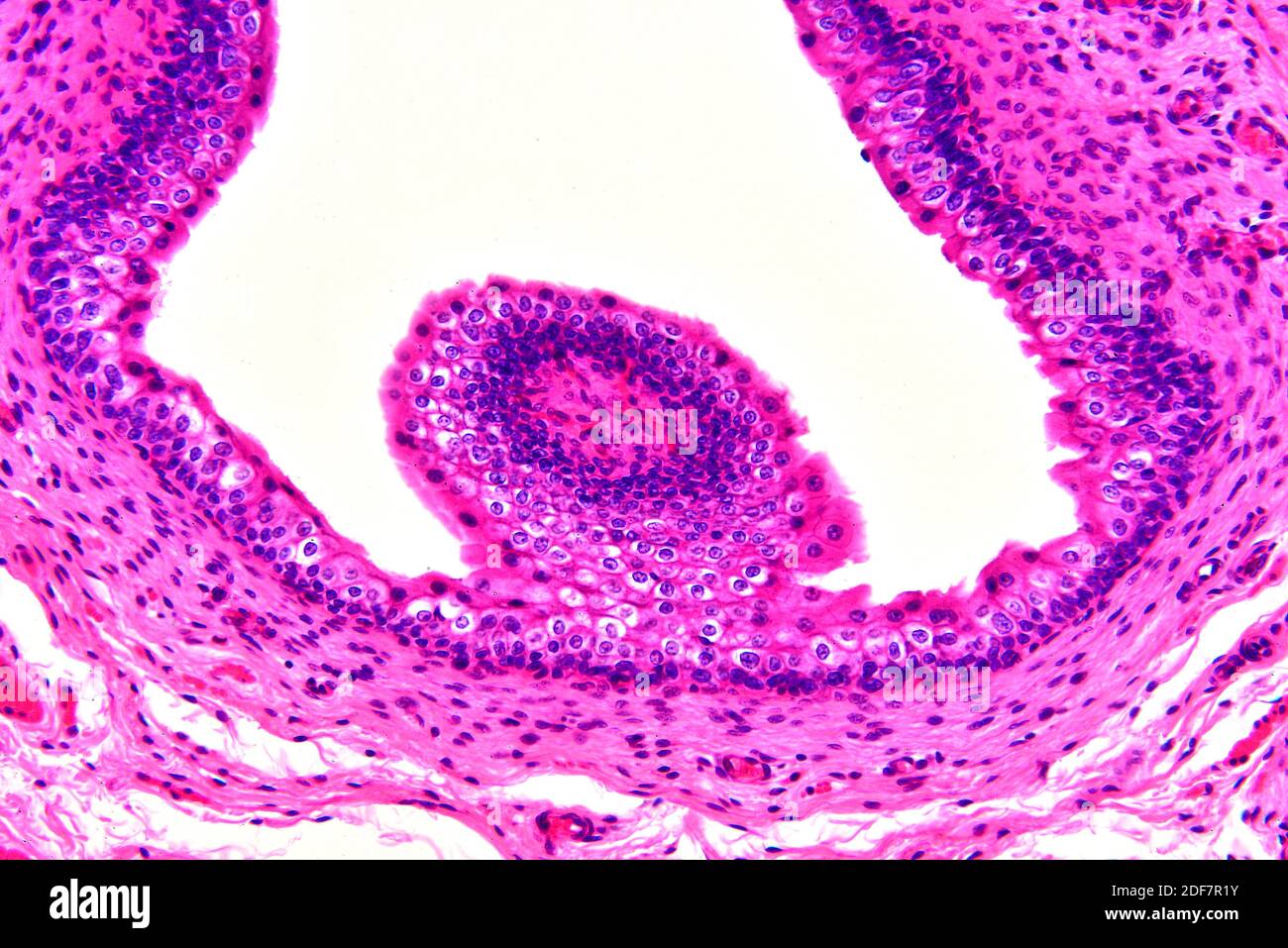
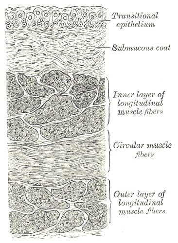
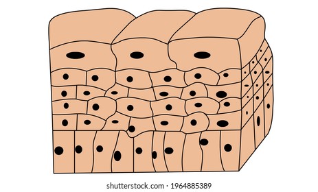
The epithelium of the urinary bladder is the transitional epithelium that consists of superficial columnar or cuboidal cells and deeper rounded or columnar cells. The layer of the transitional epithelium of a urinary bladder may change with the contracted and stretched conditions.
Stratified Or Compound Epithelial Tissues Sciencetopia
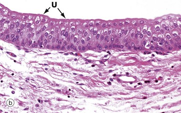
Mucous membrane (mucosa) Transitional epithelium; lines the bladder, ureters, and urethra. Epithelial layer. Contains no blood or lymphatic vessels. Basement membrane. Lies beneath the epithelial layer; single layer of cells separating the epithelial layer from the lamina propria; a sheet of extracellular material serving as a filtration ...

Transitional Epithelium Diagram Quizlet
Transitional epithelium 400x human urinary bladder transitional epithelium is a stratified tissue in which the cells are all have a fairly round shape when the organ it lines is not distended stretched out. Epithelial Tissues Basicmedical Key In this image you may find transitional epithelium of the bladder diagram. Transitional epithelium ...

Functions Of Transitional Epithelium Tissue Video Lesson Transcript Study Com
Link of Instagram:https://instagram.com/knowing_anatomy?igshid=12mmn11n2pxe6Transitional epithelium is a stratified tissue made of multiple cell layers, wher...

Transitional Epithelium Diagram Quizlet
This type of epithelium will provide more protection than a single layer, but it can't absorb. Transitional epithelium is found along almost all of the urinary tract.

Transitional Epithelium Diagram Quizlet
Start studying Transitional Epithelium. Learn vocabulary, terms, and more with flashcards, games, and other study tools.
Epithelial Tissue Definition Types Functions Examples
The development of rat transitional epithelial cells grown on conventional non-permeable surfaces was compared with development on permeable collagen supports. On glass or plastic surfaces, cells grew as expanding nomolayer sheets. Once confluent, growth ...

Selective Harvest Of The Urinary Bladder Transitional Epithelium By Download Scientific Diagram
4 Best Images of Transitional Epithelium Diagram - Transitional Epithelial Tissue Labeled, Transitional Epithelium and Transitional Epithelial Tissue Diagram / cleanri.com. See 4 Best Images of Transitional Epithelium Diagram. Inspiring Transitional Epithelium Diagram template images.
Transitional Epithelium Connective Tissue Cell Png 1116x2459px Epithelium Anatomy Area Cell Connective Tissue Download Free
The transitional epithelium of the urinary tract is lined by a layer of glycosaminoglycans (GAGs) that function to prevent microbial and crystal adherence to the bladder epithelium and minimize the movement of urine solutes and proteins through the bladder epithelium.

Illu Transitional Epithelium Transitional Epithelium Clipart Full Size Clipart 3936785 Pinclipart
Ciliated Pseudostratified Columnar Epithelium (Diagram) Transitional Epithelium (Diagram) Simple Squamous Epithelial Tissue (Image) ... Transitional Epithelium (Image) Goblet Cell (Image) The Arrow indicates _____ OTHER SETS BY THIS CREATOR. Endocrine Hormones-Intro 42 Terms.

Transitional Epithelium Photograph By Kateryna Kon Science Photo Library
Similarly, the number of cell layers ... epithelium and only the basal layer of cells rests on the basal lamina. Pseudostratified (pseudo- = “false”) describes tissue with a single layer of irregularly shaped cells that give the appearance of more than one layer. Transitional describes ...

Study Notes
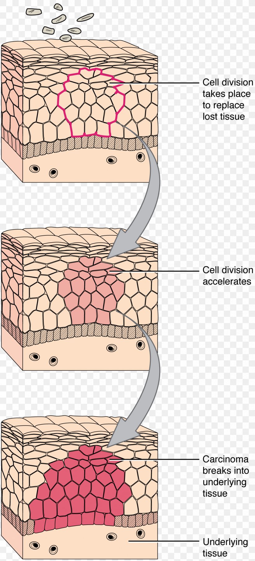
Transitional epithelium can act on osteogenic progenitor cells located intraskeletally, as demonstrated in two studies on repair of fractured bones. Pieces of bladder dispersed in polyurethane sponge or in protein-free bone matrix accelerate repair of fractured cranial bones in guinea pigs and rats, while urinary bladder mucosa accelerates ...

Transitional Epithelium Images Stock Photos Vectors Shutterstock
We are pleased to provide you with the picture named Transitional Epithelium Of The Bladder Diagram.We hope this picture Transitional Epithelium Of The Bladder Diagram can help you study and research. for more anatomy content please follow us and visit our website: www.anatomynote.com. Anatomynote.com found Transitional Epithelium Of The Bladder Diagram from plenty of anatomical pictures on ...
Transitional Epithelium High Resolution Stock Photography And Images Alamy
Epithelium / ˌ ɛ p ɪ ˈ θ iː l i ə m / is one of the four basic types of animal tissue, along with connective tissue, muscle tissue and nervous tissue.It is a thin, continuous, protective layer of compactly packed cells with little intercellular matrix.Epithelial tissues line the outer surfaces of organs and blood vessels throughout the body, as well as the inner surfaces of cavities in ...
Transitional Epithelium Definition Structure Functions Location Examples Phd Nest
Find transitional epithelium stock images in HD and millions of other royalty-free stock photos, illustrations and vectors in the Shutterstock collection. Thousands of new, high-quality pictures added every day.
Epithelial Cells Simple Stratified Teachmephysiology
Transcribed image text: et-labeling Activity: Transdtional epithelium of the urinary bladder Part A Drag the labels onto the diagram to identify structures associated with the transitional epithelium of the urinary bladder. Rest Help Connective tissue and smooth muscle layers (empty bladder Stretched bladder Epithelium (not stretched Basement membrane (empty bladder Basement membrane ...

Relaxed Transitional Epithelium Diagram Quizlet
Epithelial Tissue Diagram. The epithelium is a complex of specialized cellular organizations arranged into sheets without significant intercellular substance. It is a thin, continuous, protective layer of cells. The epithelial tissues perform many functions including protection from abrasion, radiation damage, chemical stress and. Think ...

The Transitional Epithelium Is Also Present In The Human Scj And Download Scientific Diagram
Other articles where transitional epithelium is discussed: epithelium: Transitional epithelium lines the urinary bladder; its appearance depends upon whether the bladder is contracted or distended.

Transitional Epithelium Of Human Bladder Magnification X100 H E Stain Cells Near The Surface Are The Pear Shaped Basement Membrane With Supporting Connec H E Stain Basement Membrane Tissue Types
Find out for more about simple epithelium.

Structural Organization Human Anatomy And Physiology Organic Chemistry Study Basement Membrane
The meaning of transitional epithelium is epithelium (as of the urinary bladder) consisting of several layers of cells which become flattened when stretched (as when the bladder is distended).

Transitional Epithelium Wikipedia
June 8, 2009 - Search the Large Images page with these keywords: simple columnar epithelium, stratified columnar epithelium, stratified squamous epithelium, pseudostratified columnar epithelium, transitional epithelium, goblet cells, serous gland, serous acini, mucous gland or mucous acini.

Transitional Epithelium Introduction Types Function
Simple Epithelium- it is composed of one layer of a cell and mostly has a secretory or an absorptive function. Compound (Stratified) Epithelium- it is made up of two or more than two layers of cells and mostly has a protective function. The glandular epithelium is made up of cuboidal or columnar cells. They are specialised for secretion.
Transitional Epithelium Definition And Function Biology Dictionary
Transitional epithelium is a stratified epithelium in which the shape of the surface cells changes (undergoes transitions) depending on the degree of stretch · When a transitional epithelium is not stretched (for example in an empty bladder) the cells of the surface layers are large and rounded.

The Urinary Bladder Lined By Transitional Epithelium Ta Resting On Download Scientific Diagram
We are pleased to provide you with the picture named Transitional Epithelium Anatomy.We hope this picture Transitional Epithelium Anatomy can help you study and research. for more anatomy content please follow us and visit our website: www.anatomynote.com. Anatomynote.com found Transitional Epithelium Anatomy from plenty of anatomical pictures on the internet.
Transitional Epithelium Youtube
Transitional epithelium is thin, elastic, and is found in the walls of the urethra of mammals. It forms the inner lining of the urinary bladder, ureters, and consists of 3 to 4 layers of cells. It allows stretching up to one or two cells thickness. In some cases, a simple epithelium may appear to be stratified. Such epithelium is known as the ...

Study Notes
Diagram 4.4: Columnar epithelium with cilia. Columnar epithelium with cilia on the free surface (also known as the apical side of the cell) lines the respiratory tract, fallopian tubes and uterus (see diagram 4.4). The cilia beat rhythmically to transport particles. Diagram 4.5: Transitional epithelium
Solved Simple Squamous Epithelium 2 Simple Cuboidal Epithelium Ch 4 Fig 3a Pg 68 Ch 4 Fig 3b Pg 68 Magnification Magnification Examp Course Hero
Examples of Transitional Epithelium: The transitional epithelium was commonly present in urinary and in the male reproductive tract in humans.These are the areas where volume and osmolarity of the organ can be changed fastly.. In the urinary system, the volume and concentration of solutes in urine are depending on a number of factors. The prostatic urethra in the male reproductive system is ...

Transitional Epithelium Of The Bladder Diagram
Transitional epithelium definition. Transitional epithelium is a type of stratified epithelium consisting of multiple layers of cells where the shape of the cell changes according to the function of the organ. The epithelium has a varying appearance as they appear cubical or round when in a relaxed state, except the apical layer which seems to be flattened when stretched.
Transitional Epithelium High Resolution Stock Photography And Images Alamy
Transitional Epithelium Images Stock Photos Vectors Shutterstock
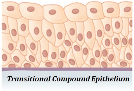
Transitional Compound Epithelium Diagram Easy Biology Class
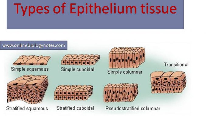
Types Of Epithelial Tissue Simple Compound And Specialized Online Biology Notes

Transitional Epithelium Of Bladder Ob Lens 10 Oc Lens 10 At The Download Scientific Diagram
Epithelia The Histology Guide

Epithelial Tissue Boundless Anatomy And Physiology
Transitional Epithelium Definition And Function Biology Dictionary

Tmsmission Electron Micrograph Of The Transitional Epithelium Of The Download Scientific Diagram

Transitional Epithelium Diagram Quizlet

Transitional Epithelium Diagram Quizlet

36 Transitional Epithelium Photos And Premium High Res Pictures Getty Images
Epithelial Tissues Basicmedical Key

How To Draw Transitional Epithelium Easy Way Youtube


Comments
Post a Comment