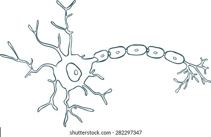
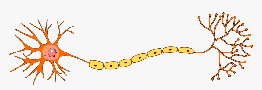
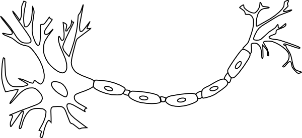
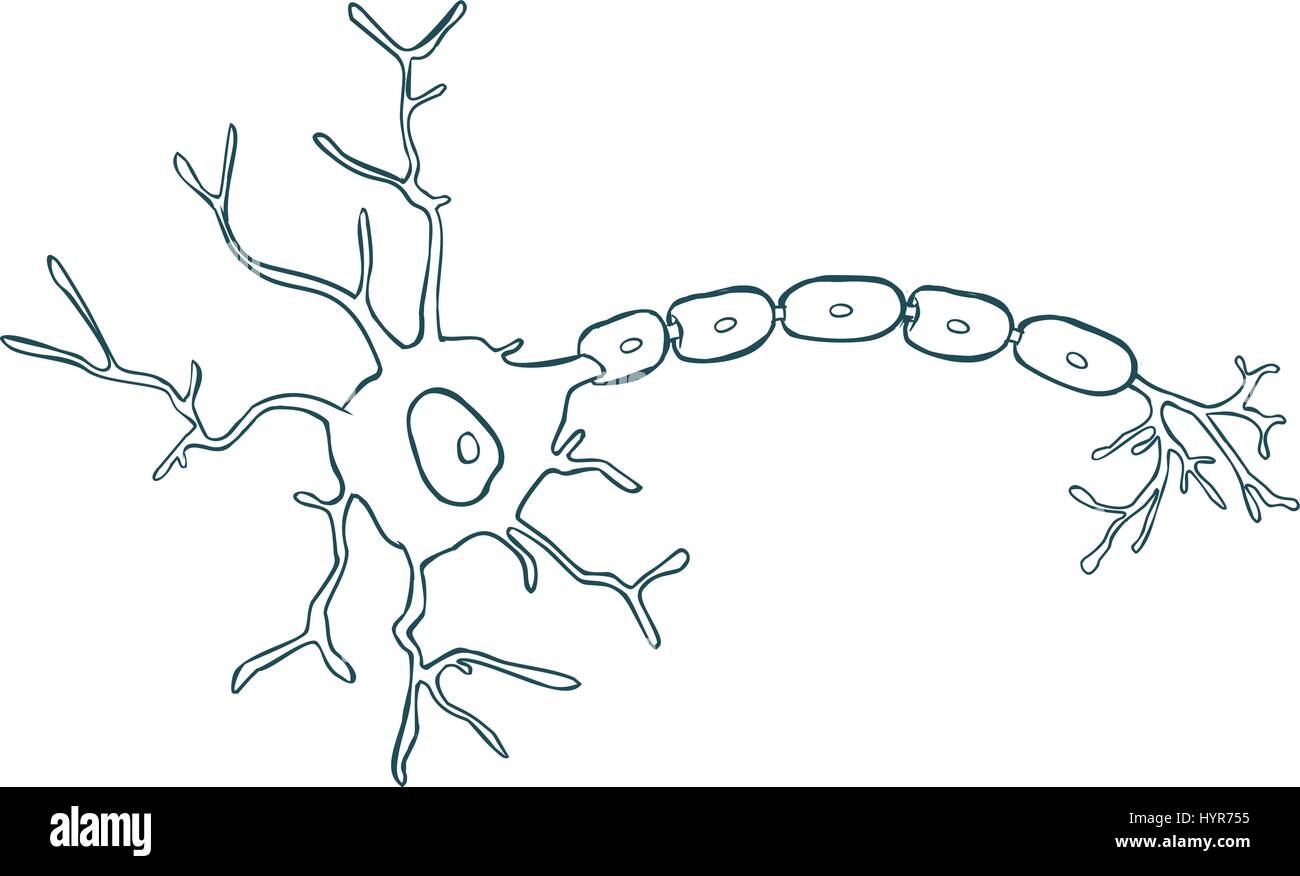
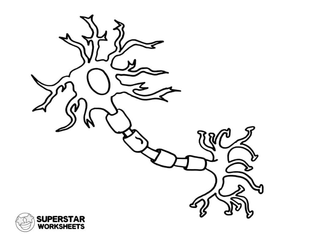
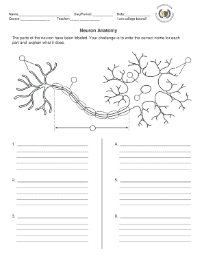
43 blank neuron diagram
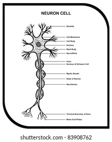
When in a resting state, the neuron is [blank_start]negatively[blank_end] charged. Upon activation by a stimulus, it temporarily becomes [blank_start]positively[blank_end] charged, creating an action potential. Answer. negatively. positively. positively. negatively. Question 9. Question. Label the diagram of synaptic transmission. Image: Unlabelled Synapse 1 … Provide the labels for the structures in a neuron shown to the left. Spinal Cord and Reflexes. In the diagram to the left, provide the labels for the structures involved in the reflex act when a person steps on a tack and jerks their leg away. Brain Anatomy. Provide the labels for the diagram on the left below and provide descriptions of the functions of each structure on the …
Dec 06, 2017 · An interactive quiz covering Spinal Cord Cross-Sectional Anatomy through multiple-choice questions and featuring the iconic GBS illustrations.

Blank neuron diagram
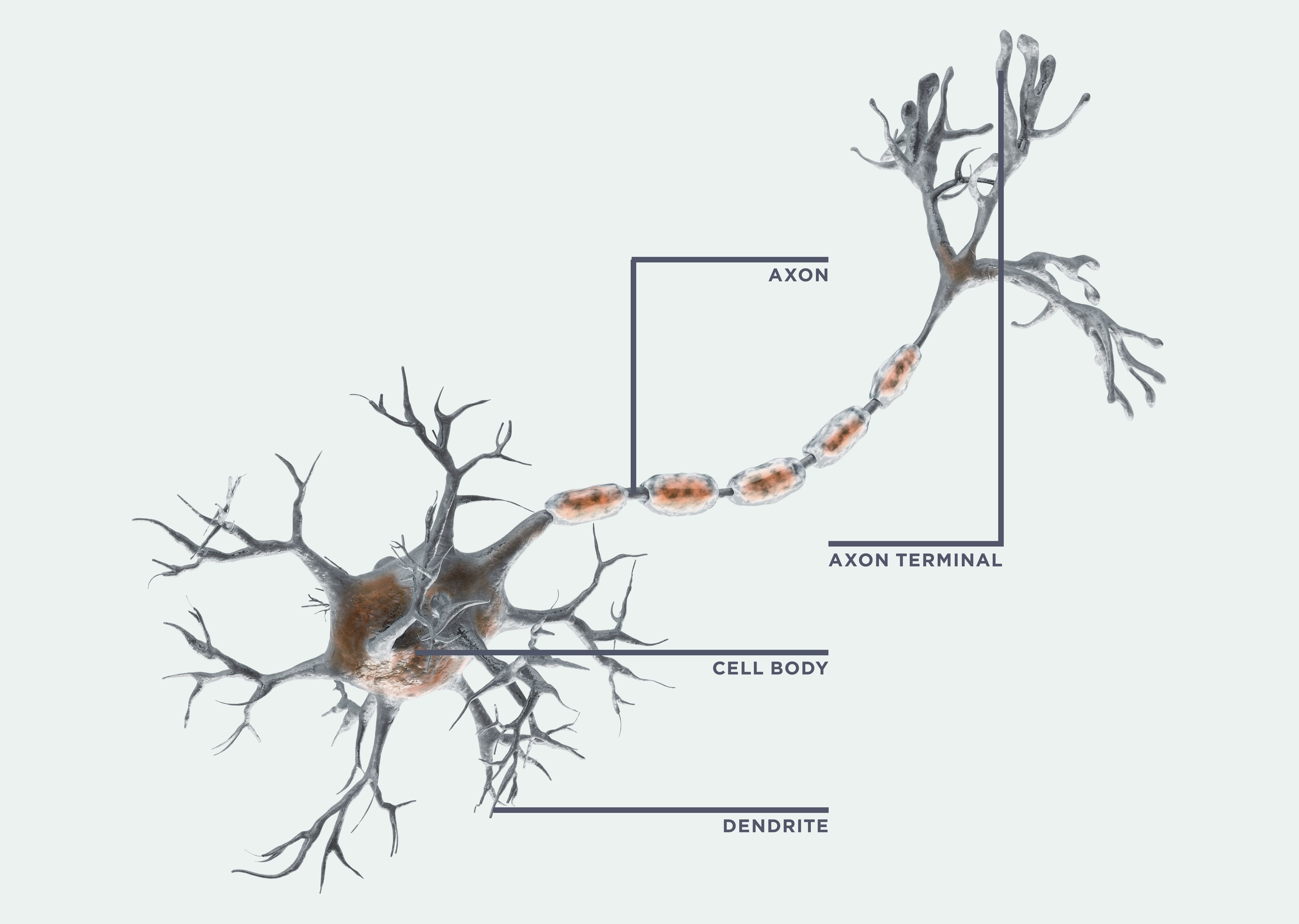
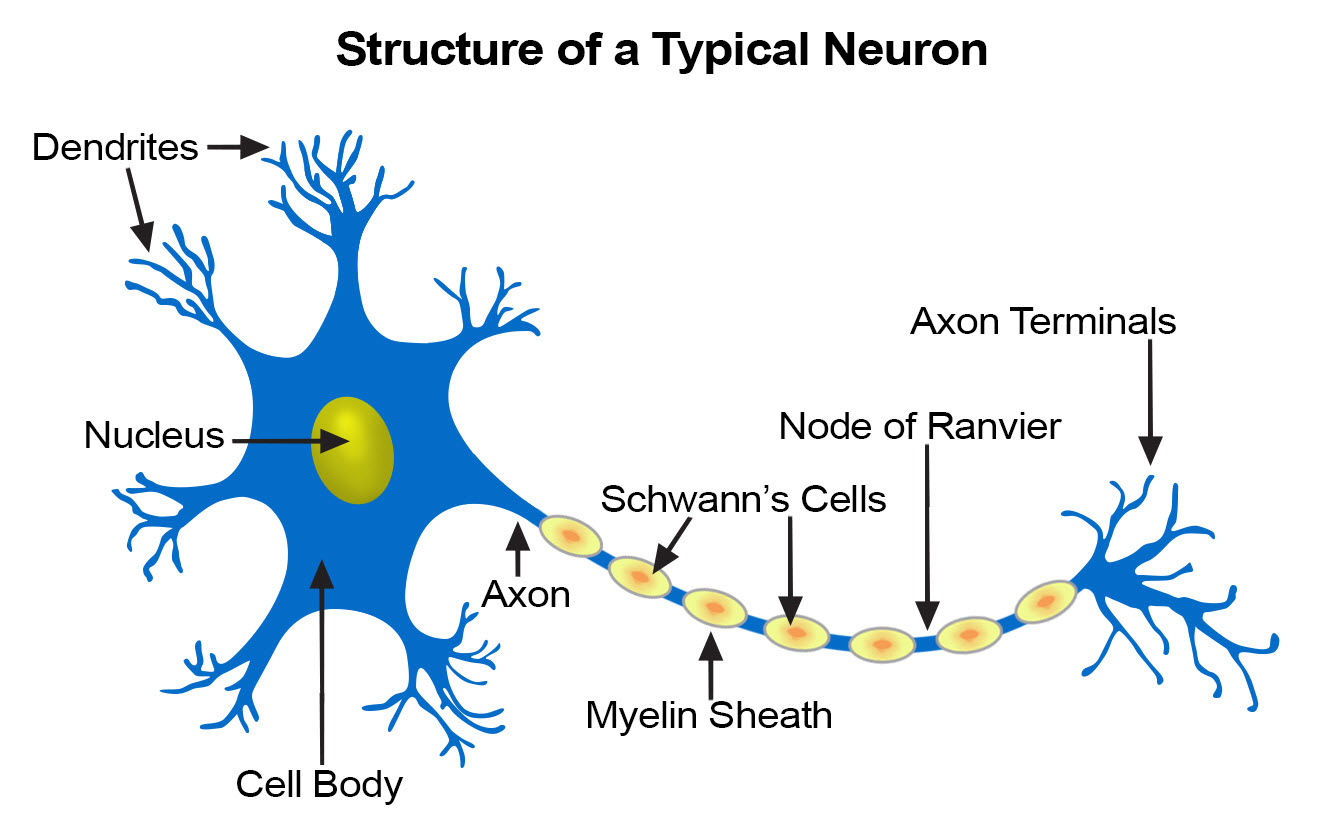
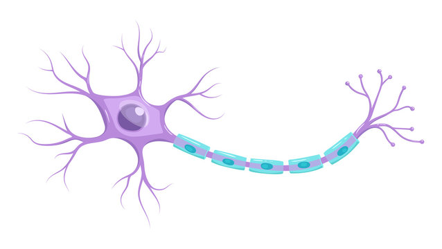
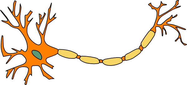
Gray matter is primarily composed of neuron somas (the round central cell bodies), and white matter is mostly made of axons (the long stems that connects neurons together) wrapped in myelin (a protective coating). The different composition of neuron parts is why the two appear as separate shades on certain scans. Each region serves a different role. Gray matter is primarily … Nov 18, 2007 · neuron fill in the blank; neuron unlabeled diagram; n ron; unlabelleddiagram of the brain; neuron images; neurons unlabelled; neuron image; cartoon brain nerve; clipart nerves; unlabelled neuron diagram; neuron without label; nueron; neuron label; blank diagram neuron; neuron draw; neuron without labels; blank neuron picture; blank structure of ... Neuron Models; Two slightly different activity sheets that I have used for in-class presentations: Activity Sheet #1; Activity Sheet #2. Outside Games. Synaptic Tag Lesson Plan; Neuron Chain Lesson Plan; Brain Freeze Tag Lesson Plan. The Research Report - for Teachers; The Research Report - for Students ; Web Page Evaluation Form. Treasure Hunts. Neuroscience for Kids …
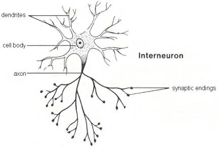
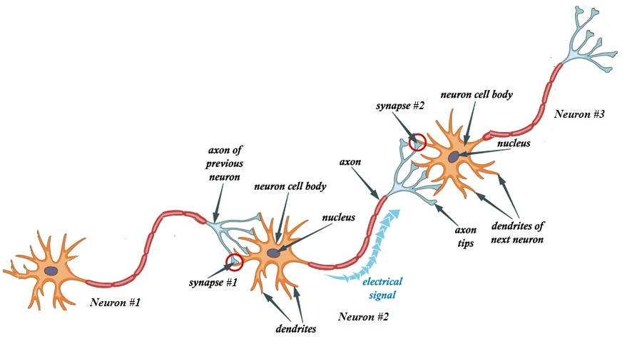
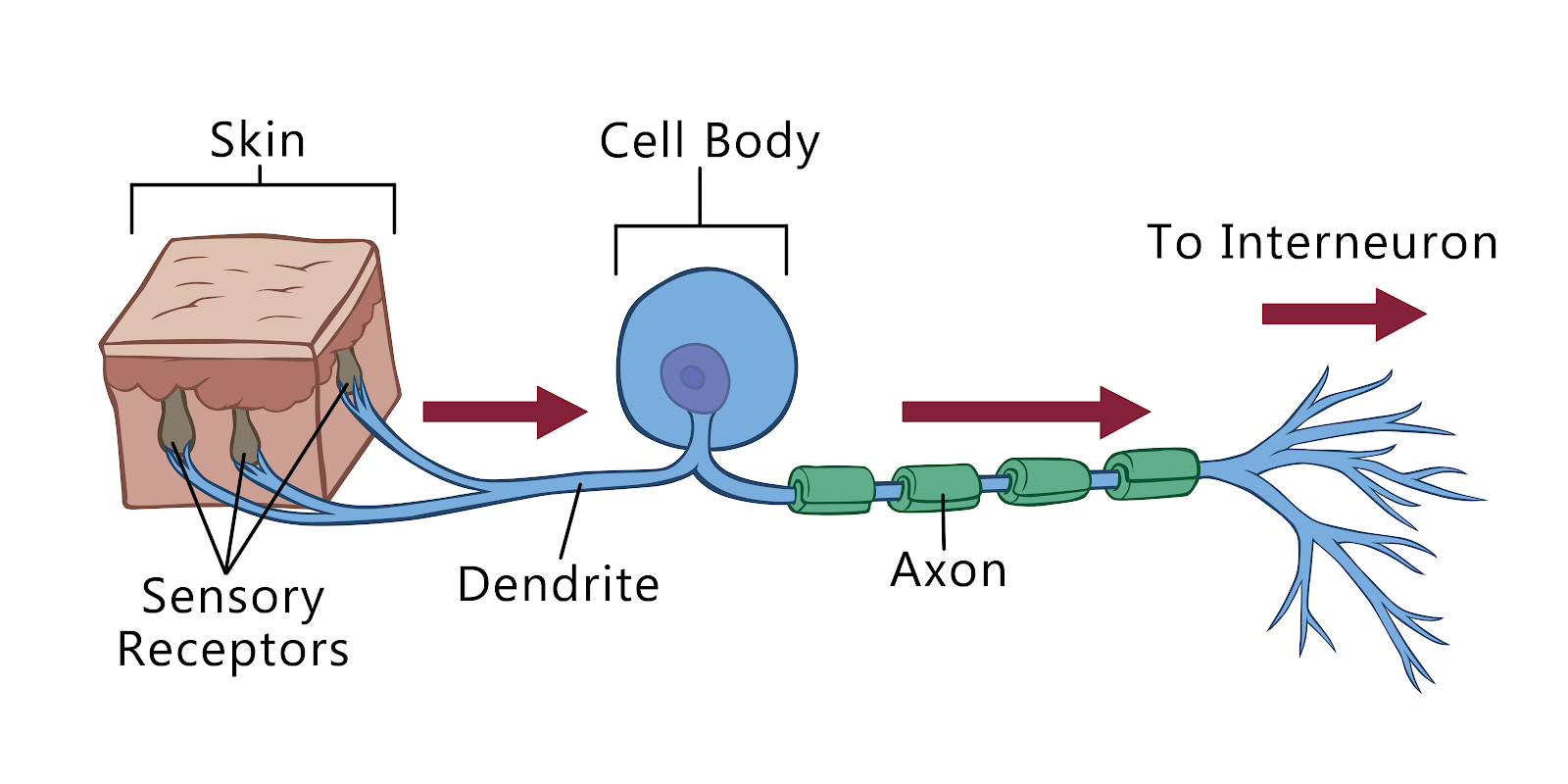
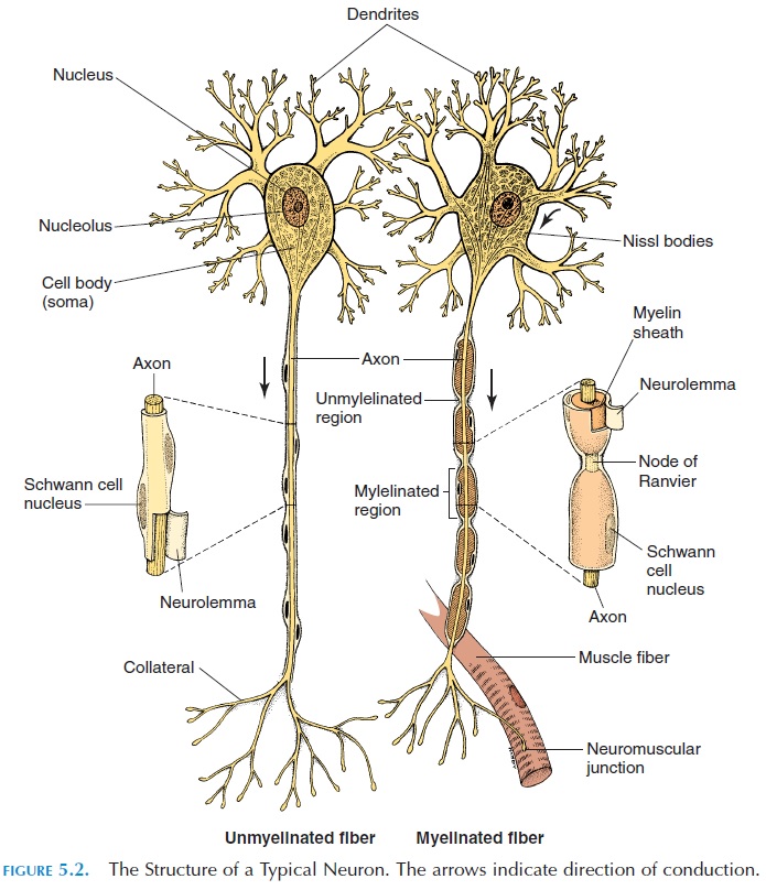
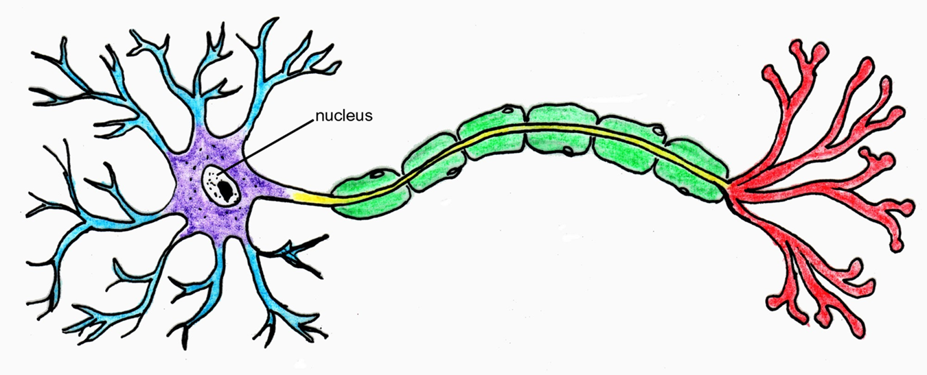
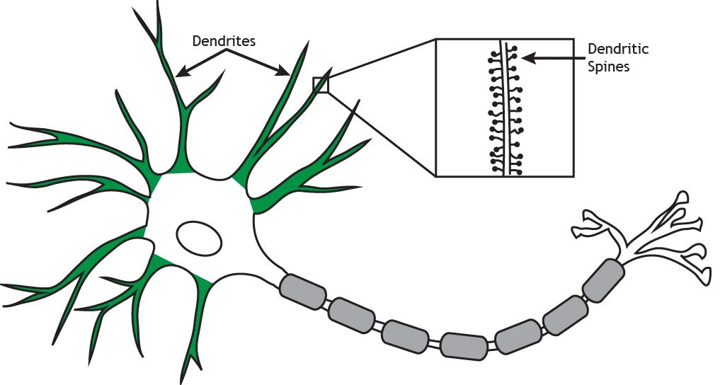
Blank neuron diagram. STARTER – Label the diagram a. b. a. d. c. e. •Transverse abdominus •external oblique •rectus abdominus •internal oblique •pectoralis major . Learning Objectives Everyone should Identify the location of skeletal muscles in the trunk and upper extremities of the body Most will Describe strengthening exercises for the specific muscle groups mentioned . RECTUS ABDOMINIS … A neuron can be compared to an electrical wire—it transmits a signal from one place to another. Glia can be compared to the workers at the electric company who make sure wires go to the right places, maintain the wires, and take down wires that are broken. Although glia have been compared to workers, recent evidence suggests that also usurp some of the signaling functions … The Overall Block Diagram of the visual process in Humans and other Chordates shown below can be used to localize the failures resulting in these diseases. In the following figure, the left and right lateral geniculate nuclei (LGN) and perigeniculate nuclei (PGN) are shown along with the superior colliculus. These are all major elements of the thalamus, a portion of what is … The dendrite receives information from another neuron’s axon at the synapse, and the axon sends information to the next neuron’s dendrites. Unlike the majority of neurons found in the CNS, an action potential in a dorsal root ganglion neuron may initiate in the distal process in the periphery, bypass the cell body, and continue to propagate along the proximal process until …
Neuron Models; Two slightly different activity sheets that I have used for in-class presentations: Activity Sheet #1; Activity Sheet #2. Outside Games. Synaptic Tag Lesson Plan; Neuron Chain Lesson Plan; Brain Freeze Tag Lesson Plan. The Research Report - for Teachers; The Research Report - for Students ; Web Page Evaluation Form. Treasure Hunts. Neuroscience for Kids … Nov 18, 2007 · neuron fill in the blank; neuron unlabeled diagram; n ron; unlabelleddiagram of the brain; neuron images; neurons unlabelled; neuron image; cartoon brain nerve; clipart nerves; unlabelled neuron diagram; neuron without label; nueron; neuron label; blank diagram neuron; neuron draw; neuron without labels; blank neuron picture; blank structure of ... Gray matter is primarily composed of neuron somas (the round central cell bodies), and white matter is mostly made of axons (the long stems that connects neurons together) wrapped in myelin (a protective coating). The different composition of neuron parts is why the two appear as separate shades on certain scans. Each region serves a different role. Gray matter is primarily …
Label Neuron Anatomy Printout Enchantedlearning Com
13 2 Neurons A Level Biology Student
Solved Let S Put It All Together Review The Steps Of An Chegg Com
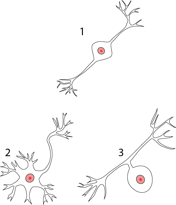
1

Biology The Nervous System A Neuron Quiz

Twitchy Nerves Literally May Explain Epilepsy Pain Npr

9 Tattoo Ideas Neurons Buffalo Tattoo Buffalo Art

Sensory Neuron The Definitive Guide Biology Dictionary

Neurofilament Light Chain Clipart Neurofilament Neuron Blank Neuron 900x450 Png Download Pngkit
Neuron Structure And Classification
Nervous System The Partnership In Education

Anatomy Of The Nervous System Microbiology
Neuron Myelin Images Stock Photos Vectors Shutterstock

Neuron Structure Quiz
Your Brain Interactive Build Your Network The Franklin Institute

Multipolar Neurons Structure And Functions
Seer Training Nerve Tissue

Lesson Worksheet Neurons Nagwa
How Strongly Does The Quantitative Fact That Most Neurons Have Many Dendrites And Only One Axon Qualitatively Correlate With Systems Mechanisms And Cognitive Abilites Quora

File Neuron Svg Wikimedia Commons
Structure Of Neuron Neuron Png Transparent Png Kindpng
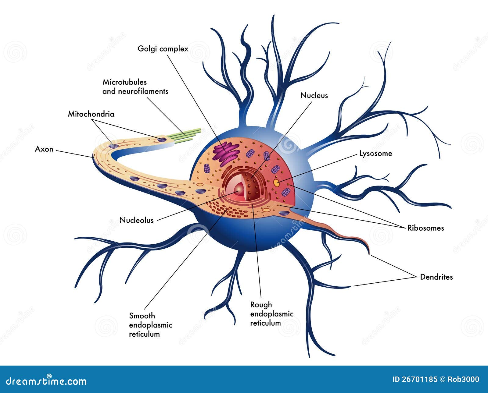
Nerve Cell Stock Vector Illustration Of Soma Golgi 26701185
Structure Of The Neuron
Neuron Black And White Clip Art At Clker Com Vector Clip Art Online Royalty Free Public Domain
White Background Vector Illustration Of A Neuron Stock Vector Image Art Alamy
Structure Of Neuron In Structure Of Neuron Fig 5 The Cell Soma And Download Scientific Diagram
67 595 Best Neuron Images Stock Photos Vectors Adobe Stock

The Nervous System And Nerve Cells Diagram Quizlet
Neuron Cell Worksheets Superstar Worksheets

Neurotransmitter Wikipedia

Diagram Quiz On Neuron Structure And Function Labeling Quiz
Nerve Cell Diagram Images Stock Photos Vectors Shutterstock
Free Neuron Cliparts Download Free Neuron Cliparts Png Images Free Cliparts On Clipart Library

The Neuron Diagram Diagram Quizlet
Neuron Anatomy Activity Fill Online Printable Fillable Blank Pdffiller

Neurons Organismal Biology

Dendrite Wikipedia

Neural Structure Quiz

Pin On Anatomy Physiology Nervous System
The Neuron Foundations Of Neuroscience
Labeled Neuron Clipart Best
Nervous System Flashcards Chegg Com

File Anatomy And Physiology Unlabeled Neuron Jpg Wikimedia Commons


Comments
Post a Comment