43 horse bone diagram
Horse Hoof And Leg Anatomy: A Guided Tour Scott J. Duggan Livestock Extension Faculty. Today's Mission Be able to visualize the skeletal anatomy of the lower leg and hoof of the horse. Develop an understanding of the causes of equine lameness and methods of treatment. horse skeleton. STUDY. Learn. Write. Test. PLAY. Match. Created by. KMadison1991 PLUS. vet tech midterm study guide. Terms in this set (7) Radius ... coffin bone... Sets found in the same folder. intestines of a dog diagram. 9 terms. KMadison1991 PLUS. oral cavity of a dog. 8 terms. KMadison1991 PLUS. cell diagram. 13 terms. KMadison1991 PLUS ...
Conjugated estrogens (CEs), or conjugated equine estrogens (CEEs), sold under the brand name Premarin among others, is an estrogen medication which is used in menopausal hormone therapy and for various other indications. It is a mixture of the sodium salts of estrogen conjugates found in horses, such as estrone sulfate and equilin sulfate. CEEs are available in the form of both natural ...

Horse bone diagram
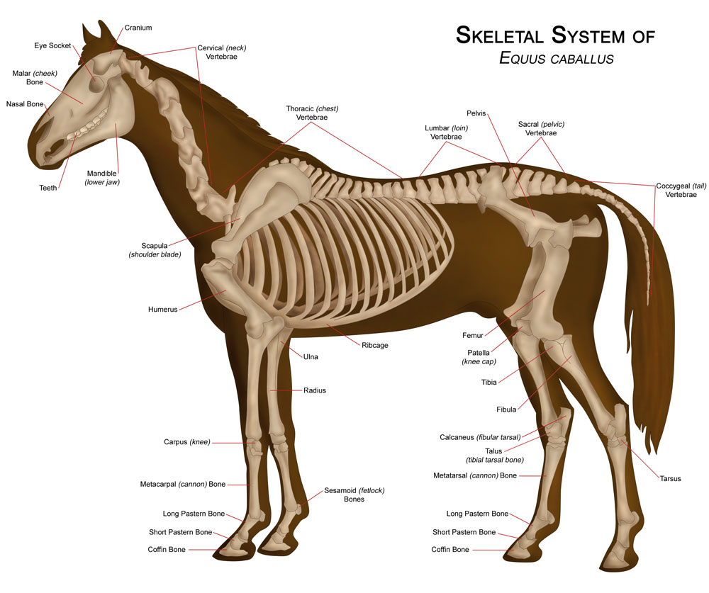
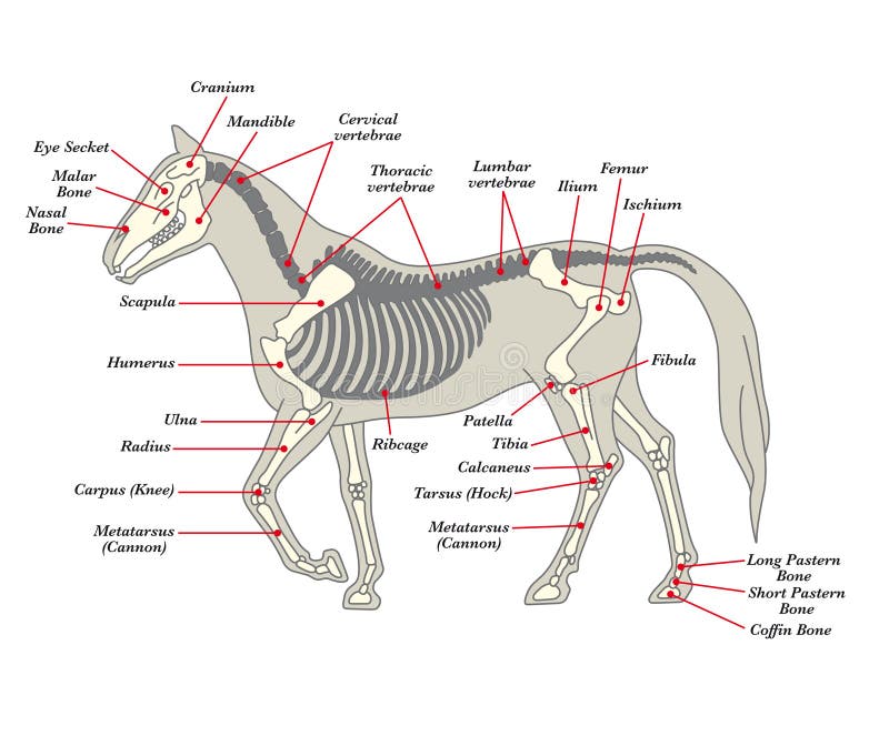
Horses have, on average, a skeleton of 205 bones. A significant difference in the bones contained in the horse skeleton, as compared to that of a human, is the lack of a collarbone. Their front limb system is attached to the spinal column by a powerful set of muscles, tendons and ligaments that attach the […] Dermal bone is from mesenchyme and ectomesenchyme of the dermis and it overlies ... (cat, horse, human) or confluent with it (wolf, rat). Crests: Notice on the wolf skull the median longitudinal sagittal crest to attach temporal muscles and a transverse nuchal crest for muscles supporting the head. THE PALATE. Modifications for breathing air: evolution of the secondary palate. The secondary ... in bone growth (endochondral ossification) affecting young horses. The cartilage is poorly affixed to the underlying bone and is readily pealed away. At left is a large lesion on the medial trochlear ridge of a weanling quarter horse. The cartilage has been removed, showing the abnormal subchondral bone. This photo was taken at necropsy.
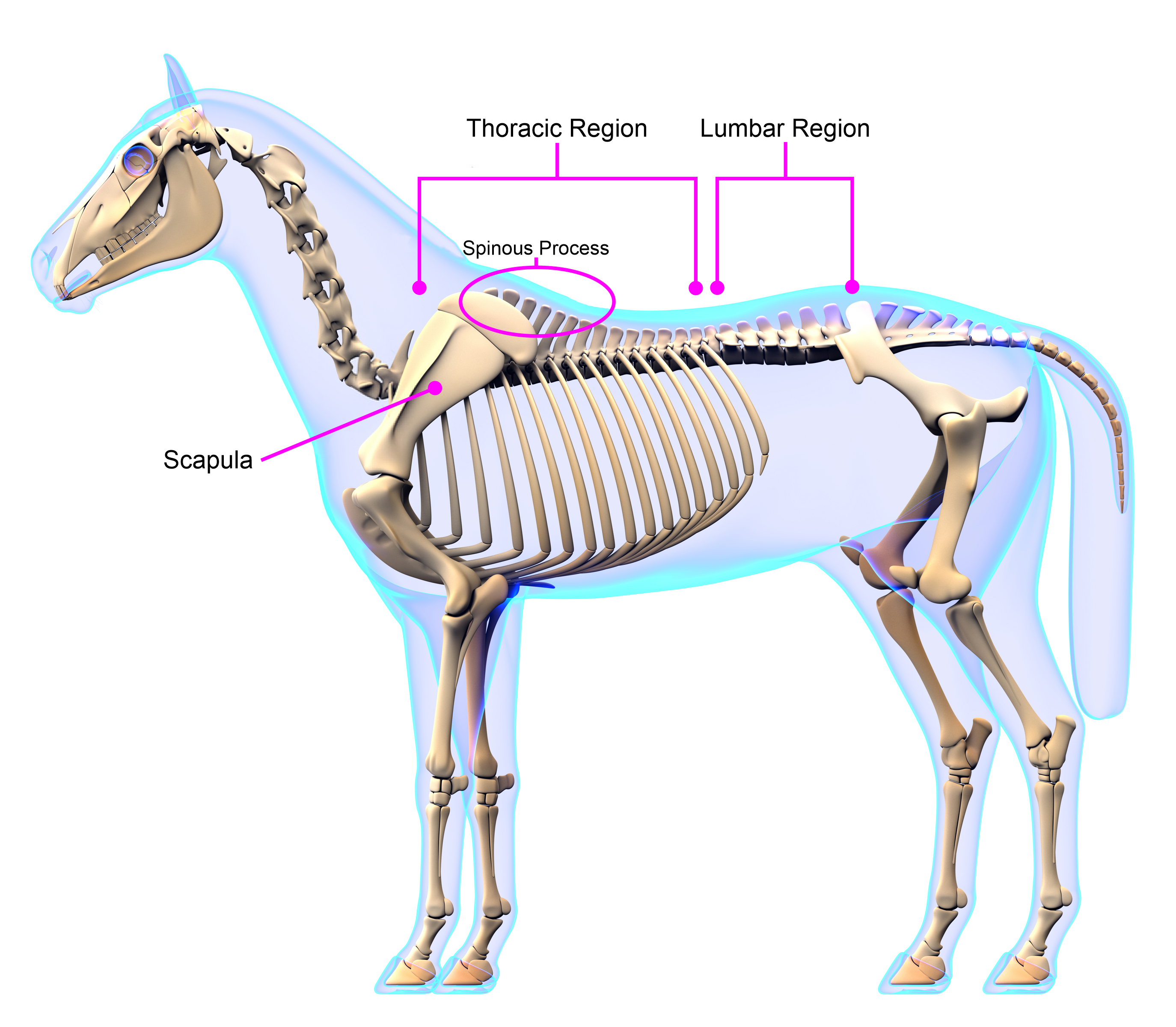
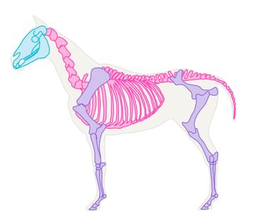
Horse bone diagram. The appendicular skeleton contains the fore and hindlimbs. The hindlimb attaches to the vertebral column via the pelvis, while the forelimb does not directly attach to the spine (as a horse does not have a collar bone), and is instead suspended in place by muscles and tendons.This allows great mobility in the front limb, and is partially responsible for the horse's ability to fold his legs up ... • Bones allow the horse movement along with the muscles, tendons and ligaments. • It is within the Bone Marrow that Red Blood cells are produced. • It is within the bones that minerals are stored for example the minerals Calcium and Phosphorous. Types of Bones The equine skeleton is made up of a combination of Flat bones, Long bones, Short By necessity, all muscles of the wrist cross the axes of rotation located at the capitate bone and therefore produce movement at the wrist. The two axes of rotation that correspond to the two planes of motion at the wrist are shown in Fig. 6.9.Flexion and extension occur about the medial-lateral axis of rotation; radial and ulnar deviation occurs about an anterior-posterior axis of rotation. The shape and health of the digital cushion will influence the angle of the Pedal Bone. "Flat footed" horses (ie, those whose pedal bones lie flat instead of being tilted slightly on their nose) often have severely atrophied digital cushions. Coriums. A corium is a vascular structure which manufactures hoof horn.
to the bone FASCI 10. A discrete bundle of muscle cells . 5. Figure 6—3 is a diagrammatic representation of a small portion of a relaxed muscle cell (bracket indicates the portion enlarged). First, select different colors for the structures listed below. Use them to color the coding circles and corre- sponding structures on Figure 6—3. Then bracket and label an A band, an I band, and a ... Equine anatomy refers to the gross and microscopic anatomy of horses, ponies and other equids, including donkeys, mules and zebras.While all anatomical features of equids are described in the same terms as for other animals by the International Committee on Veterinary Gross Anatomical Nomenclature in the book Nomina Anatomica Veterinaria, there are many horse-specific colloquial terms used by ... Horse rear leg anatomy Horse rear legs. The horse leg anatomy in the rear includes the bones of the pelvis (the ilium, ischium and pubic bones), femur, tibia, fibula, metatarsus and the phalanxes. It also includes the joints of the hip, stifle, hock, fetlock, pastern, and coffin. Hind limbs Jan 17, 2014 - free printable Horse parts diagram, farm animals parts diagram
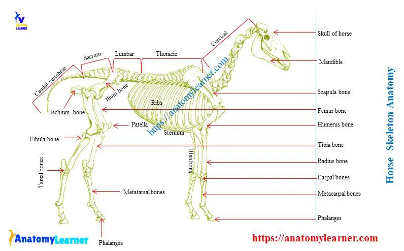
Mamenchisaurus means 'Mamenchi lizard', from the Chinese Pinyin mǎ (马 'horse') and mén (门 'gate'), while chi is a transliteration of xī (溪 'stream' or 'brook'), combined with the suffix -saurus (from Greek sauros meaning 'lizard'). The intention was to name the genus after the place where its fossil was first found. Basic Horse Anatomy for Equine Owners. Get the basics on horse anatomy that every horse owner needs. Diagrams, illustrations and charts will help you understand how your horse is put together. From equine skeletal anatomy to body parts and teeth. Develop a better understanding of where leg injuries occur, and the inner workings of the horse hoof. The horse skeleton is the rigid framework of the body that consists of bones, cartilages, and ligaments.There are two hundred and five bones found in horse skeleton.In this long article, I will discuss the osteological features of all bones from the horse skeleton anatomy labeled diagram. I will also try to cover all possible inquiries on the horse skeletal system at the end of this article. Osteology of horse. This is the basic and most important system of horse anatomy that you might not skip.You should learn all of the osteological features of horse bones. If you have good knowledge of general osteological features of animal bones (like - cow, goat, or sheep), you may easily compare the horse bones with them.
Forever Horses Anatomy Of The Equine Hindleg
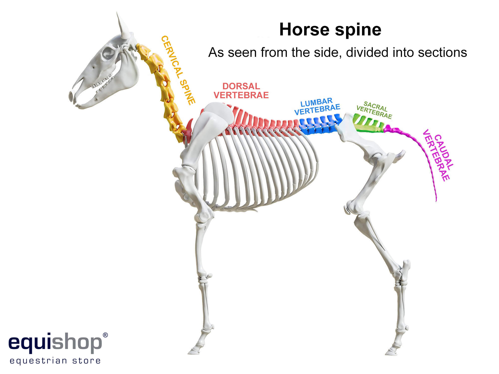
Skeleton of a horse: large hoofed and maned domestic animal of the ungulate family.Raised by humans for pulling loads and for transportation. Atlas: first bone of the neck. Cervical vertebrae: bones of the neck. Thoracic vertebrae: bones that form the dorsal part of the thoracic cage. Lumbar vertebrae: the bones of the lumbar region of the back.
15 Horse Anatomy Quiz Trivia Questions For Horse Lovers
The head or skull includes the skull roof (a set of bones covering the brain, eyes and nostrils), the snout (from the eye to the forward-most point of the upper jaw), the operculum or gill cover (absent in sharks and jawless fish), and the cheek, which extends from the eye to the preopercle.
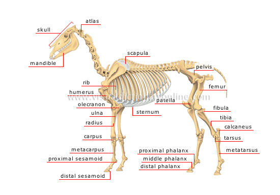
Animal Kingdom Ungulate Mammals Horse Skeleton Of A Horse 1 Image Visual Dictionary Online
We are pleased to provide you with the picture named Anatomy Of The Horse Head.We hope this picture Anatomy Of The Horse Head can help you study and research. for more anatomy content please follow us and visit our website: www.anatomynote.com. Anatomynote.com found Anatomy Of The Horse Head from plenty of anatomical pictures on the internet.
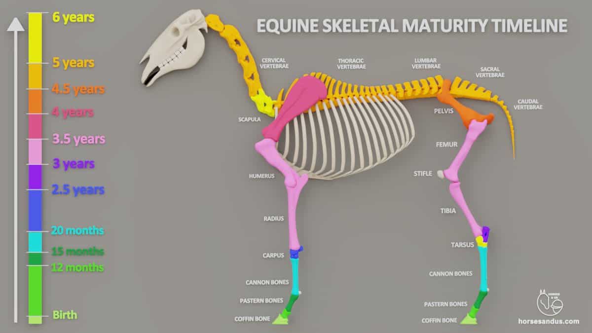
Equine Skeletal Development Charts Animation
07.09.2021 · Dog leg anatomy. First, you might have a basic idea of the different bones of the forelimb and hindlimb of a dog. Now I will provide you the few information on the other bones of dog leg anatomy with their unique features. The front leg of a dog consists of the clavicle, scapula (arm), radius and ulna (forearm), carpals, metacarpals, and phalanges (forepaw).

Horse Skeletal System 3 Diagram Quizlet
Bird anatomy, or the physiological structure of birds' bodies, shows many unique adaptations, mostly aiding flight.Birds have a light skeletal system and light but powerful musculature which, along with circulatory and respiratory systems capable of very high metabolic rates and oxygen supply, permit the bird to fly. The development of a beak has led to evolution of a specially adapted ...
Skeleton Of A Horse Visual Dictionary
Appendicular skeleton — bones of limbs, including scapula & os coxae(hip bone) Heterotopic bones — os penis [ carnivore; rodent ] os cardis [ cattle ] Shape: Long bones — length greater than diameter Short bones — approximately equivalent dimensions Flat bones — e.g., scapula, os coxae, many bones of skull
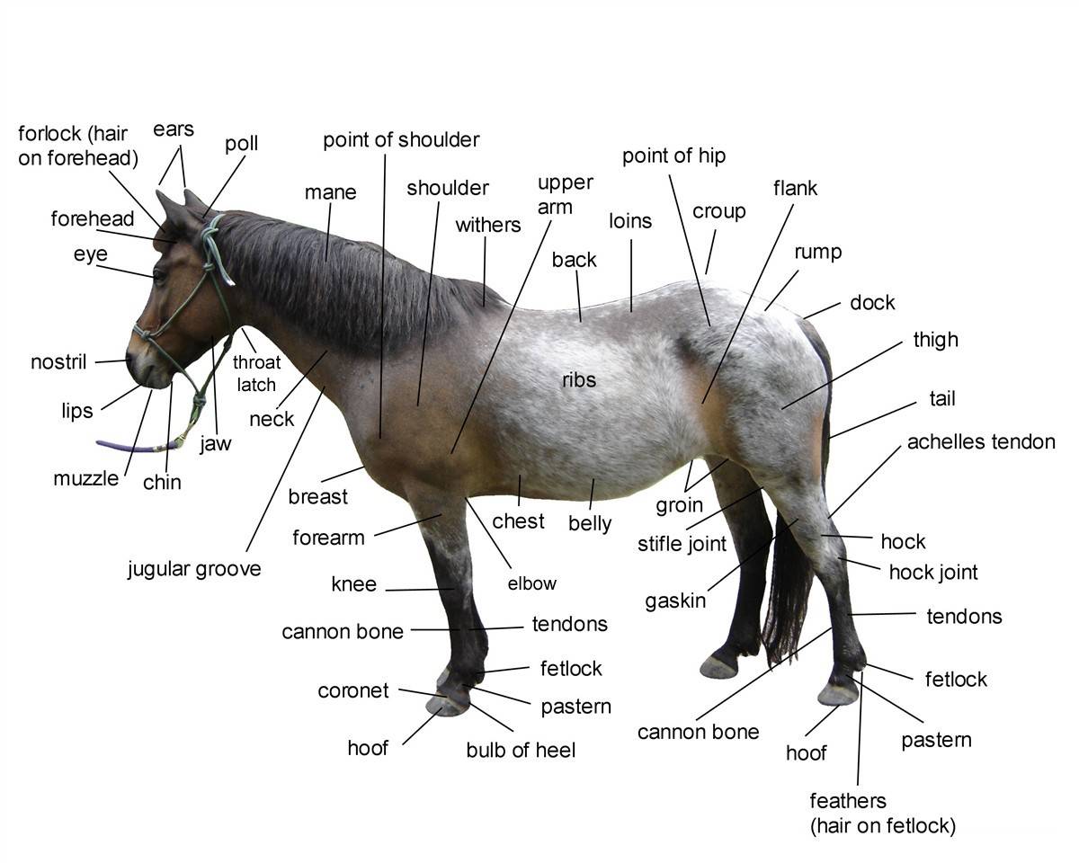
Diagram Of Horse Body Parts
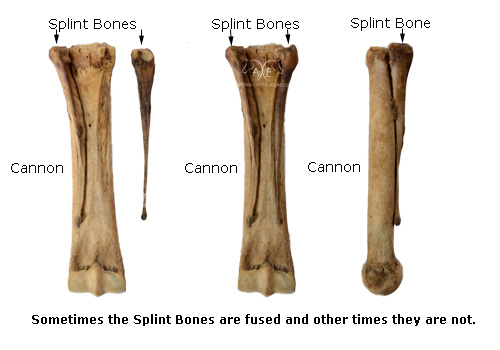
This bone extends from beneath the structures of the knee to the fetlock joint below. Along the cannon bone runs a smaller bone, called the splint bone. In most light horse breeds a cannon bone circumference that is greater than 8 inches is desirable. This means the horse has a sturdy bone mass to carry a load and withstand work.

Skeletal System Of The Horse Wikipedia
Functional Assessment of Cancer Therapy-Bone Marrow Transplant Version 4 Questionnaire Test Name: C179940: Functional Assessment of Cancer Therapy-Bone Pain Version 4 Questionnaire Test Code: C179939: Functional Assessment of Cancer Therapy-Bone Pain Version 4 …

Skeletel System Oct Canine Labrador Puppy Training Mastiffs
Jun 26, 2020 · The pelvis is a ring of bone at hip level, made up of several separate bones. A pelvic fracture is a break in any one of those bones. Some pelvic fractures involve breaking more than one of the bones, and these are particularly serious as the bones are more likely to slip out of line.
Horse Anatomy Diagrams Of Horse Body Parts Equishop Equestrian Shop
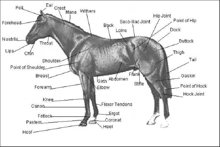
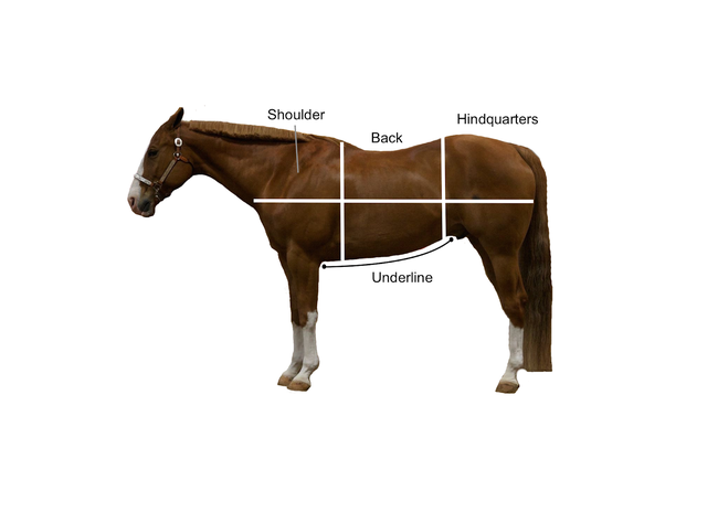
In the Horse Diagram below you can clearly see these main parts along with a example explanation below. Equine conformation evaluates the degree of correctness of a horse's bone structure, musculature, and its body proportions in relation to each other
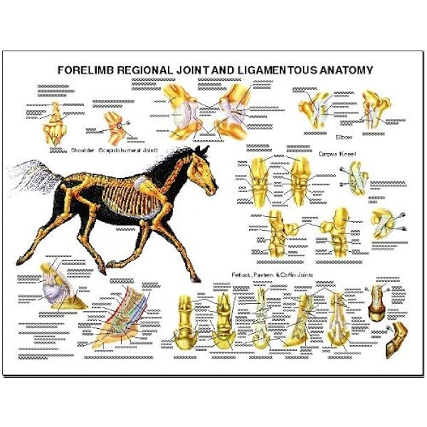
Equine Forelimb Regional Joint Bone Anatomy Chart Horse Science Prints Amazon Com Industrial Scientific
The 3rd carpal bone is by far the largest bone in this row. The 4th carpal bone is approximately 1/2 the width of the 3rd. The 2nd carpal bone is quite small and in the radiograph is less visible than in the diagram due to positioning. THE NAVICULAR BONE Oblique radiographic projections are routinely included in the evaluation of the navicular ...

Skeleton Horse Different Bones Stock Vector Image By C Lesniewski 244762022
It arrives with a clearly labeled diagram for easy assembly.A non-slip product is a safe and practical addition for bathing areas. The button top texture maximizes traction, helping to prevent slips even if wet. Premade layouts with textured surfaces include 10x10 ft, 10x12 ft, and 12x12 ft of coverage area. Installing the pieces goes fast. Durable super density foam portable stall mats are a ...
7 Facts About Your Horse S Skeleton Horse Illustrated
May 29, 2021 · The humerus bone of a cat is comparatively long and less twisted. It is the most robust bone in the cat skeleton and posses a round head. You will find a supracondylar foramen on the distomedial aspect of the cat humerus bone. But in some cat species, this supracondylar foramen may absent.

The Art Of Equine Skeletal And Muscular Systems Skeletal And Muscular System Horse Anatomy Horse Therapy
To summarize it: the movements of the horse's shoulder are going to be fairy limited compared to ours, due to the fact that the joint is against the rib cage. The main ones are going to be flexion/extension, where the joint is going to slide against the ribcage either forward or back. The flexion happens in stance phase, extension during ...

The Horse S Skeleton Hind Limbs Youtube
A series of anatomy charts to help you understand the parts of your horses's body. Whole Body Anatomy (75Kb) Skeleton (90Kb) Internal Organs (70Kb) Lower Limb Structure (1.3Mb) Hoof Cross Section (60Kb) Hoof Ground Surface (95Kb) Skull & Jaw (1.5Mb) Why not take a look at our downloadable VetEq Notes?
The Distal Limb Bones Of The Equine
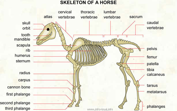
The caudal/coccygeal vertebrae make up the tail bone. A horse may have 18 - 22 individual caudal/coccygeal vertebra. C1 (Atlas) C2 (Axis) Lecture - Equine Skeletal System Return to Table of Contents Appendicular Skeleton The appendicular skeleton consists of the bones in the limbs. A horse's appendicular skeleton
Components Of The Musculoskeletal System Of Horses Horse Owners Merck Veterinary Manual
The small intestine is the 'work horse' of digestion, as this is where most nutrients are absorbed. Peristalsis is also at work in this organ, ...
1
Here are numerous simple short stories. These short stories are for all people to read. We all enjoy reading short stories. After the introduction of computer and the invention of internet, many people come to internet to spend their leisure time.
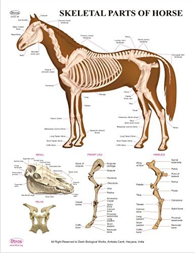
Dbios Educational Digital Printed Skeletal Parts Of Horse Anatomy Wall Charts Buy Online In Guernsey At Desertcart 67130694
Knees, stifle joint, the hock, cannon bone, fetlock and pasterns are all subject to a lot of stress on hard working horses. Proper hoof care, good exercise management and good conformation (how the horse is built) all play a factor in how well a horse will hold up to strenuous working conditions.

Horse Life And Love All About The Skeleton And Bones
Talking about Horse Anatomy Worksheets Printable, we have collected some related images to complete your references. horse body parts diagram, horse skeleton diagram and animal nervous system diagram are some main things we want to present to you based on the gallery title.
External Equine Anatomy All Things Equine
Horse Skeleton Diagram Bone Up on Your Horse's Framework. Take a look at this drawing of a horse skeleton. You are looking at about 205 bones that make up the equine skeletal anatomy. The more you study this picture the better understanding you will have of how a horse is built and how he moves.

External Anatomy Of The Horse With Easy To Read Chart Pdf Download My Life Is Better With Horses
In the cross section diagram above P3 appears to sit at a fairly steep angle to the ground plane. However this is because you are viewing the bone when it is cut in half. So the bottom edge is actually not shown in a cross section diagram. See photos below that show the full bone and a cross section of the bone when viewed from the side.
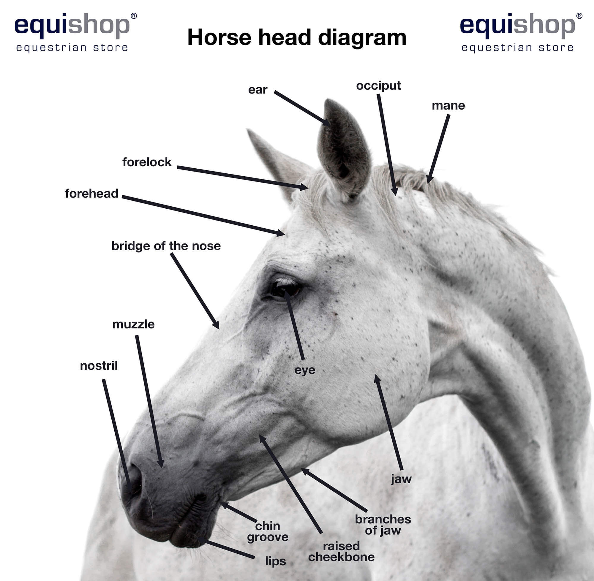
Horse Anatomy Diagrams Of Horse Body Parts Equishop Equestrian Shop
Horse - Bones of the cranium (Occipital bone, Temporal bone, Parietal bone...) Vertebral column (Labeled atlas of veterinary anatomy: equine osteology on illustrations) Horse - Skeletal system: Thoracic skeleton, Ribs, Costal cartilage, Sternum. Sternum-Veterinary anatomy (Horse):Manubrium of sternum, Cartilage of manubrium, Sternebrae, Xiphoid ...
Skeleton Of A Horse With The Different Bones Stock Vector Illustration Of Engraving Mammal 139290713
Directional Terms, Skeletal, and Muscle Introduction. These horse anatomy diagrams are a great overview and introduction to the vast study of equine anatomy.. These diagrams should explain and show you some of the basics. That way if you need to talk to a vet, or do a correct drawing, you'll have a solid foundation.

Horse Leg Skeletal Diagram Diagram Quizlet
About this Quiz. This is an online quiz called horse skeletal system. There is a printable worksheet available for download here so you can take the quiz with pen and paper.

Horse Anatomy Drawing Step By Step Drawing Guide By Dawn Dragoart Com
We are pleased to provide you with the picture named Horse leg muscles and skeleton structure diagram.We hope this picture Horse leg muscles and skeleton structure diagram can help you study and research. for more anatomy content please follow us and visit our website: www.anatomynote.com. Anatomynote.com found Horse leg muscles and skeleton structure diagram from plenty of anatomical pictures ...
Why Saddle Fit Matters The Anatomy Under The Perfect Fit Flair Strips
There are several diagrams in this resource covering basic body anatomy and basic hoof anatomy. After each diagram, there will be a glossary of terms used in the diagram to provide more clarity. Let's start out looking at a diagram showing basic horse anatomy.

The Long And Short Of It Part 2a Back Hooves
in bone growth (endochondral ossification) affecting young horses. The cartilage is poorly affixed to the underlying bone and is readily pealed away. At left is a large lesion on the medial trochlear ridge of a weanling quarter horse. The cartilage has been removed, showing the abnormal subchondral bone. This photo was taken at necropsy.
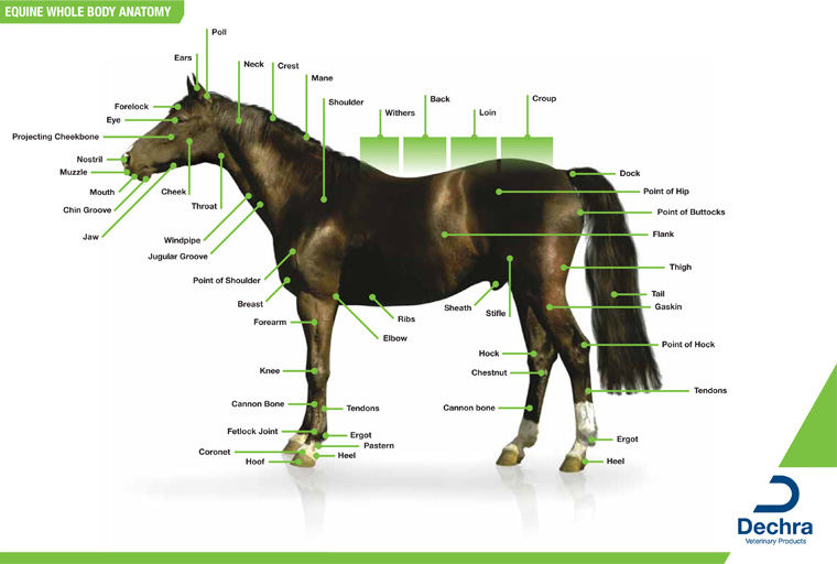
Downloads Anatomy Charts Dechra Veterinary Products
Dermal bone is from mesenchyme and ectomesenchyme of the dermis and it overlies ... (cat, horse, human) or confluent with it (wolf, rat). Crests: Notice on the wolf skull the median longitudinal sagittal crest to attach temporal muscles and a transverse nuchal crest for muscles supporting the head. THE PALATE. Modifications for breathing air: evolution of the secondary palate. The secondary ...

Equine Anatomy Wikipedia
Horses have, on average, a skeleton of 205 bones. A significant difference in the bones contained in the horse skeleton, as compared to that of a human, is the lack of a collarbone. Their front limb system is attached to the spinal column by a powerful set of muscles, tendons and ligaments that attach the […]
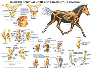
Equine Hind Limb Anatomy Chart Wc02b
How To Draw Animals Horses Their Anatomy And Poses

A P Chapter 7 Horse Skeleton 1 3 Diagram Quizlet

Equine Skeletal System Poster

Amazon Com Equine Hindlimb Regional Joint Bone Anatomy Chart Horse Toys Games
Conformation Of The Horse
Horse

Free Vector Science Horse Skeletal System
1

Horse Skeleton Flashcards Quizlet
Horse Skeleton Anatomy Osteological Features Of Bones From Equine Skeletal System Anatomylearner The Place To Learn Veterinary Anatomy Online

The Horse Skeleton Has About 205 Bones That Make Up The Equine Skeletal Anatomy Description From Viral Country I Searched Horse Anatomy Horses Horse Healing

Saddle Fit And Horse Anatomy Easy Fit Saddles


Comments
Post a Comment