43 mitochondria diagram labeled
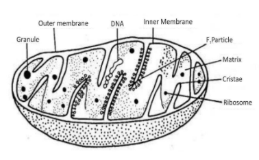
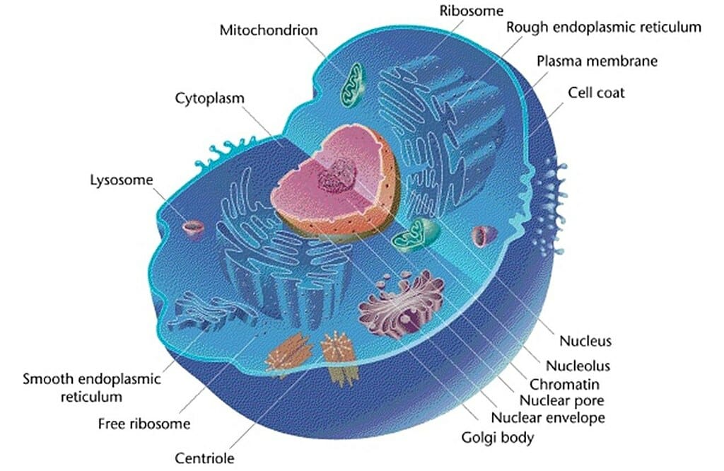
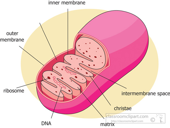
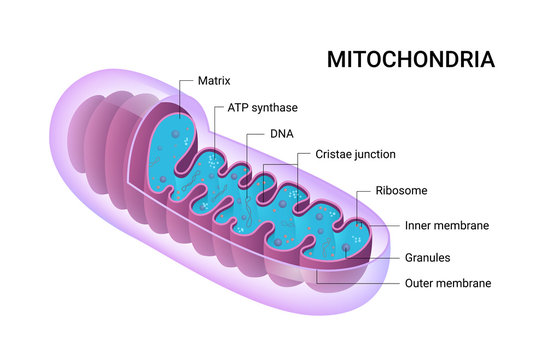
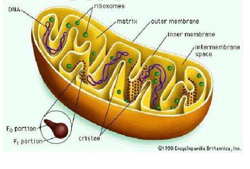
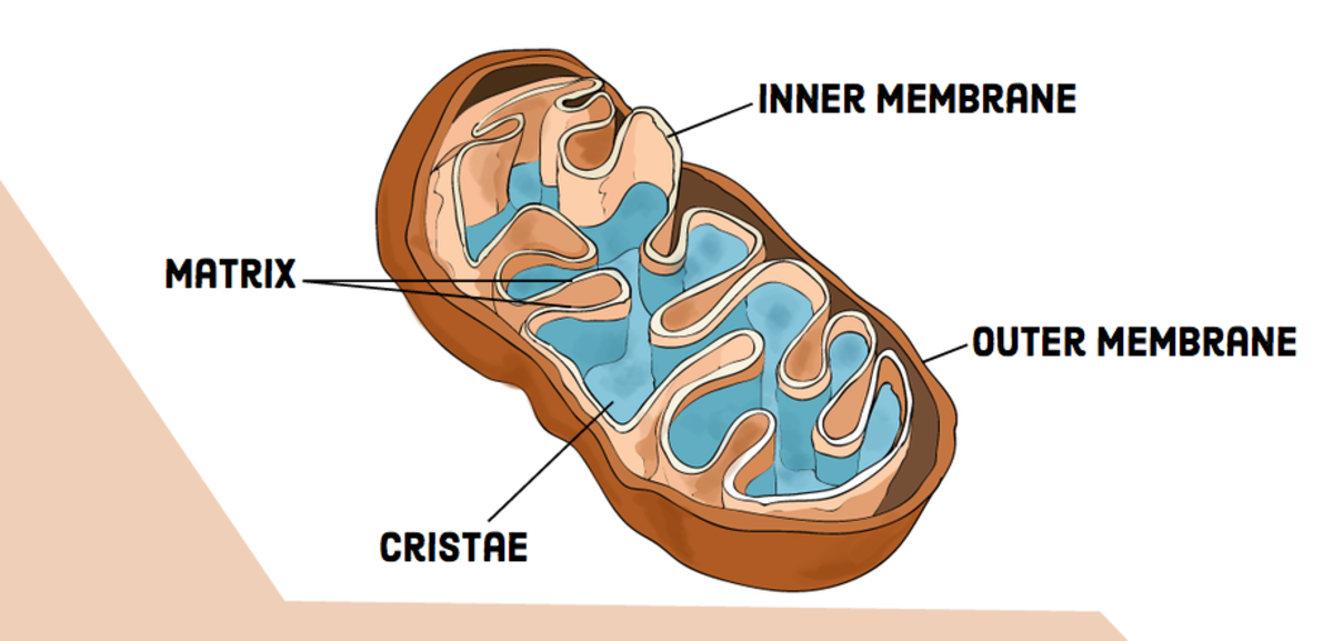
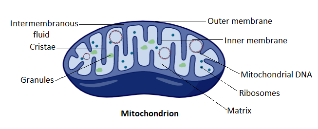
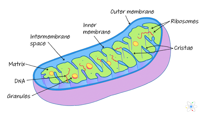
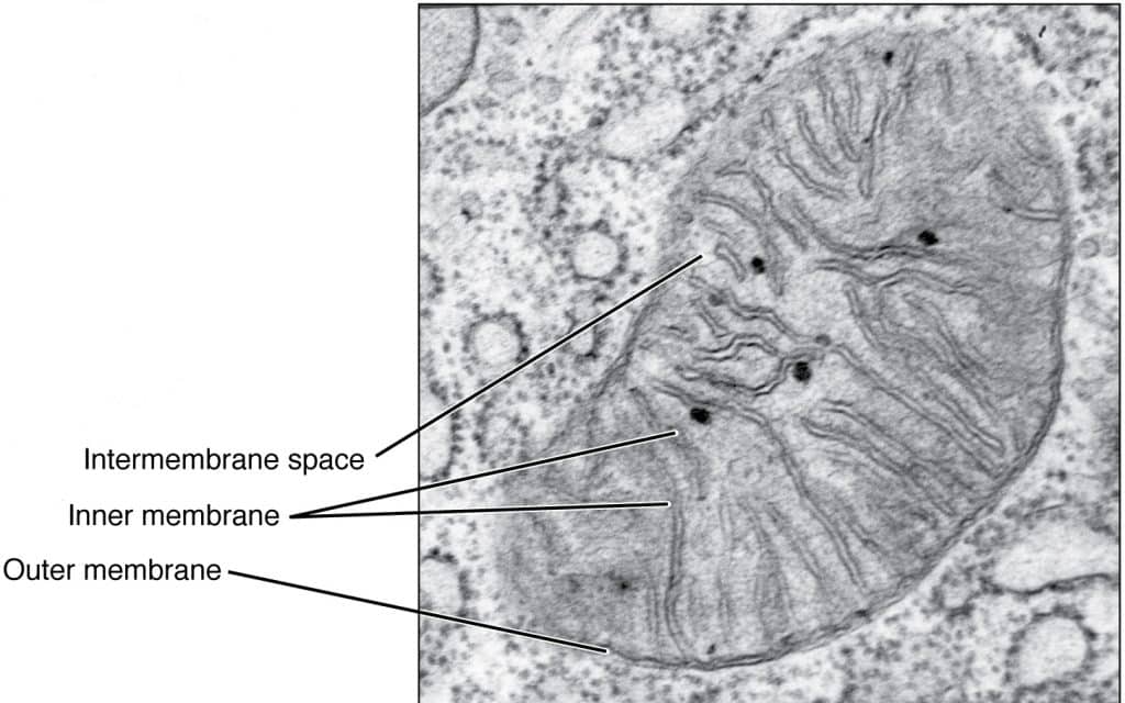
Mitochondria have a double-membrane structure and contain many substructures including enzymes, ribosomes and mitochondrial DNA (mtDNA). Structure of ... Figure: Well labelled diagram of mitochondria Well labelled Diagram of mitochondria and their parts a) Mitochondrial outer membrane. Mitochondrial outer and inner membranes are closer to each other at a particular region called contact site or contact zones.
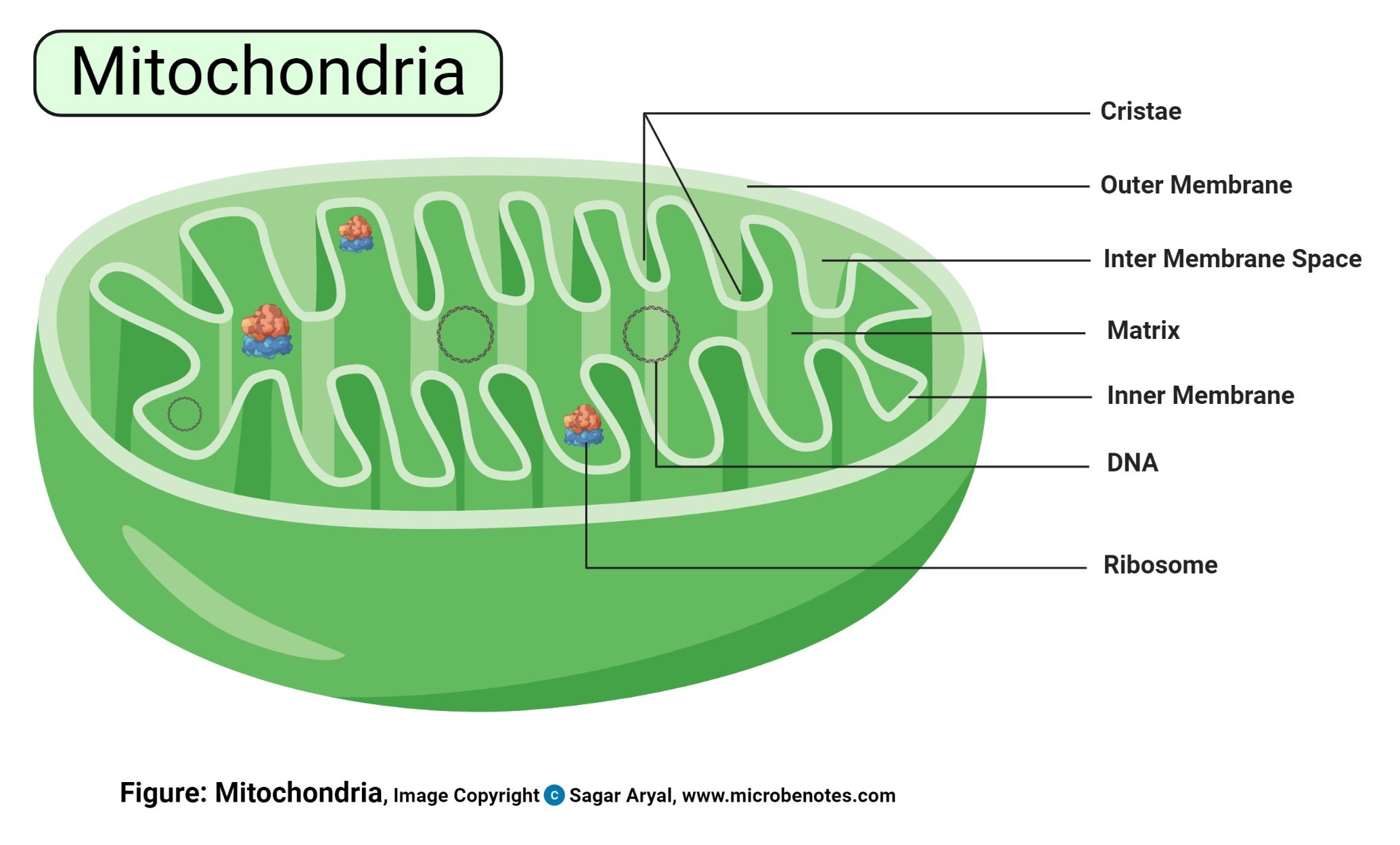
The Mitochondria are organelles that act like a digestive system which takes in nutrients, breaks them down, and creates energy rich molecules for the cell. The biochemical processes of the cell are known as cellular respiration.Here you have some diagrams of the Mitochondria Structure and Parts.
Mitochondria diagram labeled
Animal Cell Diagram Mitochondria Labeled. Tuesday, April 27th 2021. | Diagram. Animal Cell Diagram Mitochondria. Animal cells have a basic structure. The mitochondrion (plural mitochondria) is a membrane-bound organelle found in the cytoplasm of eukaryotic cells. The structure of the mitochondrion is adapted to the function it performs AND Annotation of a diagram of a mitochondrion to indicate the adaptations to its ... 3,421 mitochondria stock photos, vectors, and illustrations are available royalty-free. See mitochondria stock video clips. of 35. 3d mitochondria organism energy icon genetic cross mitochondries mitochondria structure mitochondrion animal cells diagram mitochondria diagram atp energy mitochondrian. Try these curated collections.
Mitochondria diagram labeled. Mitochondria are double membrane-bound cell organelles responsible for the supply and storage of energy for the cell. The oxidation of various substrates in the cell to release energy in the form of ATP (Adenosine Triphosphate) is the primary purpose of mitochondria. Structure of Mitochondria This will also help you to draw the structure and diagram of mitochondria. 1. Mitochondria are commonly called the "Power house" of the cell. 2. Benda (1897) was the first to coin the term mitochondrion. 3. Usually, mitochondria are 0.5 to 1 n in diameter and 3-6n in length. Oxidative Phosphorylation of Mitochondrion (With Diagram) Linkage of phosphorylation to oxidative metabolism was first proposed in the 1930s. The first evidence was the observation that phosphate was removed from the medium when TCA cycle intermediates were metabolized by cells. Subsequently, it was shown that the disappearance of inorganic ... How Many Membranes Are Present In Mitochondria. Mitochondria have generally spherical or rod-shaped or filamentous structures. The inner membrane separates the mitochondrial matrix from the intermembrane space. A bacteria diagram in actual fact facilitates us to profit more approximately this unmarried cell organisms which have neither membrane-bounded nucleolus or organelles like mitochondria ...
Mitochondria Diagram Unlabeled. mitochondrion the mitochondrion plural mitochondria mitochondrion ultrastructure interactive diagram a mitochondrion has a double membrane printable animal cell diagram - labeled unlabeled and blank printable animal cell diagram to help you learn the organelles in an animal cell in preparation for your test or quiz 5th grade science and biology Hi /r/biology. I'm currently enrolled in a first year biology class and while I do great at most of the class, many slides that I encounter in labs leave me scratching my head. Real life cells are very different from the well organized and labeled diagrams in textbooks. I have some questions about identifying parts of cells. For [cells like this](http://i.imgur.com/idloiLK.jpg), I can obviously make out chloroplasts and the cell wall, but I'm at a loss as to where everything else is. How would... The fusion fission cycle of mitochondria. C). Internal structure. D). MtDNA, structure and packaging. ... labeled mitochondria showing the looping of the reticulum throughout its length. transitioning of mitochondria between punctate and reticulum states through alternating fission and fusion is now known to be critical to A WELL LABELED DIAGRAM OF MITOCHONDRIA. Leave a Comment / By Manoj Saxena. A WELL LABELED DIAGRAM OF MITOCHONDRIA.
Mitochondria are the "power houses" of cells. Eukaryote cells, meaning animal cells (more advanced organisms) are the type of cells that have them, and they need mitochondria to function properly. This quiz has tags. Click on the tags below to find other quizzes on the same subject. Start studying Mitochondria Labeling. Learn vocabulary, terms, and more with flashcards, games, and other study tools. Structure of Mitochondria. Mitochondria are mobile, plastic organelles that have a double-membrane structure. It ranges from 0.5 to 1.0 micrometer in diameter. It has four distinct domains: the outer membrane, the inner membrane, the intermembrane space, and the matrix. The organelle is enclosed by two membranes—a smooth outer membrane and a ... Mitochondria structure label workbook diagram 1. Cellular respiration is the process that releases energy from food in the presence of oxygen. Explain, in general terms, how carbohydrates are oxidized by glycolysis and krebs cycle to practice: Diagram and describe how eukaryotic cellular respiration works.
Draw A Neat Diagram And Label The Following Diagram Class 9 Biology Cbse
Mitochondria (singular: mitochondrion) are organelles within eukaryotic cells that produce ... This diagram shows the structure of a mitochondrion.
Animal Cell Definition Organelles Structure Parts Functions Labeled Diagram Worksheet
Download mitochondria-diagram-labeled : Filetype: Size: EPS/Vector format Available for Purchase: PNG with Transparent Background (Members) GIF with transparent Background (Members) Large JPG (Members) Medium JPG (Members) Free Download: 87 Kb: To Purchase Clipart as Vector File contact support@classroomclipart.com

437 Mitochondria Vector Images Mitochondria Illustrations Depositphotos
Click here to get an answer to your question ✍️ Draw a neat diagram and label the following diagram: MitochondriaWhich reaction is operated in ...1: Cristae, Matrix, Ribosome2: Nerve, Muscle, Golgi apparatus1 answer · Top answer: respiration process is operated in mitochondria.

Animal Cell Definition Organelles Structure Parts Functions Labeled Diagram Worksheet
As the video plays, (remember: you can pause the video at any time) label everything you see labeled in the video on your own diagram of a mitochondria and a phospholipid bilayer. Note: the narrator uses a variety of colors, which is a good idea to use, especially if you are a visual learner or a color learner.

29 Labeled Mitochondria Diagram Illustrations Clip Art Istock
Structure of the Mitochondria. Mitochondria are membrane-bound organelles enclosed by a double membrane. They have a smooth outer membrane enclosing the organelle and a folded inner membrane. The folds of the inner membrane are called cristae, the singular of which is crista, and the folds are where the reactions creating mitochondrial energy ...
Mitochondrion Definition Structure And Function Biology Dictionary
The mitochondrion is a double-membraned, rod-shaped structure found in both plant and animal cell. Its size ranges from 0.5 to 1.0 micrometre in diameter. The structure comprises an outer membrane, an inner membrane, and a gel-like material called the matrix. The outer membrane and the inner membrane are made of proteins and phospholipid layers ...
31 Draw And Label Mitochondria Labels Design Ideas 2020
The diagram of Mitochondria is useful for both Class 10 and 12. It is one among the few topics having the highest weightage of marks and is majorly asked in the examinations. Read More: Mitochondria. Diagram Of Mitochondria. The diagram below shows the structure and functions of the mitochondria.

Labeling A Mitochondria Diagram Quizlet
Learn about Mitochondria a topic of Biology in details explained by subject experts on vedantu.com. Register free for online tutoring session to clear your doubts. Here you will understand the structure of Mitochondria using Labelled Diagram of Mitochondria.
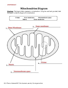
Mitochondrion Cellular Respiration Diagram Worksheet By A Thom Ic Science
Mitochondria occupy a substantial portion of the cytoplasmic volume of eucaryotic cells, and they have been essential for the evolution of complex animals. Without mitochondria, present-day animal cells would be dependent on anaerobic glycolysis for all of their ATP. When glucose is converted to pyruvate by glycolysis, only a very small fraction of the total free energy potentially available ...

Biology4kids Com Cell Structure Mitochondria
#mitochondria #biologydiagram #cellbiologyA beautiful drawing of a Mitochondria . And it will teach you draw Mitochondria very easily. Watch the video and p ...

Mitochondria Labeling Diagram Quizlet
Read, Answer, Color, Label: Mitochondria. Mitochondria are the powerhouses of the cell because they "burn" or break the chemical bonds in glucose to release energy to do work in a cell. Remember that this energy originally came from the sun and was stored in chemical bonds by plants during photosynthesis.

Solved Label Structures On The Mitochondrion Below Then Chegg Com
This BiologyWise article the structure and function of mitochondria with the help of a labeled diagram. The study of cell organelles and their functions covers individual responsibilities of each subunit of a cell - each organelle is a structure within a cell that has a specific function.
Solved Label Two Parts Of The Mitochondria Label Where Each Step Of Aerobic Cellular Respiration Occurs Use Arrows To Show What Goes Into Each Pa Course Hero
〔 〕〕 〔 〕 The story goes like this: Earth is captured by a technocapital singularity as renaissance rationalitization and oceanic navigation lock into commoditization take-off. Logistically accelerating techno-economic interactivity crumbles social order in auto-sophisticating machine runaway. As markets learn to manufacture intelligence, politics modernizes, upgrades paranoia, and tries to get a grip. The body count climbs through a series of globewars. Emergent Planetary Commercium trashes the...

Mitochondrial Ribosome Wikipedia
Functions of Mitochondria. The most important function of the mitochondria is to produce energy. The simpler molecules of nutrition are sent to the mitochondria to be processed and to produce charged molecules. These charged molecules combine with oxygen and produce ATP molecules. This process is known as oxidative phosphorylation.

How To Draw The Diagram Of Mitochondria Easy Steps By Step For Beginners Youtube
Note: I take Pure Chem + Bio. All Physics tips are courtesy of my classmates and this community ([https://www.reddit.com/r/SGExams/comments/qqr4pn/o\_levels\_pure\_physicscombined\_science\_p1\_tips/](https://www.reddit.com/r/SGExams/comments/qqr4pn/o_levels_pure_physicscombined_science_p1_tips/)) ​ 3rd last exam day till the end. Might be your last day for some. Either way, hope you had a good rest yesterday. Pure Physics starts at 8am, so make sure to head to school earlier. &a...

Mitochondrion Cellular Respiration Diagram Worksheet Cellular Respiration Biology Lessons Biology Notes
Cellular respiration diagram mitochondria. The second worksheet has students identify the main reactants and products of cellular respiration as they relate to the mitochondrion. Atp is the energy currency of cells and is produced inside the mitochondria. ... The first is a simple worksheet that has students label the main parts of a mitochondrion.
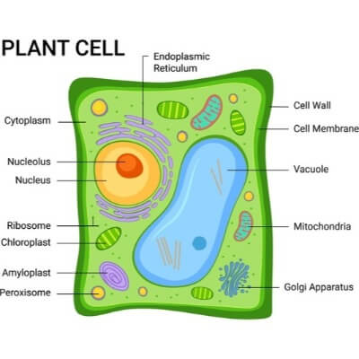
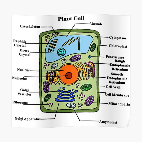
Plant Cell Facts Facts Just For Kids
Find mitochondria diagram stock images in HD and millions of other royalty-free stock photos, illustrations and vectors in the Shutterstock collection.
Science Clipart Mitochondria Diagram Labeled Classroom Clipart
How Many Membranes Are Present In Mitochondria. It has many folds that form a layered structure called cristae, and this helps in increasing the surface area inside the organelle. The cristae and the proteins of the inner membrane aids in the production of ATP molecules.

Mitochondria Functions Location Diagram And Structure Jotscroll
Given below is a well- labeled diagram of a mitochondria to help in better understanding of its structure. Structure of Mitochondria. What are Chloroplasts? Besides plant cells, chloroplasts are also found in algae. They are located in the cytosol of a cell, and contain chlorophyll to absorb solar energy.

Lysosomal Mediated Degradation Of Mitochondria A Mitochondria Are Download Scientific Diagram
3,421 mitochondria stock photos, vectors, and illustrations are available royalty-free. See mitochondria stock video clips. of 35. 3d mitochondria organism energy icon genetic cross mitochondries mitochondria structure mitochondrion animal cells diagram mitochondria diagram atp energy mitochondrian. Try these curated collections.

Mitochondria Form Function And Disease
The structure of the mitochondrion is adapted to the function it performs AND Annotation of a diagram of a mitochondrion to indicate the adaptations to its ...

Muscle Membrane Vector Illustration Labeled Scheme With Myofibril Disc Zone Line And Band Anatomical Diagram With Mitochondria Sarcoplasm Reticulum And Nucleus Art Print Barewalls Posters Prints Bwc59793239
Animal Cell Diagram Mitochondria Labeled. Tuesday, April 27th 2021. | Diagram. Animal Cell Diagram Mitochondria. Animal cells have a basic structure. The mitochondrion (plural mitochondria) is a membrane-bound organelle found in the cytoplasm of eukaryotic cells.

Cellular Respiration Mitochondria Diagram Quizlet

How To Draw Mitochondria Diagram Easy And Well Labelled Diagram Ncert Power House Of The Cell Youtube
49 Best Cristae Images Stock Photos Vectors Adobe Stock
33 Draw And Label A Mitochondria Labels Design Ideas 2020

Mitochondria Structure Functions And Diagram Studiousguy
Labeled Plant Cell Diagram Poster By Bundabear Redbubble
1
Cell Organelles Definition Structure Functions Labeled Diagram

Structure Function Of Mitochondria 2019 21 Cie A Level Biology Notes

Assignment 5 Page 3
Compare And Contrast Chloroplasts And Mitochondria Owlcation

Mitochondria Parts Free Vector Eps Cdr Ai Svg Vector Illustration Graphic Art
1

35 Label The Mitochondria Labels For Your Ideas

1 696 Mitochondria Stock Photos And Images 123rf
Label The Diagram And Write Down The Details Of The Class 11 Biology Cbse

Printable Animal Cell Diagram Labeled Unlabeled And Blank
Mitochondria Definition Structure Function With Diagram

Mitochondria Definition Structure Function With Diagram
Mitochondria Structure Function Teachmephysiology
3


Comments
Post a Comment