38 rat muscles diagram
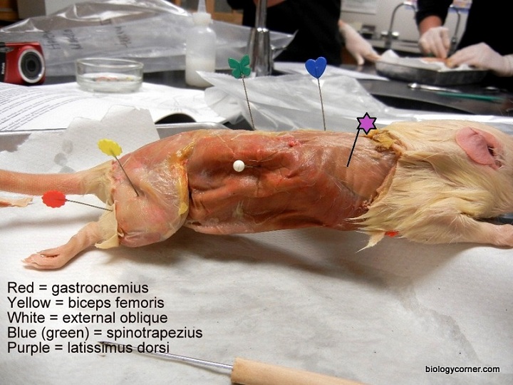
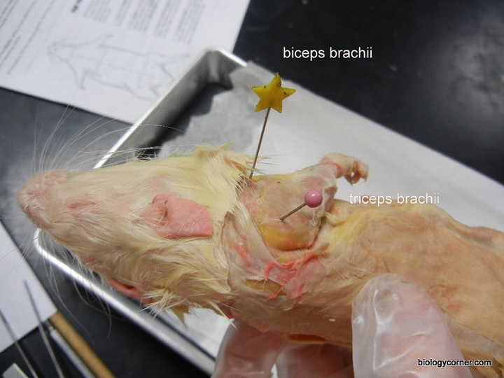
Rat Muscles. Biceps brachii. triceps brachii. Spinotrapezius. latissimus dorsi. Located on the anterior side of the upper arm (flexes lower ar…. located on the sides and back of upper arm (extends lower arm) located across the dorsal thoracic region of rat (moves scapul…. Start studying Rat Muscles- Labeling. Learn vocabulary, terms, and more with flashcards, games, and other study tools.
Anatomy. A basic explanation of the rat's reproductive system is useful information for both the breeder and the rat owner. The purpose of the reproductive system is to ensure the continuation of the species. Rats have shown a remarkable ability to survive and thrive. This, in part, is due to their ability to reproduce often and to give birth ...
Rat muscles diagram
Part 1 of 2. Shows the dissection of muscles of the face, neck, chest, back and upper limb. Figure 8. Rat skull, showing the forward placement of the attachment points of the masseter muscle on the upper jap. The medial masseter muscle passes through the eye socket, right next to the eye, and attaches on the muzzle. This is the muscle that "boggles" the eye when the rat bruxes. Rat Skeletal Muscle Tissue Sections. Skeletal muscle exhibits an alternating light and dark striped appearance when viewed under a light microscope and is, consequently, often referred to as striated muscle. This type of muscle is typically under conscious control and comprises the majority of the body's muscle mass.
Rat muscles diagram. Use the lines on the diagram to cut a similar pattern, avoiding the genital area. Gently peel the skin from the muscles, using scissors and a probe to tease away muscles that stick to the skin. Muscles are attached to bones by connective tissue called tendons that adhere to spines, knobs, and ridges on bones. You will need to refer to the rat ... To determine the extent to which the rat abdominal wall resembles that of human, 10 adult male Sprague-Dawley rats were killed and formalin-fixed for architectural and morphological analyses of the four abdominal wall muscles (rectus abdominis, external oblique, internal oblique, and transversus abdominis). Veins (see diagram page 9) Your rat specimen has been double injected with latex to help you identify veins and arteries. Veins carry used blood (blue) back to the heart and lungs. The lungs re-oxygenate the blood and the heart pumps it back to the rest of the body. In the human body, these veins are not the same bright blue that you see in ... This is an online quiz called Rat Muscles Dorsal . There is a printable worksheet available for download here so you can take the quiz with pen and paper. Your Skills & Rank. Total Points. 0. Get started! Today's Rank--0. Today 's Points. One of us! Game Points. 13. You need to get 100% to score the 13 points available.
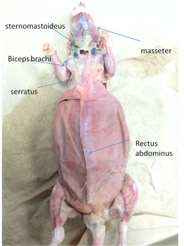
You should encounter two brownish muscles attached to the skin (the cutaneous maximus in the trunk area, and the platysma in the neck area). You will probably have to cut these muscles close to the skin before the skin can be removed. MUSCULAR SYSTEM The diagram below illustrates the muscles of the ventral surface of the rat. Be able to ... Rat is a common rodent of the order Rodentia.It belongs to the class Mammalia of the subphylum Vertebrata and is often studied as a representative organism for mammals. As the structural organization of rats and humans are similar, it is widely used in medical research to evaluate the effects of medications, before releasing them for human use. The majority of muscles in the leg are considered long muscles, in that they stretch great distances. As these muscles contract and relax, they move skeletal bones to create movement of the body. a playlist by sethjones. •. 60,265 plays. too few (you: not rated) Several pictures in the text book of Rat Anatomy. Play All Use in Tournament Branch. Rat Muscles Dorsal by shari23 31,351 plays 13p Image Quiz. Rat Muscles Ventral by shari23 18,968 plays 13p Image Quiz. Rat Skeleton by shari23 22,526 plays 34p Image Quiz.
Biology 129 - Review of Rat Muscles. Select one of the following regions: Ventral Head and Neck Muscles ; Superficial Shoulder and Back Muscles 5 Movement of the skeleton requires the coordinated activity of muscles. (a) The diagram below shows the arrangement of some muscles around the human knee joint. hamstring muscles patella quadriceps muscles (i) Put a cross in the box next to the row that correctly describes the hamstring and quadriceps muscles when flexing (bending) the knee. (1) Download scientific diagram | Rat's gastrocnemius muscle applied to the CCl 4. PAS from publication: The effect of melatonine on rats gastrocnemius muscle applied with carbon tetrachloride (CCl 4 ... Rat Dissection Manual. Rats are a great, economical choice for a dissection specimen for beginning anatomy students. Preserved rats are inexpensive, easily skinned, and have highly-developed muscles that are very comparable to those of humans. This Google Slides presentation is designed for students to refer to as they dissect through the ...
Closeup of skeleton pelvic model
The dynamic passive response of the left gastrocnemius medialis muscle of thirty male Wistar rats was studied as a function of muscle dimensions and absolute and relative amount of connective tissue. Values of the absolute active and passive length-force curves (active force, passive force, active working range) correlated well (coefficients of correlation in a range of 0.62-0.92) with ...
Rat Dissection Step 6
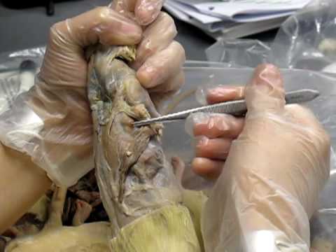
*** Study the diagram to learn the bones of the rat. ... You will carefully remove the skin of the rat to expose the muscles below. This task is best accomplished with scissors and forceps where the skin is gently lifted and snipped away from the muscles. You can start at the incision point where the latex was injected and continue toward the tail.
Scientific Illustration | Rat musculature
The floating ribs are not drawn. 31. Right scapula and clavicle. 32. Thoracic cage, formed by 13 thoracic vertebrae, 13 pairs of ribs and the sternum. 33. Lateral aspect of right humerus, radius and ulna. 34. Flexor surface of right humerus.

The Piman Rattaya Throne Hall in The Grand Palace
A3D Rat Anatomy LESSON 2.2 MUSCLES Like rats, human muscles only pull. They come in pairs that complement each other. And they are bulkier near the trunk, thinner near the extremities. Compare the legs and you see that the human upper leg emerges from the body. The upper leg of a rat is buried in

Histological analysis in rat skeletal muscle contusion. a ...
1. Observe the interior of the rat for any veins and arteries. Veins carry used blood (blue) back to the heart and lungs. Arteries carry oxygenated blood to the muscles and organs that need it. The arteries in your rat should be stained red for easy identification. Use Figures 3 and 4 to help you locate the major veins and arteries.

Rat Anatomy
Muscle roles within a given movement (classification of involved muscles): — agonist = prime mover or principal muscle(s) executing the particular joint movement — antagonist = muscle(s) that oppose the action of the agonist on the joint(s) — synergist = muscle(s) that assist the agonist; e.g., fixators stabilize distant joints. Muscle ...

Image from page 62 of "The butterfly book;" (1898)
Review of Rat Anatomy These pages will show you pictures of parts of a dissected rat with structures identified by numbers. To quiz yourself, see if you can identify the numbered parts. You can then check your answers by looking at another page which will show the same picture with all the parts labled.

Rat Dissection And Anatomy - Biology 1105 with Bracey at ...
You will carefully remove the skin of the rat to expose the muscles below. This task is best accomplished with scissors and forceps where the skin is gently lifted and snipped away from the muscles. You can start at the incision point where the latex was injected and continue toward the tail. This page shows images of how a rat is skinned and what the exposed muscles look like.

Label the White Rat Muscles
The rat is a typical mammal. Formerly guinea pig (Cavia sp.) (Fig. 19.1) were used for dissection in most of the undergraduate and postgraduate colleges in Indian Universities. Of late, due to prevailing high cost, guinea pig is being replaced by rat. Four species of rats are common in India, of which three are wild.

Histological sections of rat muscle tissue obtained in 1 ...
You will carefully remove some of the skin of the rat to expose the muscles below. You will only need to remove the skin from the areas indicated under muscular system on the check list. This task is best accomplished with scissors and forceps where the skin is gently lifted and snipped away from the muscles. Use the lines on the diagram above as a

Image from page 67 of "The butterfly book [microform] : a popular guide to a knowledge of the butterflies of North America" (1898)
The dominant muscle in the upper chest is the pectoralis major. This large fan-shaped muscle stretches from the armpit up to the collarbone and down across the lower chest region on both sides of ...

Image from page 42 of "Principles of education" (1910)
Rat Skeletal Muscle Tissue Sections. Skeletal muscle exhibits an alternating light and dark striped appearance when viewed under a light microscope and is, consequently, often referred to as striated muscle. This type of muscle is typically under conscious control and comprises the majority of the body's muscle mass.

Transplantation procedure of rat cremaster muscle. (A) The ...
Figure 8. Rat skull, showing the forward placement of the attachment points of the masseter muscle on the upper jap. The medial masseter muscle passes through the eye socket, right next to the eye, and attaches on the muzzle. This is the muscle that "boggles" the eye when the rat bruxes.
Frontiers | Connections of the superior colliculus to ...
Part 1 of 2. Shows the dissection of muscles of the face, neck, chest, back and upper limb.

Image from page 68 of "The butterfly book; a popular guide to a knowledge of the butterflies of North America" (1904)

Schematic representation of a cross-section of the rat ...

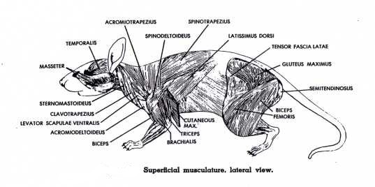
Rat Muscles - Lateral View - PurposeGames

The rat's muscle anatomy. The rat's muscle anatomy is ...

A Color atlas of sectional anatomy of the rat | Anatomy ...

Muscular System - Rat Dissection

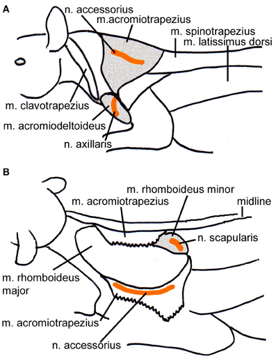
Anatomy and innervation of the iliococcygeus and the ...
Rat Dissection Step 4

Rat Muscle Anatomy - Anatomy Drawing Diagram
Rat muscle dissection - YouTube
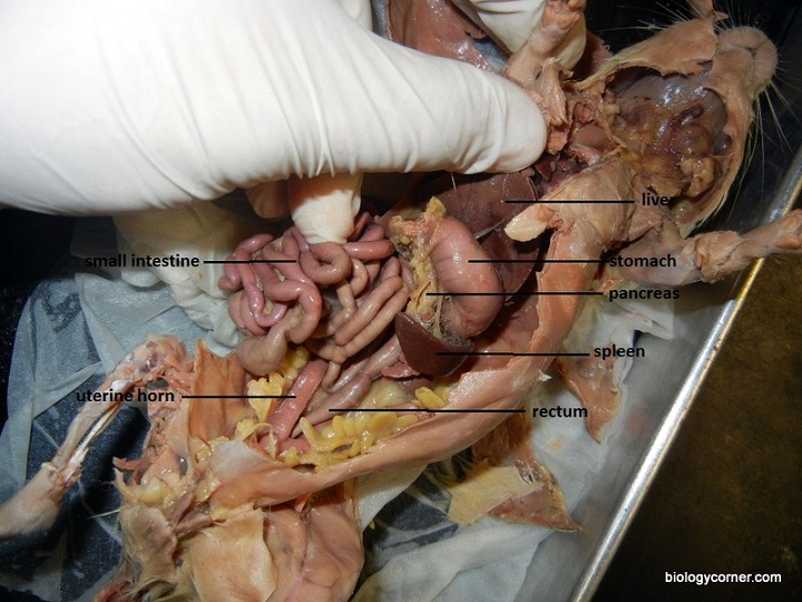
Muscular - Pregnant Rat Dissection

Image from page 66 of "The butterfly book; a popular guide to a knowledge of the butterflies of North America" (1898)

Rabbit Muscles - Yahoo Image Search Results | Muscle ...

Muscles - Pregnant Rat Dissection

Image from page 66 of "The butterfly book : a popular guide to a knowledge of the butterflies of North America" (1899)

External Anatomy Of A Rat - Anatomy Drawing Diagram

Sailing into sunset

Mouse Muscle Anatomy - See more about Mouse Muscle Anatomy ...

Image from page 853 of "Comparative animal physiology" (1950)

Rat Dissection Review for Teachers | Common Sense Education
Rat Dissection Step 4
+Labelled+Black+Heavier-01.jpg?format=1500w)
Rat Anatomy Diagram - Explore Organs & Anatomy Diagram
Virtual Rat Dissection Step by Step

Image from page 407 of "Biology; the story of living things" (1937)


Comments
Post a Comment