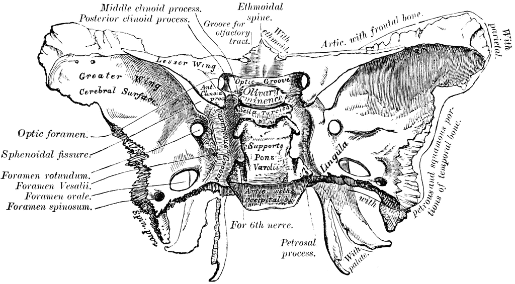
38 sphenoid bone diagram
Aug 12, 2019 · Sphenoid sinuses: The sphenoid sinuses are located in the sphenoid bone near the optic nerve and the pituitary gland on the side of the skull. ... Diagram of sinus infection. Sphenoid Bone - Definition, Location & Function - Human Anatomy | Kenhub.
Sphenoid (1) Ethmoid (1) Total number of bones=8. Frontal Bone. This bone forms the forehead, the roof of the orbital cavity (eye socket), and the root of the nose. A newborn has a frontal bone that consists of two parts, separated by the frontal suture. However, the parts fuse to form a single bone, by the time a child is eight years old ...

Sphenoid bone diagram
The body of the sphenoid bone is the midline cubical portion of the sphenoid bone, hollowed by the sphenoid air sinuses. Gross anatomy The body has superior, inferior, anterior, posterior... The sphenoid bone of humans is homologous with a number of bones that are often separate in other sphenoid bone — noun butterfly shaped bone at the base of the skull • Syn: ↑sphenoid, ↑os... Sphenoid bone - Read online for free. Sphenoid bone components: Body: pneumatized to a variable degree 2 wings: lesser and greater wings 2 pterygoid processes: Surfaces of the body: A-The...
Sphenoid bone diagram. Jan 27, 2015 · The sphenoid sinuses are located in the sphenoid bone near the optic nerve and the pituitary gland on the side of the skull. There are … 33F. I have had chronic on/off sinus problems for years that includes chronic post nasa drip. Recently, an mri revealed I had “moderate to severe” right sphenoid and right maxillary sinusitis. I took 10 days of augmentin. It still comes back periodically and makes me dizzy (similar to vertigo) tension headache and very tired. It also makes my right eye painful as it’s on the right side. My ENT put a camera up my nose and said my mucus drainage is clear and I do not have an active infection. M... The sphenoid bone is located at the central skull base and is commonly considered the most An understanding of the sphenoid bone anatomy enables the prediction of intra- and extracranial... The occipital condyles are undersurface protuberances of the occipital bone in vertebrates, which function in articulation with the superior facets of the atlas vertebra.. The condyles are oval or reniform (kidney-shaped) in shape, and their anterior extremities, directed forward and medialward, are closer together than their posterior, and encroach on the basilar portion of the …
Hey everyone. I'm looking to sculpt a dinosaur skeleton, and I remember there are some documents floating around that show each bone (for specific species) in three different views. There's a specific name for the type of document and I can't recall what it is. It's similar to a blueprint, in that I THINK they're handdrawn. Anyone have any good resources? 27M Currently having what I believe to be Sphenoid Sinus issues. CT came back normal. Symptoms: Consistent headache on the top left area. Left eye complications Dizzy Memory issues I responded well to the first day of taking Claritin, but only the first day. Nasal spray will cause a burning sensation that travels through my nose to the point that is experiencing complications. Is it possible something that is not Sphenoid related? What issues could be present that a CT would not detect? The sphenoid bone is an unpaired bone of the neurocranium. It is situated in the middle of the skull towards the front, in front of the basilar part of the occipital bone. The sphenoid bone is one of the seven bones that articulate to form the orbit. Sphenoid bone is a butterfly-shaped cranial bone that is located in the middle of the skull between Sphenoid bone primarily consists of a centrally positioned body, which surrounds and protects the...
Hello everyone, I've been having migraines at the area of the Sphenoid bone (directly behind the brow). It goes between the two hemispheres, started very sharply at the left side but now it travels back and forth. Can anyone name which parts of the brain are cased directly behind the Sphenoid bone? I've tried finding illustrations but couldnt find any that correlate specifically what it covers. I know it's either the prefrontal cortex or the temporal lobe, and if it was the prefrontal cortex I c... So I thumb pushed the lower part of the sphenoid bone you know the one near the end of the hard palate Like a total idiot and it made a lot of cracks and now it hurts I feel like I fractured it Gender: male Age: 14 Height: 5'3 Weight:48 kgs I don't take any medications Sutures connect cranial bones and facial bones of the skull. Develop a good way to remember the cranial bone markings, types, definition, and names including the frontal bone, occipital bone, parieta. Sphenoid Bone - Anterior Aspect. Back to the Skull. This diagram is from Grant's Method of Anatomy.
The 25+ best Sphenoid bone ideas on Pinterest | Palatine ...
Nov 08, 2021 · The shoulder joint (glenohumeral joint) is a ball and socket joint between the scapula and the humerus.It is the major joint connecting the upper limb to the trunk. It is one of the most mobile joints in the human body, at the cost of joint stability.
Closeup of skeleton foot model
Known as: Sphenoidal bone, Sphenoid Bones, Os sphenoidale. Expand. An irregular unpaired bone situated at the SKULL BASE and wedged between the frontal, temporal, and occipital bones...
Lateral wall of the nasal cavity: Anatomy and diagrams ...
The sphenoid is the most complex bone in the human skeleton. It contributes to the formation of the orbits and the base of the skull. It also acts as a gateway between facial structures and the brain.

Image from page 294 of "Journal of anatomy" (1867)
Skull diagram of Champsosaurus, showing the pterygoid bone The pterygoid is a paired bone forming part of the palate of many vertebrates, behind the palatine bones . It is a flat and thin lamina, united to the medial side of the pterygoid process of the sphenoid bone, and to the perpendicular lamina of the palatine bone.

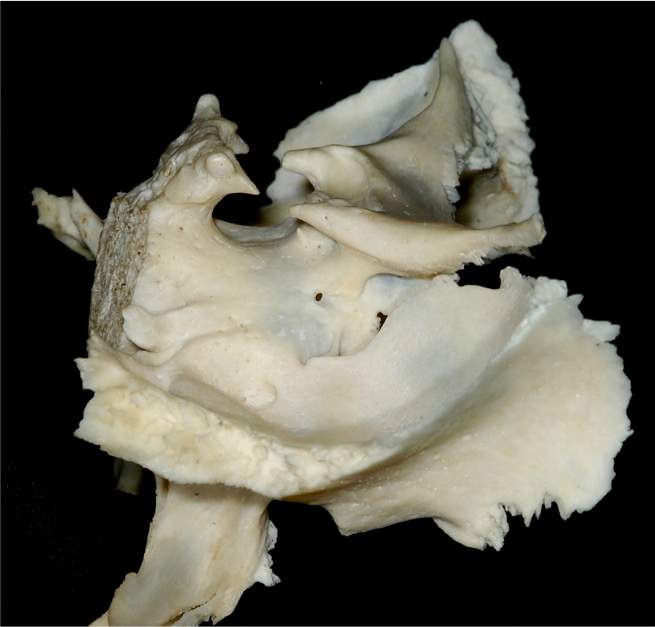
Sphenoid bone (extracted). Sphenoid sinuses see -anterior ...
Asking this because I'm trying to remember a game I saw at a childhood friend's birthday party. This was a long time ago, I think around 2007, so my memory is quite hazy. I've never been into sports video games, but I distinctly remember this one game I was watching with my friends at a birthday party, and I was really squeamish (always have been haha) and felt grossed out seeing the character riding a skateboard and whenever he fell from a great height it would show blood and the character bein...

(A,B) left greater wing of sphenoid bone fracture with a ...
Hi all, I'm finding that in the standard neti pot rinse the frontal and maxillary sinuses are targeted pretty well, but congestion still remains in the sphenoid sinuses. There isn't much information online about this— does anyone have tips on how to rinse these?

Anterior view of the sphenoid bone of a normal fetus and ...
The sphenoid bone is one of the seven bones that make up the orbit (the space that holds the eyeball), and helps make up the floor of the middle cranial fossa, the butterfly-shaped depression at...

-Schematic of the sphenoid bone in a CBCT coronal section ...
I had a sinus infection and I took an antibiotic by my neurologist medication such as Levofloxacin (I think it’s spelled) it took some head pressure I had on the back of my head by the Sphenoid Bone. Left some side effects that kinda went away.. (it’s been 16 days since I finish the medicine) but now for the past 1 week I’m feeling a wet/cold sensation on my head (top of head) but I’m not sure why. I’m trying to keep calm and with Head Paresthesia as well the Neurologist says is because of all t...

Anatomy Made Easy : Inferior View Of The Skull
The sphenoid bone is one of the eight bones that comprise the cranium - the superior aspect of the It contains the sphenoidal sinuses , which are separated by a septum - meaning that the sphenoid...

Photography. Anterior view of the sphenoid bone. This ...
Sphenoid bone is made up of several projections and features, allowing you to view it in many The sphenoid bone is joined by an interconnection of bones. These include Palatines Zygomatic...

Skull Anatomy - Medical Art Library
Sutton JB. On the development and morphology of the human sphenoid bone. (1885) Proc. Zool. Soc. 308: 577-. By J. Bland Sutton, F.B.C.S., Lecturer on Comparative Anatomy at the Middlesex. Hospital Medical School. (Received May 25, 1885.) (Plate XXXV.)
Os sphenoidale | definition of os sphenoidale by Medical ...
Title

Doctors Gates: Anatomy of sphenoid bone(Pic & vid)
The sphenoid bone is an unpaired bone situated in the middle of the cranial base. It articulates with the adjacent temporal, parietal, frontal, occipital, ethmoid, zygomatic, palatine, and vomer bones and...
Closeup of skeleton hand model
Sep 12, 2021 · Sphenoid Bone: It is an unpaired and irregular bone that lies below the frontal bone and forms the depression or cavity called sella turcica in which the pituitary gland is present. It forms the base of the skull, forms walls and floors of the orbit , and spans the width of the head to articulate with other bones and provide rigidity to the ...
Sphenoid Bone - Location, Function and Anatomical Structure
Figure 9.4 The sphenoid bone. Watch a video of the Sphenoid Bone >Study Area>Pre-Lab Videos Cranial bone Important markings Description Occipital (1) Figures 9.1, 9.2, 9.3, and 9.6 N/A Forms the posterior aspect and most of the base of the skull. Foramen magnum Large opening in the base of the bone, which allows the spinal cord to join

Features Noted by the HSCA Radiologists
Does the sphenoid bone have anything to do with mewing ? I mewed assymetrically which made me assymetrical and dumb ass me pushed my left lower part of my sphenoid so hard up since it was a lot down in my developed side that it hurted for like 2 months and now it's really up ,upper than my right side ,it doesn't hurt now though

Sphenoid bone, superior view with labels - Axial Skeleton ...
sphenoid bone clipart etc, sphenoid bone location structure function teachmeanatomy, file rotation sphenoid bone gif wikimedia commons, sphenoid bone anatomy youtube, sphenoid bone osteology.
Lateral View of the Sphenoid Bone | Neuroanatomy | The ...
Fissure narrow slit through bone, e.g., superior orbital fissure on sphenoid bone Fovea a shallow pit on the bone, e.g., fovea on head of femur Lines roughened ridge on the bone, e.g., linea aspera on femur Meatus outlet, opening in temporal bone, e.g., auditory meatus Fossa deeper pit in single bone or formed by several bones, e.g.,
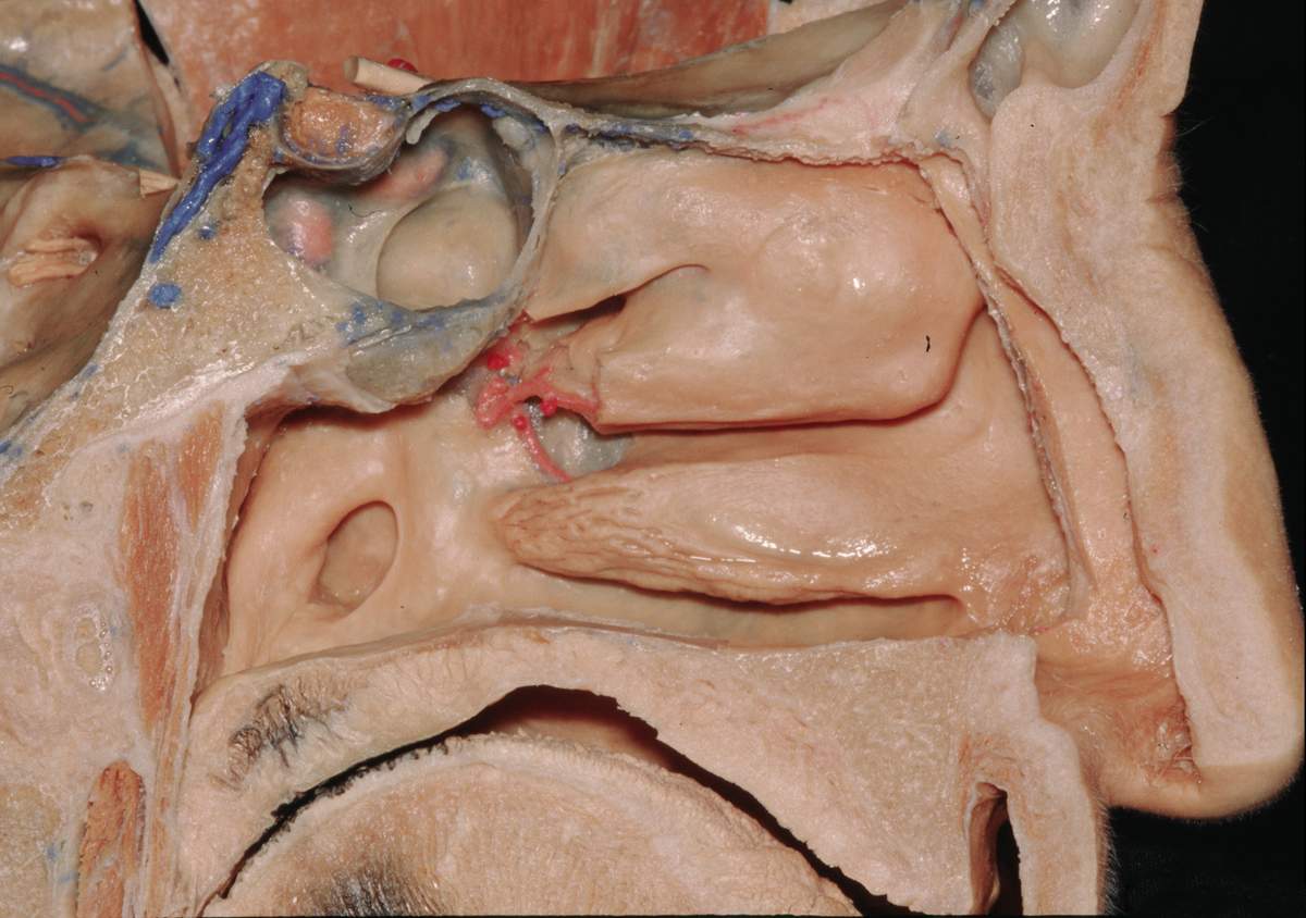
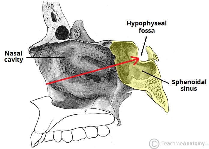
Sagittal View of Left Nasal Cavity | Neuroanatomy | The ...
Reddit, please help. I don't know what sub reddit to post this to, maybe someone can point me in the right direction. There is a tool I will be using at work that I wish to purchase. They have sets available but not enough for everyone. I work in an orthotics manufacturing lab. The thing I'm looking for is a set of foot bone diagrams, unlabled, in every size available for men and women. Sizes range from 34 to 48 in adults. I appreciate any and all help

sphenoid bone labeled - Google Search | Anatomy bones ...
Axial Skeleton (80 bones) Skull (28) Cranial Bones. Parietal (2) Temporal (2) Frontal (1) Occipital (1) Facial Bones. Maxilla (2) Zygomatic (2) Mandible (1)

Sphenoid Bone Cross Section : Cross Section Of Camel S ...
The sphenoid bone[note 1] is an unpaired bone of the neurocranium. It is situated in the middle of the skull towards the front The sphenoid bone is one of the seven bones that articulate to form the orbit.

Sphenoid bone (extracted) -view from above. | Download ...
My original post: https://www.reddit.com/r/AskDocs/comments/rc2mhs/i_dont_understand_whats_happening_to_me_and/?utm_source=share&utm_medium=ios_app&utm_name=iossmf Age: 30 Sex: female Height: 5’4 Weight: 205 lbs Medical conditions: hyperlipidemia, fatty liver Medications: augmentin, vitamin d, fish oil. So my symptoms have definitely improved, and I’ve been on antibiotics during that improvement. I’m finishing them up today. I checked out my CT- everything was normal but I had “muc...

I wanna go home
Background: Sphenoid sinuses are pneumatic spaces within the body of the sphenoid bone. Their development begins in the prenatal life and continues until the adulthood.

Vesalius' images of the skull and the sphenoid bone. The ...
Sep 30, 2021 · The frontal bone, the parietal bone, the greater wing of the sphenoid bone, and the squamous part of the temporal bone meet at the pterion, forming the floor of the temporal fossa. The following videos, articles, and quizzes will cover everything you need to know about the temporal region of the skull, so make sure to check them out!

The Sphenoid Bone - Human Anatomy
The zygomatic bone articulates with the frontal bone, sphenoid bone, and paired temporal bones, and maxillary bones. Development [ edit ] The zygomatic bone is generally described as ossifying from three centers—one for the malar and two for the orbital portion; these appear about the eighth week and fuse about the fifth month of fetal life.
Sphenoid Bone | ClipArt ETC
Sphenoid and Ethmoid bones. PhD., Dr. David Lendvai Department of Anatomy, Histology and •neurocranium (8) - UNPAIRED: frontal, occipital, sphenoid, ethmoid bones PAIRED: temporal...

I’ve always loved how late afternoon light scatters diamonds and fireworks across the water.
Hey y’all, I’m new to reddit and new to this sub, I am wondering if anybody on here has also dislocated their Sphenoid bone, (it’s a bone in your face happened when I had my tbi and is now an ongoing issue) iv never met anyone who has done it and I’m in constant pain 😅 hoping there is someone out there with the same situation? Or if you know anything about it or tips on helping the pain cuase I can’t afford to put it back in place every other day lol. Hope everyone is headache free today and is ...

easyhumanatomy: Sphenoid: easy and brief discussion
It is an unpaired bone situated at the base of the skull. Its resemblance is of the shape of a butterfly or bat with outstretched wings.Parts It is composed of the following 7 parts:A body.
Sphenoid Bone from Behind | ClipArt ETC
I thumb pushed the sphenoid bone and now it hurts really badly ,it also made some cracking noises when I did

Image from page 249 of "Radiography and radio-therapeutics" (1917)
My original post: https://www.reddit.com/r/AskDocs/comments/rc2mhs/i_dont_understand_whats_happening_to_me_and/?utm_source=share&utm_medium=ios_app&utm_name=iossmf Age: 30 Sex: female Height: 5’4 Weight: 205 lbs Medical conditions: hyperlipidemia, fatty liver Medications: augmentin, vitamin d, fish oil. So my symptoms have definitely improved, and I’ve been on antibiotics during that improvement. I’m finishing them up today. I checked out my CT- everything was normal but I had “muc...
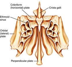
Ethmoid bone | definition of ethmoid bone by Medical ...
Start studying sphenoid bone - diagram. Learn vocabulary, terms and more with flashcards, games and other study tools.
Sphenoid Bone - Location - Structure - Function ...
Sphenoid Bone Human Anatomy. Your model is disabled. For more details go to Edit properties.

Sphenoid Bone - Location - Structure - Function ...
Sphenoid bone (sagittal section) - Yousun Koh. Sphenoid bone: want to learn more about it? Our engaging videos, interactive quizzes, in-depth articles and HD atlas are here to get you top results...

Image from page 313 of "The anatomy of the domestic fowl " (1918)
Sphenoid bone ex situ & in situ. To better depict the position of this bone, the picture above shows the skull with the right zygomatic bone removed. Ventral aspect of sphenoid bone.
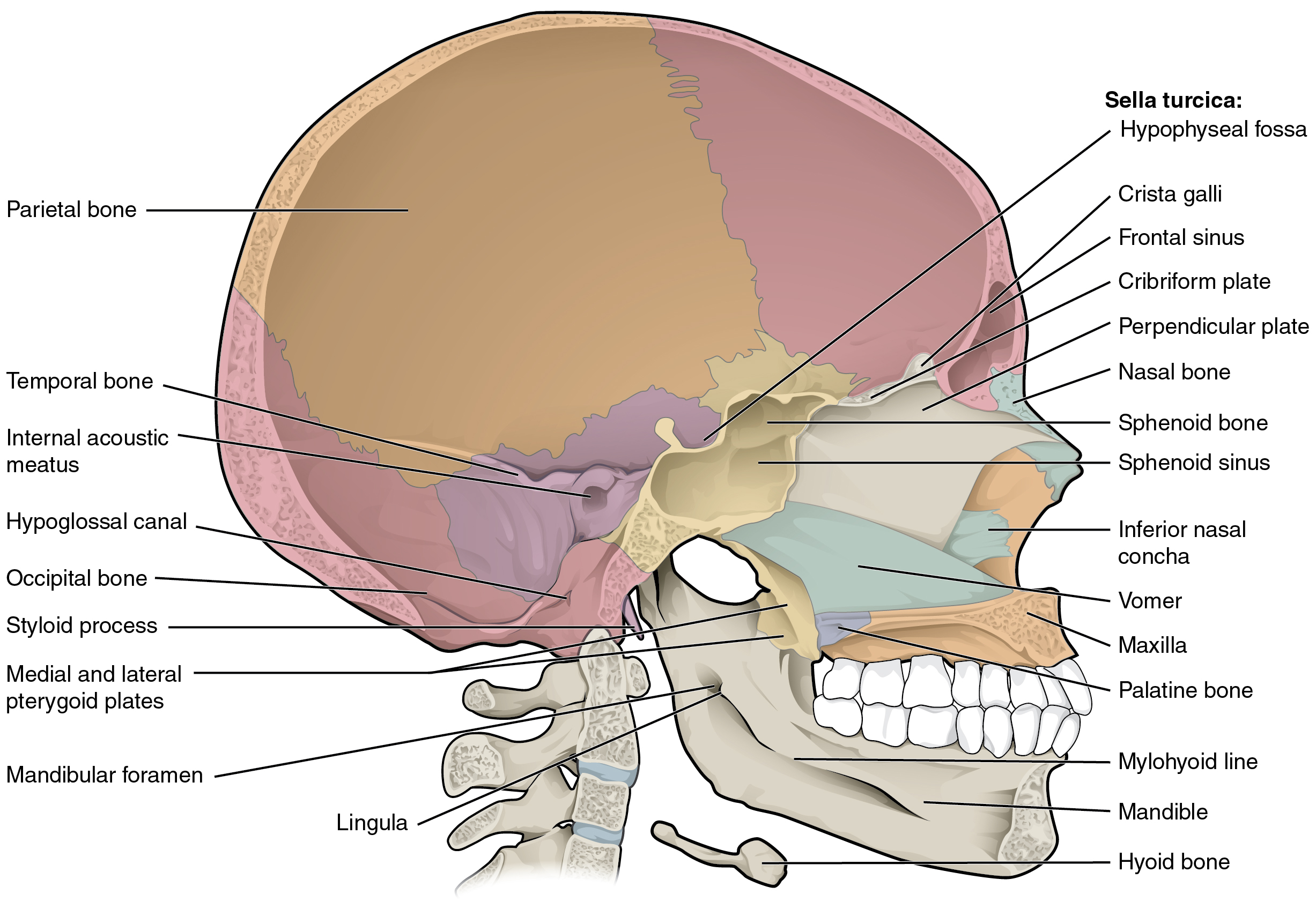
7.2 The Skull - Anatomy and Physiology
Sphenoid bone, superior view with labels - Axial Skeleton Visual Atlas, page 31. This is Page 31 of a photographic atlas I created as a laboratory study All other diagrams and illustrations used in…
Sphenoid bone: Anatomy, function and development | Kenhub
The unpaired sphenoid bone is often referred to as bat-or butterfly-shaped. The sphenoid bone body sits behind the nasal cavity with various protrusions that play important roles in the construction...

Sphenoid Bone - Anatomy, Structure, Location, Function ...
Sphenoid bone location and sphenoid bone fracture. The space inside the body is the sphenoidal sinus, which drains into the nasal cavity (see Figure 3). The sella turcica (sella = saddle; turcica...

Image from page 617 of "Biology of the vertebrates : a comparative study of man and his animal allies" (1949)
The sphenoid bone forms part of the hind wall of the orbits (upper aspect of the greater sphenoid wings) The sphenoid bone in cats at the level of the pituitary fossa is usually not thicker than 5 mm.


Comments
Post a Comment