38 wiggers diagram cardiac cycle
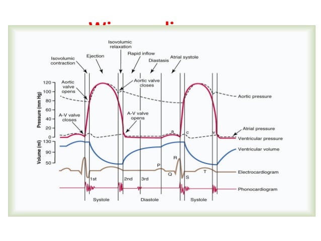
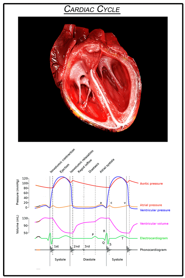
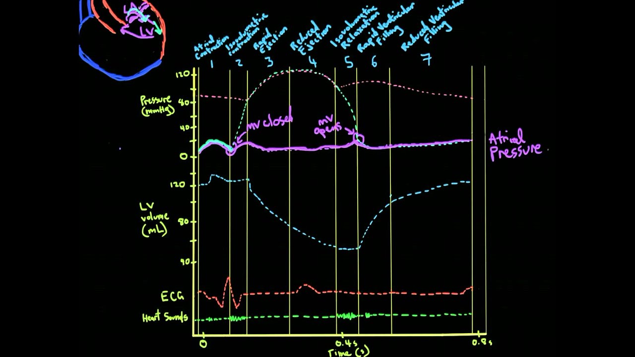
How to draw Wigger’s Diagram of Cardiac Cycle? Summary of Cardiac Cycle Opening and closing of valves When the valve opens, different compartments act as a single chamber (atrio-ventricle or aorto-ventricle). For a blood to flow, pressure in “giver” must be higher then that in “receiver”. Pressure difference opens or closes the valve: The Wiggers diagram is a synchronous tracing of aortic pressure, left atrial pressure, left ventricular pressure, left ventricular volume, and EKG throughout the cardiac cycle.Which of the following is the best description the events that occur during diastole on a Wiggers diagram, during the course of S2 on the phonocardiogram - marking the beginning and end of isovolumetric relaxation?
Anybody who asks for a discussion of ventricular cavity pressure and how it contrasts aortic pressure must surely be asking for a Wiggers diagram. The most prominent visually appealing part of that diagram is the top section where the ventricular cavity pressure triumphantly overcomes aortic pressure in systole.

Wiggers diagram cardiac cycle
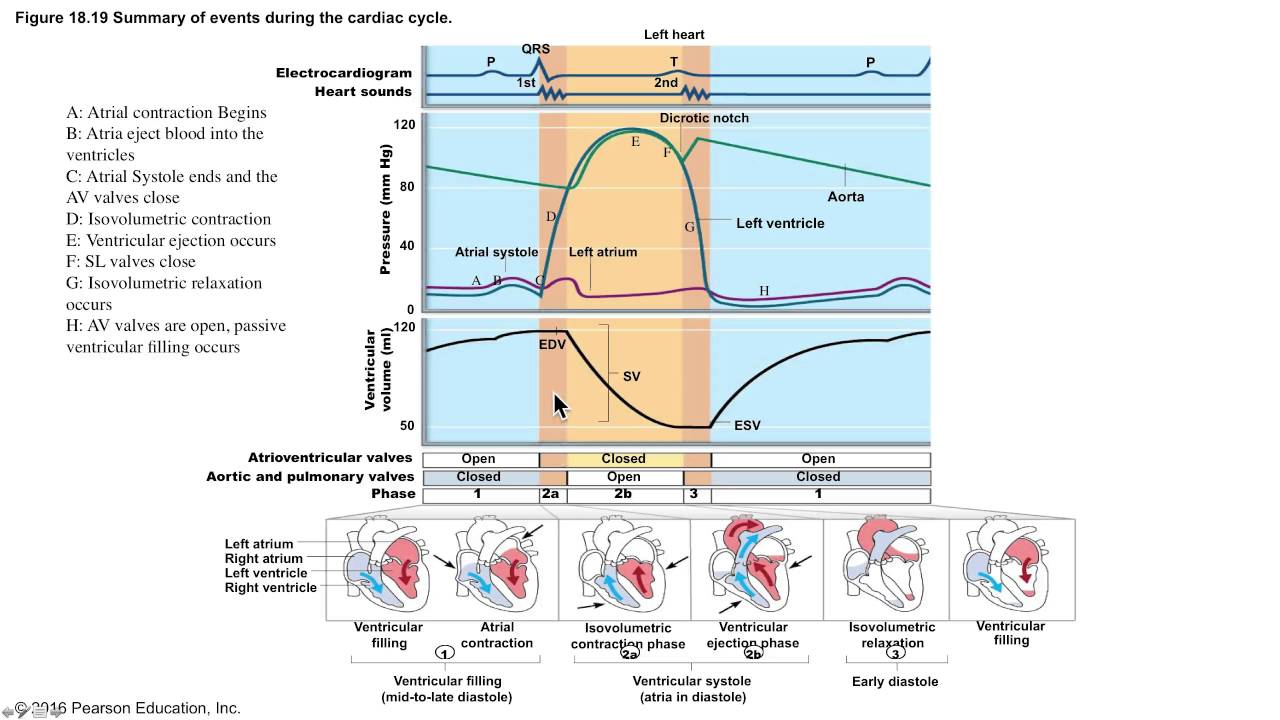
The events of the cardiac cycle are summarized in the Wigger 's Diagram (Fig 4). The Wigger's diagram (Fig 4) shows the relationship between the ECG, the pressure, and volume changes in the left ventricle and aorta. Note that there are two heart sounds (lub-dub). The first is associated These didactic lectures usually use PowerPoint slides, with pictures and animations. For instance, the Wiggers diagram is often used to teach the concept of the cardiac cycle in a cardiovascular physiology class. Teaching the cardiac cycle to students can be challenging for many teachers. Consider the Wiggers diagram, for example. The cardiac cycle and Wiggers diagram A Wiggers diagram illustrate events and details of the cardiac cycle with electrographic trace lines, which depict (vertical) changes in a parameter's value as time elapses left-to-right.
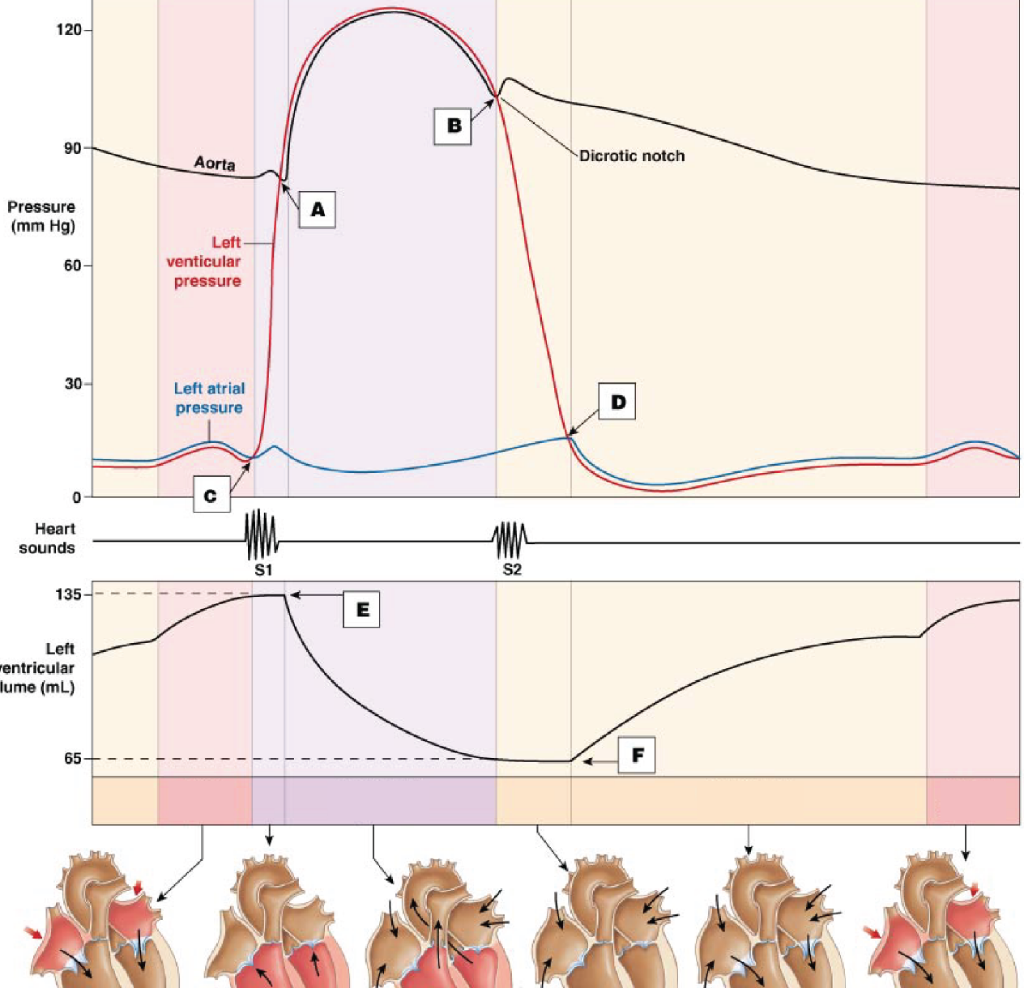
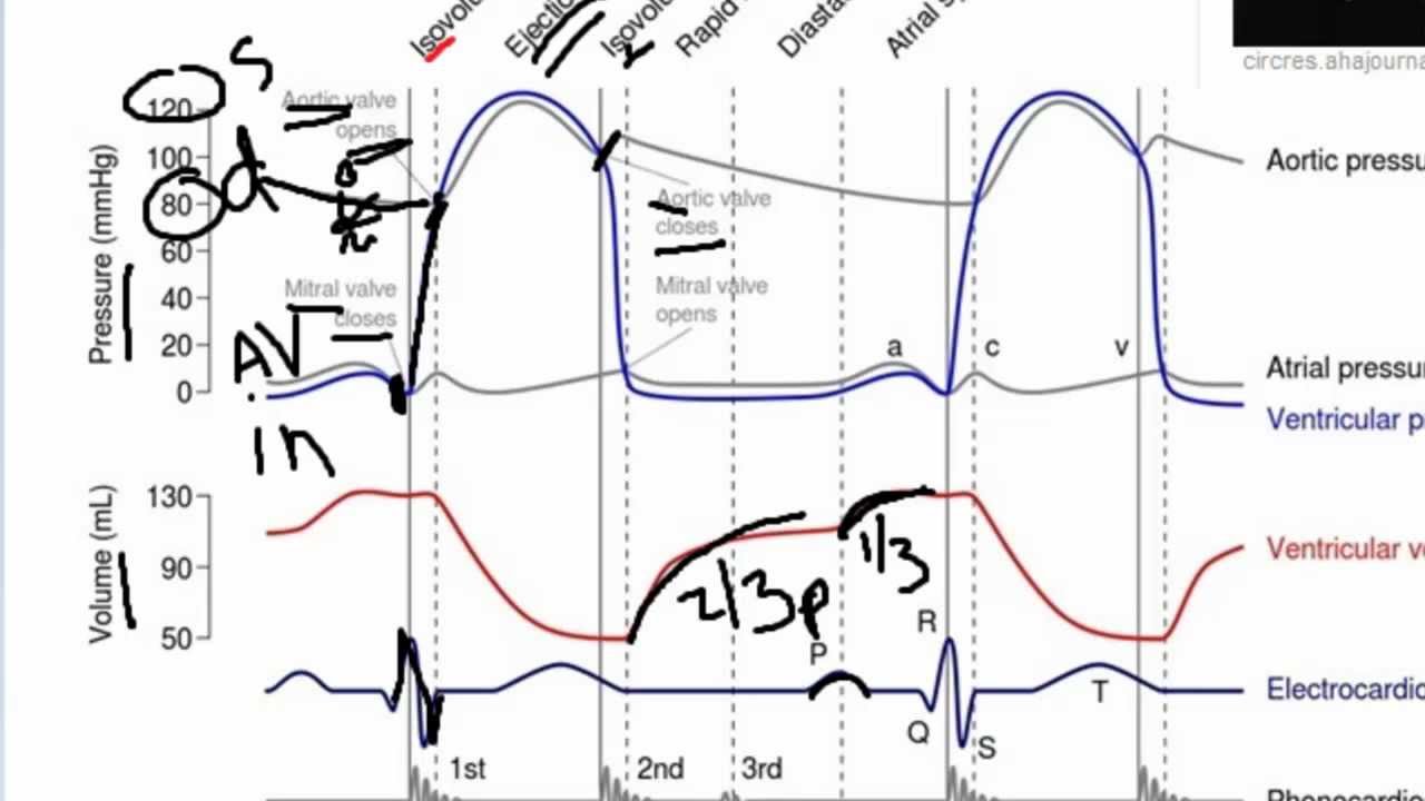
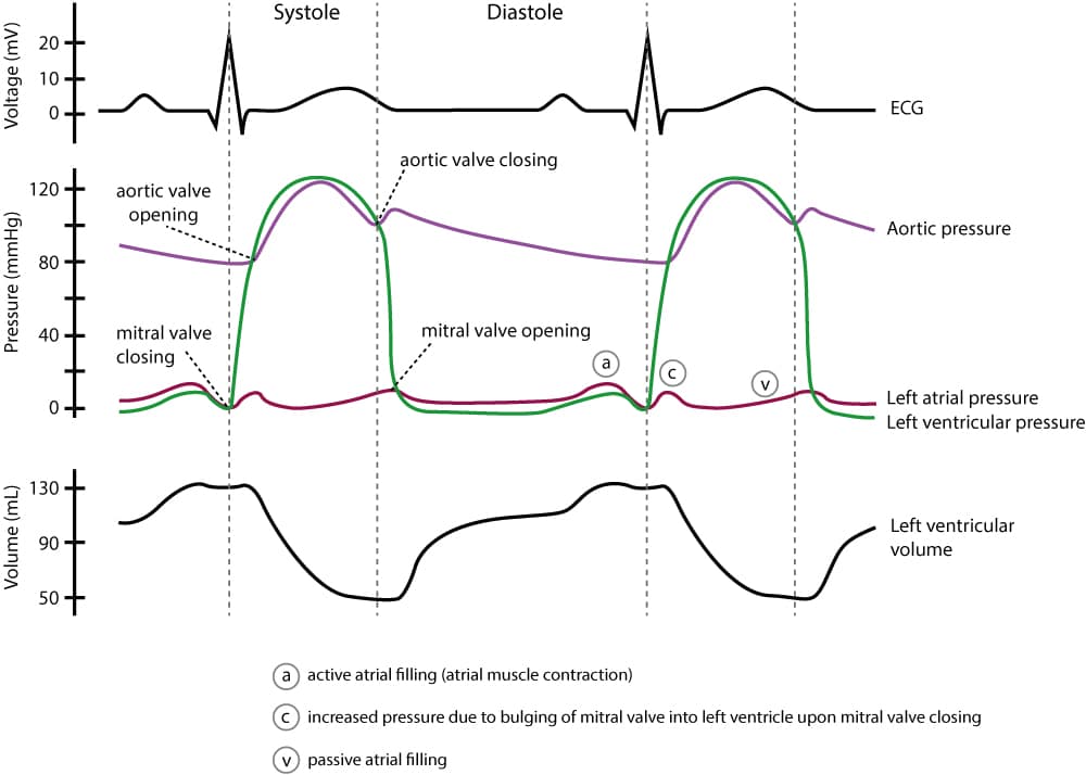
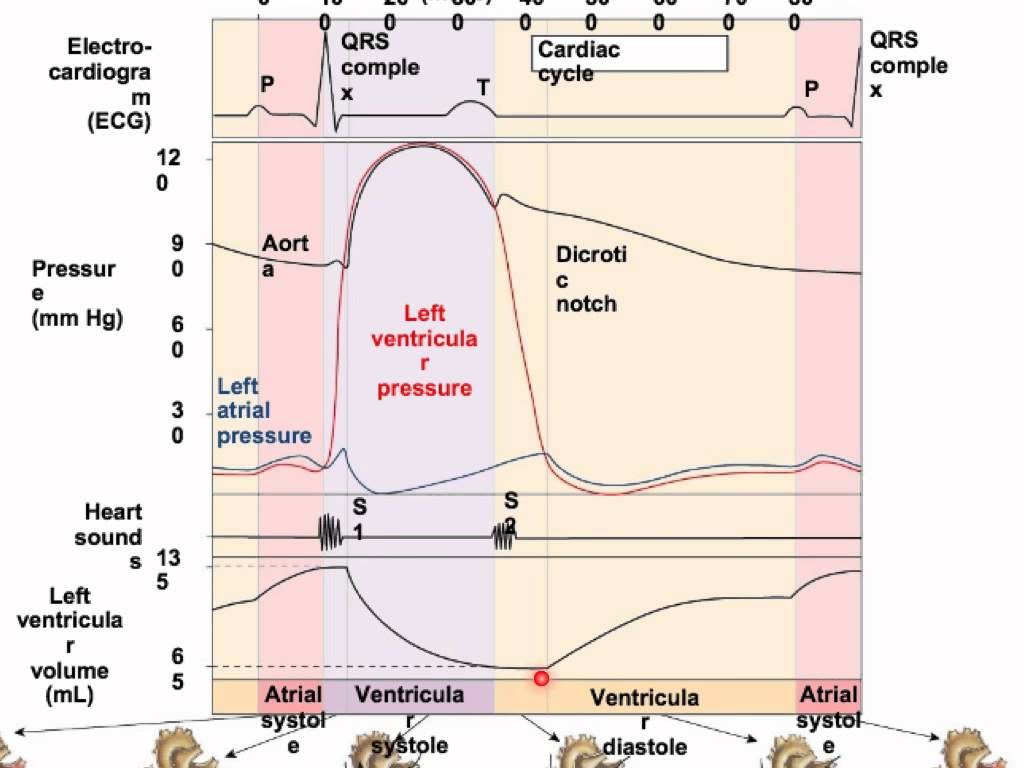
Wiggers diagram cardiac cycle. Description. Wiggers Diagram.svg. English: A Wiggers diagram, showing the cardiac cycle events occuring in the left ventricle. In the atrial pressure plot: wave "a" corresponds to atrial contraction, wave "c" corresponds to an increase in pressure from the mitral valve bulging into the atrium after closure, and wave "v" corresponds to passive ... 7. WIGGERS DIAGRAM EVENTS IN CARDIAC CYCLE DETERMINANTS OF MYOCARDIAL PERFORMANCE PRESSURE VOLUME LOOP PHYSIOLOGICAL VS PATHOLOGICAL HYPERTROPHY 8. Cardiac cycle • The cardiac cycle describes pressure,volume and flow phenomena in the ventricles as a function of time. • Similar for both LV and RV except for the timing,levels of pressure. 9. A Wiggers diagram, named after its developer, Carl Wiggers, is a standard diagram that is used in teaching cardiac physiology. In the Wiggers diagram, the X-axis is used to plot time, while the Y-axis contains all of the following on a single grid: Blood pressure Aortic pressure Ventricular pressure Atrial pressure Ventricular volume Basically, a "Wiggers Diagram" is a description of the events which take place over the cardiac cycle and which a plotted on a time scale. Yes, it's the Wiggers diagram, not Wigger's diagram or Wiggers' diagram, because a guy called Wiggers was responsible for the development of its most important components.
This is well-illustrated on a Wiggers diagram where the QRS complex on the ECG directly precedes ventricular systole (represented on the diagram by increased ventricular pressure). Function. Concerning the events of the cardiac cycle, it is important to compartmentalize their sequence. The cardiac cycle - The “Wiggers diagram ” Prof. J Swanevelder 4 - 4 Cardiac valves The cardiac valves open and close passively in response to the changes in pressure gradient across them. These valves control the sequence of flow between atria and ventricles, and from the ventricles to the pulmonary and systemic circulations. Valve timing in relation to the ventricular pressure curve is ... Wiggers diagram records are most commonly used in cardiac care units. The primary benefit of the Wiggers diagram is that it shows several aspects of the cardiac cycle together over time so that doctors can ensure the heart is beating properly. In a healthy heart, electrical impulses from the nervous system control the four chambers. Wigger's diagram: This seemingly complex diagram (Don't Panic!) is actually a very useful tool to learn the components of the cardiac cycle and how they relate to cardiac exam findings, which I have indicated in blue. To start, identify the aortic, LV and LA pressure lines and talk yourself through the sequence of
The P wave is a summation wave generated by the depolarization front as it transits the atria. Normally the right atrium depolarizes slightly earlier than left atrium since the depolarization wave originates in the sinoatrial node, in the high right atrium and then travels to and through the left atrium. A Wiggers diagram, named after its developer, Dr. Carl J. Wiggers, is a standard diagram that is used in teaching cardiac physiology. In the Wiggers diagram, the . Wiggers Diagram Explained Health Education, Ultrasound, Physiology, . Ekg en 3D Más Heart Anatomy, Ekg Leads, Nursing Students, Ekg Interpretation. The cardiac cycle diagram shown ... The Wright table is introduced as a novel tool for teaching and learning the cardiac cycle. It supplements the nearly 100-yr-old Wiggers diagram, which is information rich but difficult for many students to learn. The Wright table offers a compact presentation of information, viewable both in terms … Draw a Wigger's diagram without prompts - Time on horizontal axis - Several variables on vertical axis What are two diagrams to evaluate cardiac function? - Pressure-Volume loops - Frank-Starling curves Phase A of cardiac cycle Atrial systole - Atria contract - Mitral valve is open - Final phase of ventricular filling - Preceded by P wave

301 Moved Permanently
A Wiggers diagram, named after its developer, Dr. Carl J. Wiggers, is a standard diagram that is used in teaching cardiac physiology. In the Wiggers diagram, the . This Pin was discovered by Cardiac Sonography. Discover (and save!) your own Pins on Pinterest. wiggers diagram explain.

USMLE Step 1: Cardiovascular Physiology flashcards | Quizlet
A Wiggers diagram, named after its developer, Dr. Carl J. Wiggers, is a standard diagram that is used in teaching cardiac physiology. In the Wiggers diagram, the X-axis is used to plot time, while the Y-axis contains all of the following on a single grid: Blood pressure. Feb 12, · BYU Physiology Winter This feature is not available right now.
Cardiac cycle & pressure wave forms copy
This is the 1st first video in the Cardiology section dedicated to an important topic.Wiggers Cardiac Diagram.We go through different curves of the Wigger's ...

Wiggers Diagram 2 - Wiggers diagram - Wikipedia | Cardiac ...
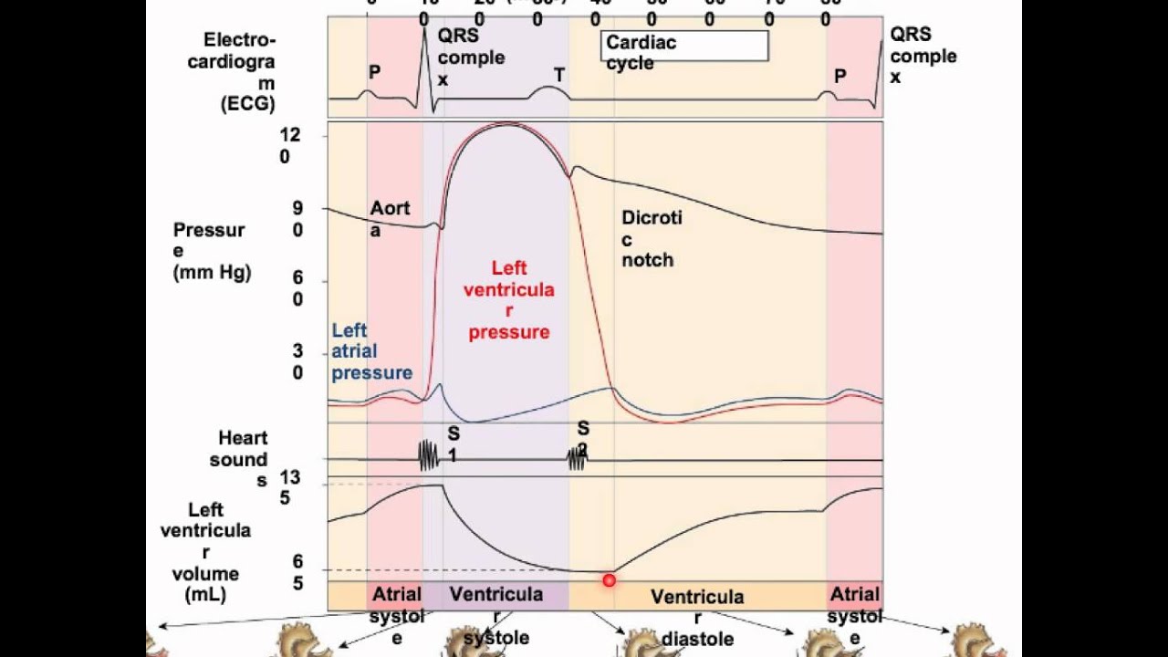
The cardiac cycle diagram shown to the right depicts changes in aortic pressure (AP), left ventricular pressure (LVP), left atrial pressure (LAP), left ventricular volume (LV Vol), and heart sounds during a single cycle of cardiac contraction and relaxation. These changes are related in time to the electrocardiogram.

Wiggers Diagram - Cardiac cycle - Wikipedia, the free ...
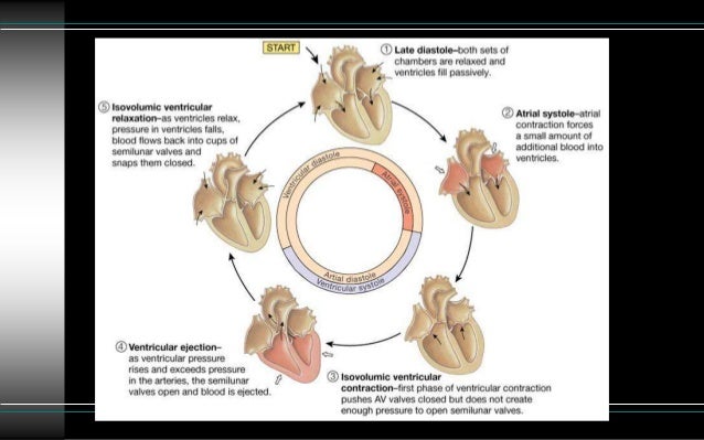
Place the following terms on this diagram: 1. Ventricular filling 2. Ventricular ejection 3. Isovolumetric contraction 4. Isovolumetric relaxation 5 Electrical Premises 1. What property of cardiac cells is critical for initiation of the electrical activity? 2. How would you ensure synchronous cardiac muscle contraction? 3.

Pin on Heart geek
Oct 28, 2021 · Wiggers Diagram. The American-born physiologist Dr. Carl J Wiggers has provided many health care students over the past 100 years with a unique tool to understand the cardiac cycle. The Wiggers diagram highlights the relationship between pressure and volume over time, along with the electrical activity of the heart. The diagram uses the left ...

A Wiggers diagram is a standard diagram used in cardiac ...
The Cardiac Cycle III: Volumes in the Left Ventricle: 6 mins: 0 completed: Learn. The Cardiac Cycle IV: Heart Sounds: 7 mins: 0 completed: Learn. The Cardiac Cycle V: The Wiggers Diagram: 10 mins: 0 completed: Learn. The Cardiac Cycle VI: The Ventricular Pressure-Volume Loop: 9 mins: 0 completed: Learn. Cardiac Output I: Definitions: 5 mins: 0 ...

Left ventricular diastolic function - ECG & ECHO
This rhythmic sequence causes changes in pressure and volume that are often seen graphically in the form of a Wiggers diagram or venous pressure tracings. Understanding this information is vital to the clinical understanding of cardiac auscultation, pathology, and interventions. Cellular

Image from page 183 of "Physiology and biochemistry in modern medicine" (1918)
Cardiac Cycle - Summary and Wigger's Diagram. Aortic valve stenosis. Summary of Cardiac Cycle How to draw Wigger's Diagram of Cardiac Cycle? Epomedicine is a source of concise notes on medical topics with relevant details and mnemonics which makes them easy to conceptualize.

The Cardiac Cycle - YouTube
Etymology. The term "bathmotropic" is derived from the Ancient Greek word βαθμός (bathmós), meaning "step" or "threshold". [citation needed]History. In 1897 Engelmann introduced four Greek terms to describe key physiological properties of the heart: inotropy, the ability to contract; chronotropy, the ability to initiate an electrical impulse; dromotropy, the ability to conduct an ...
Cardiac cycle physiology_4_dpt
what's the wigger's diagram it is a diagram that shows the sequence of electrical and mechanical events from the beginning of one heartbeat to the next. parts - left atrial pressure - ventricular pressure - aortic pressure - ventricular outflow (measurement of the volume leaving the ventricle per unit time)

Wiggers Diagram Cardiac Cycle
This diagram remains the most commonly used model to teach the cardiac cycle today. A typical Wiggers diagram is shown in Fig. 1. Fig. 1. The Wiggers diagram. From top to bottom, the lines show: 1) aortic pressure, 2) ventricular pressure, 3) atrial pressure, 4) electrocardiogram, 5) mitral and aortic valve opening and closing, and 6) heart sounds.

The Cardiac Cycle | Deranged Physiology
A Wiggers diagram shows the changes in ventricular pressure and volume during the cardiac cycle. Often these diagrams also include changes in aortic and atrial pressures, the EKG, and heart sounds. Diastole starts with the closing of the aortic valve (the second heart sound).

grayscale photo of people walking on snow covered road
A description of the cardiac cycle using the Wiggers Diagram.
Systole og diastole - Anafys.dk
The cardiac cycle and Wiggers diagram A Wiggers diagram illustrate events and details of the cardiac cycle with electrographic trace lines, which depict (vertical) changes in a parameter's value as time elapses left-to-right.
Wiggers Diagram Made Easy
These didactic lectures usually use PowerPoint slides, with pictures and animations. For instance, the Wiggers diagram is often used to teach the concept of the cardiac cycle in a cardiovascular physiology class. Teaching the cardiac cycle to students can be challenging for many teachers. Consider the Wiggers diagram, for example.
Wiggers Diagram Heart Sounds - Data Diagram Medis
The events of the cardiac cycle are summarized in the Wigger 's Diagram (Fig 4). The Wigger's diagram (Fig 4) shows the relationship between the ECG, the pressure, and volume changes in the left ventricle and aorta. Note that there are two heart sounds (lub-dub). The first is associated

green and white labeled box

Image from page 249 of "American journal of physiology" (1898)
Cardiac Cycle Broken Down - YouTube

Wiggers diagram. The diagram shows the aortic, atrial and ...

Cardiac Cycle Drawing at GetDrawings | Free download
Explanation Of Wiggers Diagram

white and blue box on white table

Wiggers Diagram showing cardiac pressures, volume and ...
WIGGERS DIAGRAM - Unmasa Dalha

Pressure Volume Loop Wiggers Diagram - Diagram Media

Cardiac Cycle Overview Tutorial | Sophia Learning
Clinical electrocardiography and ECG interpretation – ECG ...

Wiggers Diagram Cardiac Cycle

Drag The Labels Onto The Wiggers Diagram To Identify ...

The Wright table of the cardiac cycle: a stand-alone ...

Drag The Labels Onto The Wiggers Diagram To Identify ...
Cardiac Cycle Drawing at GetDrawings | Free download

Wiggers diagram shows events of the cardiac cycle for the ...

Wiggers diagram | Lazım | Pinterest | Diagram

Wiggers Diagram Cardiac Cycle

12 InClassCardioDiagramkaukpepple - Wiggers Diagram ...


Comments
Post a Comment