40 eye diagram ap psychology
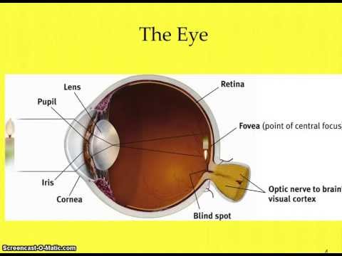
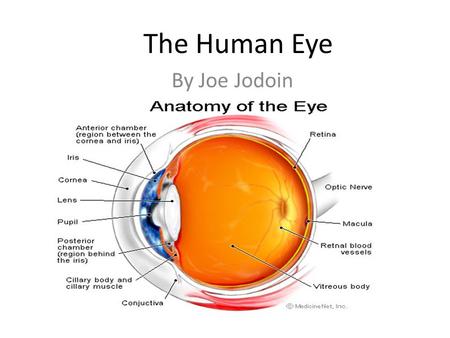
Start studying AP Psychology: The Eye. Learn vocabulary, terms, and more with flashcards, games, and other study tools. AP Psychology - Unit 4 Assignment ... Draw a diagram of the eye. Label and explain the function of the iris, lens, pupil, cornea, retina, fovea, blind spot, and optic nerve. 5. What is transduction and how does this process occur in the photoreceptors of the eye and cochlea of the ear? 6. Draw a diagram of the ear.
Ap psychology eye diagram quizlet. This structure controls our bodys general arousal and our ability to focus and it is a. Barrons AP Psychology 9th Edition. There are two major types of hearing loss. Begins to focus the light by bending it toward a central focal point pupil the adjustable opening in the center of the eye through which light ...
Eye diagram ap psychology
Lens Transp arent eye structure that focuses light rays falling on retine Optic Chiasm Where optic nerves cross Optic Disk Hole in retina where optic nerves exit eye Optic Nerve Axons that connect eye to brain Photor ‐ eceptor Rods and cones Pupil Opening in iris that allows light to pass to back of eyes Retina Neural tissue at back of eye that AP Psychology Eye diagram. Acetylcholine ACha neurotransmitter that causes contraction of skeletal muscles helps regulate heart muscles is involved in memory and also transmits messages between the brain and spinal cord. Instead it is made up of two separate segments fused together. 2- Complete the Unit 2 Progress Check on AP Classroom. Start studying AP Psychology Eye Ear Review. Looking for human brain diagram that will help you understand the anatomy of the brain. It consists of the following parts. A human eye is roughly 23 cm in diameter and is almost a spherical ball filled with some fluid. Chemicals released in synaptic gap received by neurons o GABA.
Eye diagram ap psychology. View Notes - AP_Eye_Diagram from AP PSYCH AP Psych at Deep Run High. Name: _ Parts of the Eye and their Functions Parts of the Eye 1. Cornea: _ _ _ _ _ 2. Pupil: _ _ _ _ _ 3. Iris: _ _ _ _ _ 4. Lens: AP Psychology Eye diagram. The light-sensitive inner surface of the eye, containing the receptor rods and cones plus layers of neurons that begin the processing of visual information. The central focal point in the retina, around which the eye's cones cluster. Nice work! Ap psychology eye ear review study guide by magicboy118 includes 21 questions covering vocabulary terms and more. Learn vocabulary terms and more with flashcards games and other study tools. This is Michael Britt and I developed the mnemonic images contained in. Unit 4 Ear diagram. Start studying AP Psych Eye Diagram. Learn vocabulary, terms, and more with flashcards, games, and other study tools.
along the back of the eye and it contains the rods, cones, bipolar and ganglion cells. Use "red tin" as your mnemonic and imagine that the back of your eye is covered with red tin. Fovea: is a spot in the eye that is directly behind the lens. There is a very high concentration of cones in this area which means that images t AP Psychology Diagram - Ear, Eye diagram: AP Psychology, Biological Psychology: Brain Diagram, Biological Psychology: Brain Diagram 69 Terms. lilmrslrobz TEACHER; Flickr Creative Commons Images. Some images used in this set are licensed under the Creative Commons through Flickr.com. [AP PSYCHOLOGY CHAPTER 4: SENSATION] The Ear & Auditory Sensation Using textbook pg 111-118, identify the terms and concepts & answer the questions associated with the auditory sensations. • Sound: A repeated fluctuation, a rising and falling, in the pressure of air, water, or some other substance called a medium. Start studying Eye diagram: AP Psychology. Learn vocabulary, terms, and more with flashcards, games, and other study tools.
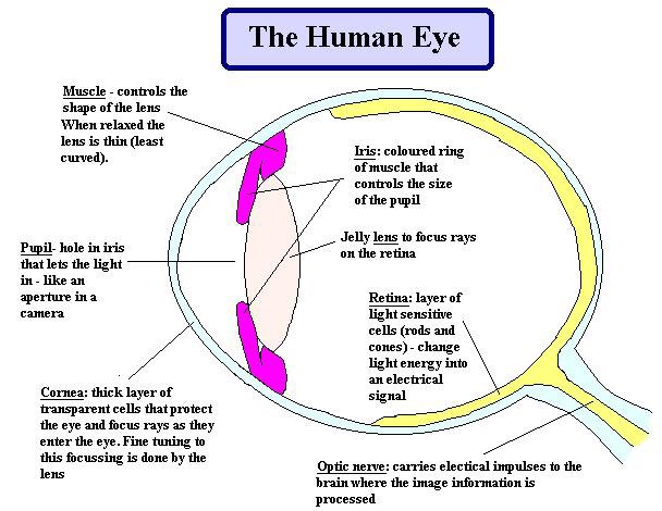
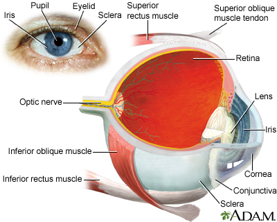
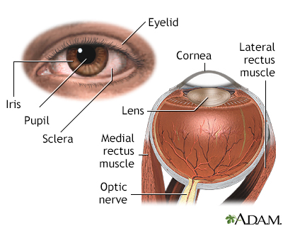
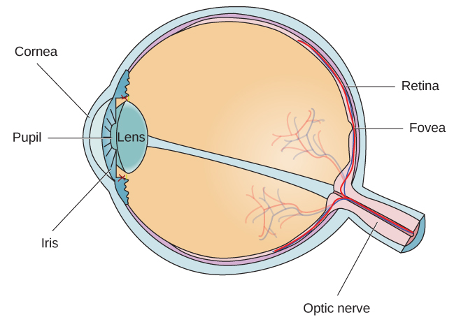
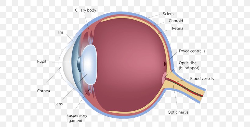
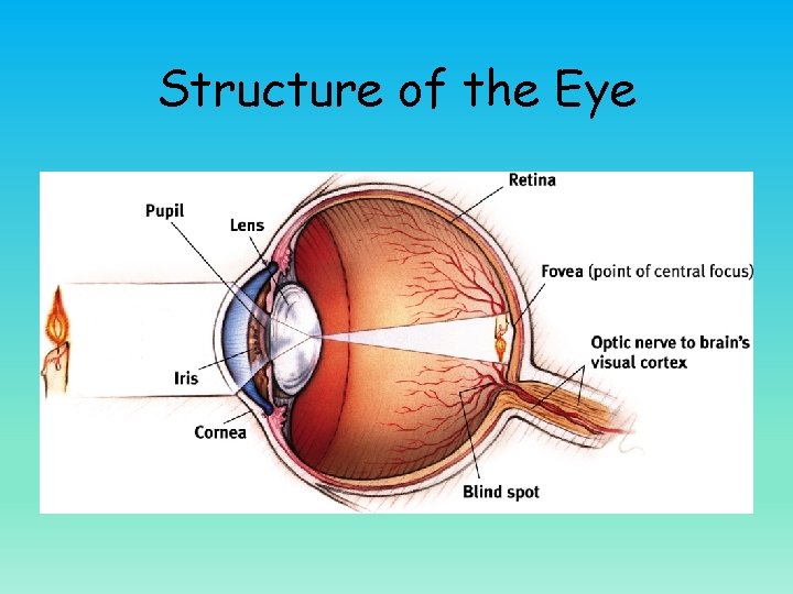
Human Eye Diagram: Contrary to popular belief, the eyes are not perfectly spherical; instead, it is made up of two separate segments fused together. Explore: Facts About The Eye To understand more in detail about our eye and how our eye functions, we need to look into the structure of the human eye. Eye Diagram Iris Muscles controls the size of the pupil. Pupil Iris opening that changes size depending on the amount of light in the environment. Cornea Bends light waves so the image can be focused on the retina. Lens Changes shape to bring objects into focus. Retina Contains photoreceptor cells. Optic Nerve Sends visual information to the brain. The Eye - Science Quiz: Our eyes are highly specialized organs that take in the light reflected off our surroundings and transform it into electrical impulses to send to the brain. The anatomy of the eye is fascinating, and this quiz game will help you memorize the 12 parts of the eye with ease. Light enters our eyes through the pupil, then passes through a lens and the fluid-filled vitreous ... 38. $2.00. Zip. This pack features high-quality anatomical diagrams of the human eye and ear and is ideal for middle school life science or high school biology students. Included are two, one-page worksheets and answer keys. The eye diagram worksheet asks students to first match identified parts of the eye with t.
Transduction Psychology Eye​: Detailed Login Instructions ...
Psychology- Eye & Ear Diagram. STUDY. PLAY. Eye Diagram. Iris. Muscles controls the size of the pupil. ... AP Psychology: Parts and Functions of the Eye and Ear. 60 terms. Tongue Taste Areas, Ear, Ear diagram, The Eye, Eye diagram. 45 terms. Chapter 29: The Senses. 69 terms. The Senses. OTHER SETS BY THIS CREATOR.
AP Psychology on Twitter: "Take a LOOK st this. Parts of the ...
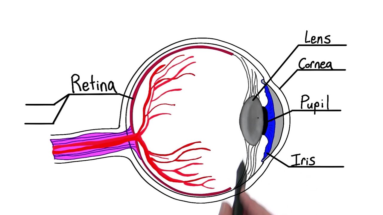
So here's a very simple diagram of an eye and as the light comes towards the eye here the first area that it's going to reach is this outer layer covering the front of the eye and it's clear. This is called the cornea. So the cornea serves two purposes.

1) Structure of the Eye - Hook AP Psychology 4B
Step Three: Transduction Psychology Definition. Ok, so how does our eye turn the light into neural impulses so that our brain can understand. Most of the process occurs on the back of the eye called the retina. The retina is the most important part of our eye (it is often referred to as the brain of the eye). First, it is important to know that ...

Parts of the Eye and their Functions - Video & Lesson ...
Virtual Learning Week #3 AssignmentsMonday, April 6 - Thursday, April 9 (No School Friday April 10, School Holiday)All work assigned on Monday at 8am and due by Thursday at 4pm. 1- Complete the Unit 1 Progress Check on AP Classroom. 2- Complete the Unit 2 Progress Check on AP Classroom.
Parts of the eye - Intro to Psychology
AP Psychology > > > > AP Seminar Modern World Blog Voices Unit 3: Sensation and Perception ... Eye Diagram Retina to Brain Diagram: F. Describe the vision process, including the specific nature of energy transduction, relevant anatomical structures, and specialized pathways in the brain for each of the senses. ... Ear Diagram I. Describe the ...

Vision - Coach Wise's AFNORTH LIONS Social Sciences
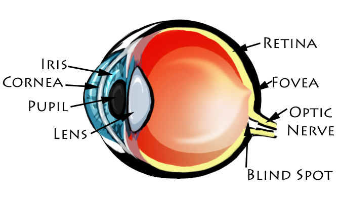
AP Psych Eye Diagram STUDY PLAY Cornea Clear, curved bulge of tissue on the front of the eye that bends light rays to begin focusing them and protects the eye (lots of nerve endings) Iris Ring of muscle tissue that forms the colored portion of the eye; surrounds the pupil and controls its size Pupil

AP Psychology: Sensation - Diagram #1 (The Eye) Diagram | Quizlet
Ultimate Study Guide for AP Psychology ... • how light waves travel through eye and get transduced • major eye anatomy • (including rods and cones - see diagrams ) • feature detectors in occipital lobe in brain • why we have a blind spot • trichromatic color theory • opponent-process color theory ...
![The Human Eye & Color Blindness [AP Psychology Unit 3 Topic 3] (3.3)](https://i.ytimg.com/vi/Mwq23JTBnN0/maxresdefault.jpg)
The Human Eye & Color Blindness [AP Psychology Unit 3 Topic 3] (3.3)
Luckily, you stumbled across this ultimate guide to the brain for AP® Psychology that we have prepared for you. In this AP® Psychology crash course review, we will provide a summary of the anatomy and function of the major areas of the brain. The brain is divided into three main parts: the forebrain, the midbrain, and the hindbrain.

20 Questions Tuesday: 419 - Vision — 20 Questions Tuesday
Where the optic nerve leaves the eye is a blind spot, as a result of the absence of receptor cells there. Image Courtesy of Myers' AP Psychology Textbook - 2nd Edition As mentioned before, feature detectors were discovered by Hubel and Wiesel in the visual cortex.

Pin on Examples Printable Label Templates
Human Eye Diagram. AP Psychology Eye diagram 17 terms Human Eye 29 terms Exercise 24 The Eye 14 terms Vision Part 1 – Anatomy of the Eye OTHER SETS BY THIS CREATOR 111 terms AP. A human eye is roughly 23 cm in diameter and is almost a spherical ball filled with some fluid. Included are two one-page worksheets and answer keys. By on april 25 2013.

Human Eye Ball Anatomy & Physiology Diagram
AP PSYCHOLOGY: Home Course Description Homework > > Projects Extra Help AP Exam wednesday 10/9. FRQ Practice HW= Module 16. thursday 10/10. DN: What is selective attention? an you give me an example? ... -Introduce Eye Diagram - use as lecture - Watch Videos and discuss Eye HW= Module 18 (Due Tuesday 10/15) Seeing.
Sensation & Perception
Start studying AP Psychology: Parts of the Eye. Learn vocabulary, terms, and more with flashcards, games, and other study tools.
Sensation & Perception
Start studying Eye diagram: AP Psychology. Learn vocabulary, terms, and more with flashcards, games, and other study tools.

AP Psychology - Sensation & Perception - Part 2 - Vision
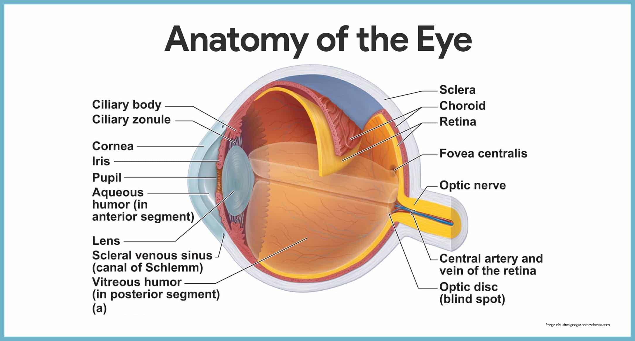
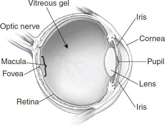
Eye Diagram Handout Author: National Eye Health Education Program of the National Eye Institute, National Institutes of Health Subject: Handout illustrating parts of the eye Keywords: parts of the eye, eye diagram, vitreous gel, iris, cornea, pupil, lens, optic nerve, macula, retina Created Date: 12/16/2011 12:39:09 PM
Special Senses Anatomy and Physiology - Nurseslabs
of light entering the eye. Lens: The lens is a clear part of the eye behind the iris that helps to focus light, or an image, on the retina. Macula: The macula is the small, sensitive area of the retina that gives central vision. It is located in the center of the retina. Optic nerve: The optic nerve is the largest sensory nerve of the eye.

Pharmaceutics | Free Full-Text | Viral-Vector-Delivered Anti ...
Parts of the Neuron. Neurons are our body's nerve cells which make up the nervous system. For a neuron to fire, or communicate with another neuron, information must first be gathered in by the dendrites of the receiving neuron. From there, the information passes through the cell body to the axon. Part of the Neuron.
Retinal detachment Information | Mount Sinai - New York
Start studying AP Psychology Eye Ear Review. Looking for human brain diagram that will help you understand the anatomy of the brain. It consists of the following parts. A human eye is roughly 23 cm in diameter and is almost a spherical ball filled with some fluid. Chemicals released in synaptic gap received by neurons o GABA.
Vision - night blindness Information | Mount Sinai - New York
AP Psychology Eye diagram. Acetylcholine ACha neurotransmitter that causes contraction of skeletal muscles helps regulate heart muscles is involved in memory and also transmits messages between the brain and spinal cord. Instead it is made up of two separate segments fused together. 2- Complete the Unit 2 Progress Check on AP Classroom.

Parts of the Eye - Pupil.
Lens Transp arent eye structure that focuses light rays falling on retine Optic Chiasm Where optic nerves cross Optic Disk Hole in retina where optic nerves exit eye Optic Nerve Axons that connect eye to brain Photor ‐ eceptor Rods and cones Pupil Opening in iris that allows light to pass to back of eyes Retina Neural tissue at back of eye that
The Eye and Ear Diagrams
How We See | Introduction to Psychology
Sunglasses to hide behind may also prevent melanoma of the ...

Epigenetic Treatment of Neurodegenerative Ophthalmic ...

What is the Pupil of the Eye? - Definition & Function - Video ...
Chapter 9: The Human Eye
International Journal of Innovative Technology and Exploring ...

AP Psychology: Chapter 5 Flashcards | CourseNotes

Parts of the Eye and their Functions - Video & Lesson ...
Net Vision Search Site Map Contact Net Vision Optic Nerve ...
2 Understanding the Epidemiology of Vision Loss and ...

VISION Eye Diagram The Retina Synethesia ESP/Psychics - ppt ...

Anatomy of the human eye and the retina with its specialized ...

Types of Eye Diseases | Study.com

AP Psych Sensation and Perception (Eye) Diagram | Quizlet
Eye Diagram - Cliparts.co

Is the mind modular?
Human Eye Diagram Retina Anatomy, PNG, 600x418px, Watercolor ...
AP Psychology Eye Diagram Project by Jennifer Kasyan | TpT
Sensation and Perception Sensation your window to the
Human Eye Iris Diagram Mammalian Eye - Parts Of The Eye ...


Comments
Post a Comment