41 cervical plexus diagram
Please accept our privacy policy Healthlineuses cookies to improve your experience and to show you personalized ads.Privacy Policy. ACCEPT More information Vagina Overview Anatomy and function Diagram Conditions Symptoms Health tips Definition When people talk about the vagina, they’re usually referring to the vulva, which is... venous plexus Medically reviewed... venous plexus includes two uterine... READ MORE Vaginal venous plexus... This human anatomy module is about the cranial nerves. It consists of 15 vector anatomical drawings with 280 anatomical structures labeled. It is intended for the use of medical students working on human anatomy, student nurses, physiotherapists, electro-radiological technicians and residents - especially those working in neurology, neurosurgery, otolaryngology - and for any physician ...
The brachial plexus (plexus brachialis) is a somatic nerve plexus formed by intercommunications among the ventral rami (roots) of the lower 4 cervical nerves (C5-C8) and the first thoracic nerve (T...

Cervical plexus diagram
The brachial plexus is a complex intercommunicating network of nerves formed by spinal nerves C5, C6, C7, C8 and T1. The brachial plexus, frequently appears in examination questions. This guide will cover the brachial plexus and includes a summary diagram. One of the best ways to memorise the brachial plexus is by drawing it. Suprascapular Nerve Block Introduction The suprascapular nerve provides sensory innervation to the glenohumeral joint (shoulder). Suprascapular nerve block is indicated for relief of acute shoulder pain e.g., after shoulder surgery and is more effective when combined with blockade of the axillary nerve. It is also useful for... Neck Cervical Plexus Block Upper Limb Upper Limb Axillary Block Infraclavicular Block Lower Limb Lower Limb... One problem with understanding this anatomy is that the traditional wiring diagram for the brachial plexus is unnecessarily complex and intimidating. Fig. 4.1 illustrates that the plexus is formed by the ventral rami of the fifth to eighth cervical nerves and the greater part of the ramus of the first thoracic nerve. In addition, small ...
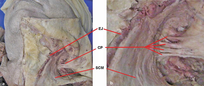
Cervical plexus diagram. In the bladder diagram above you can see the structure of the urinary bladder. In this image you will find 1st cervical vertebrae atlus cervical plexus 7th cervical vertebrae 1st thoracic vertebrae brachial plexus spinal dura mater filaments of spinal nerve roots 12th thoracic vertebra 1st lumber vertebra. Human Anatomy Diagram Organs Back View. Cervical nerves are spinal nerves that arise from the cervical region of the spinal cord. These nerves conduct motor and sensory information via efferent and afferent fibers, respectively, to and from the central nervous system. While classified as peripheral nerves, the motor cell body resides in the anterior horn of the spinal cord. There are eight pairs of cervical nerves, denoted C1 to C8 ... The cervical portion of the spine is an important one anatomically and clinically. It is within this region that the nerves to the arms arise via the, and where... structures.Cervical spine vertebrae Typical cervical vertebrae: C3-C6 Atypical cervical vertebrae: The atlas (C1), axis (C2) and C7 The bony component of the... Normal cervical plexus and its anatomical markers. A Anatomic diagram of the CP.B Longitudinal view of the C3-C5 roots (arrows), which appear as tubular hypoechoic structures with echogenic walls and a fibrillar texture.C The C7 vertebra was used as an anatomical marker to identify the CP. Arrows point to cervical nerve C7 (transverse view, arrow) and the PT.
The in the thorax, runs in front of the left common carotid artery and across the left side of the arch of the aorta, to the superficial part of the cardiac plexus. Diagram of the cervical sympathetic. (Testut.) ( ) The ( ) ramify upon the common carotid artery and upon the external carotid artery and its branches, forming... The brachial plexus sections branches teachmeanatomy. And over the middle of robert taylor's right leg (fig. The brachial plexus is formed by the five posterior cervical and the first dorsal nerves. This figure is inverted to a right side image. For branches and labelling, follow the video or picture. This figure is inverted to a right side image. When the woman isn’t ovulating, the cervical mucus thickens and serves as a barrier to keep sperm out of the uterus. During childbirth, the cervix thins out and... If dysplasia isn’t diagnosed and treated, cervical cancer, which is usually caused by the human papilloma virus, begins to spread. Definition (UWDA) Subdivision of neural tree (organ) which primarily consists of cell bodies of neurons located outside the neuraxis (brain and spinal cord); together with a nucleus and its associated nerve, it constitutes a neural tree (organ). Examples: spinal ganglion, trigeminal ganglion, superior cervical ganglion, celiac ganglion ...
270 anatomical structures of the spinal cord were labeled, subdivided into different chapters: The first image shows the different segments of the spinal cord (cervical, thoracic, lumbar, sacral and coccygeal segments), the emergence of spinal nerves (cervical, thoracic, lumbar and sacral nerves and coccyx at the level of the cauda equina and ... The cervical part of dog spine anatomy includes the cervical vertebrae, thick intervertebral discs, part of the spinal cord, and cervical spinal nerves. Now, I will describe the anatomical facts of dog cervical vertebrae with a labeled diagram. In most mammals, you will find seven cervical vertebrae in their vertebral column. The brachial plexus represents a complex network of nerves formed from the ventral rami of the lower cervical nerves (C5-C8) and the greater portion of the ventral ramus of the first thoracic nerve... The anterior distribution includes the cervical plexus (C1–C4) and brachial plexus (C5–T1). The muscles innervated by the cervical nerves are the sternohyoid, sternothyroid, and omohyoid muscles. A loop of nerves called ansa cervicalis is also part of the cervical plexus. Thoracic nerve branches exit the spine and go...
Pin on Medicine Notes
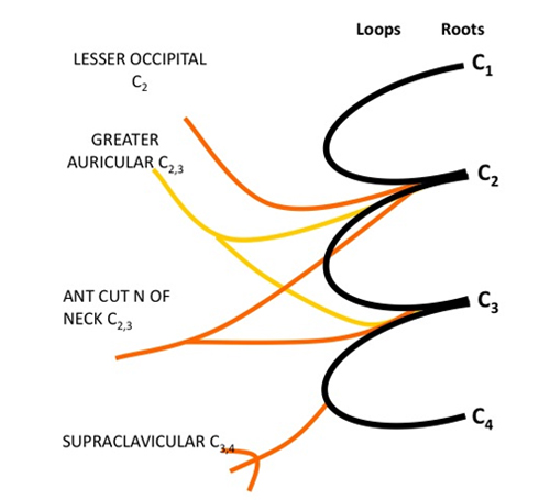
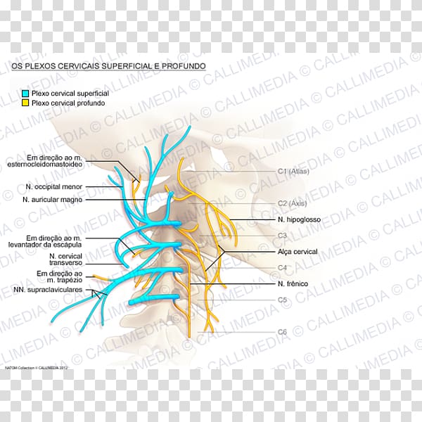
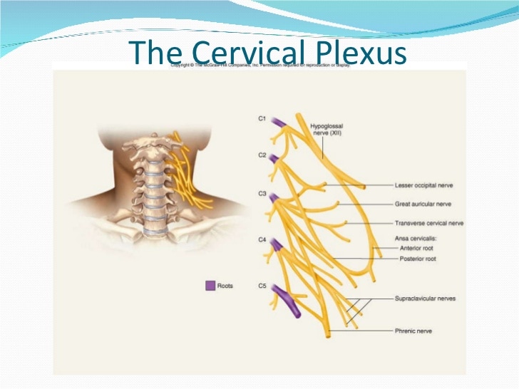
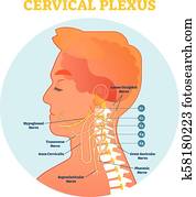
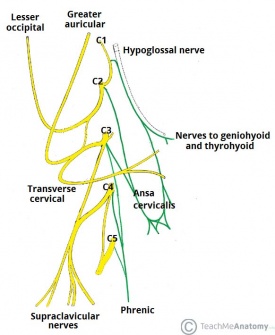
The cervical plexus is a network of nerve fibres that supplies innervation to some of the structures in the neck and trunk.. It is located in the posterior triangle of the neck, halfway up the sternocleidomastoid muscle, and within the prevertebral layer of cervical fascia. The plexus is formed by the anterior rami (divisions) of cervical spinal nerves C1-C4.

External carotid artery | Radiology Reference Article ...
Triangular vertebral foramen Two cervical vertebrae that are unique. C1 and C2 (called the atlas and axis... In contrast to the cervical vertebrae, the vertebral foramen of thoracic vertebrae is .There are five lumbar... However, like the cervical vertebrae, they have a -shaped vertebral foramen. Their spinous processes are...
Life in Overdrive !: Peripheral Nervous Systems : Spinal ...
brachial plexus originate in the neck, in the cervical spine. The nerves re-grow from the neck down the arm. This healing will occur at a rate of 1 mm per day or 1 inch per month. A mixed or incomplete recovery may occur if the nerves do not fully reattach at their original motor and sensory targets. Full recovery will occur...
place to be
Upper Cervical Muscles. Here are a number of highest rated Upper Cervical Muscles pictures on internet. We identified it from reliable source. Its submitted by dispensation in the best field. We acknowledge this nice of Upper Cervical Muscles graphic could possibly be the most trending subject with we ration it in google lead or facebook.

Image from page 81 of "Dissection of the dog as a basis for the study of physiology" (1888)
Sacral Plexus Diagram. Here are a number of highest rated Sacral Plexus Diagram pictures upon internet. We identified it from well-behaved source. Its submitted by government in the best field. We acknowledge this kind of Sacral Plexus Diagram graphic could possibly be the most trending subject similar to we allowance it in google lead or facebook.

Image from page 427 of "Anatomical technology as applied to the domestic cat; an introduction to human, veterinary, and comparative anatomy" (1882)
The sacral plexus is formed from the ventral rami of nerves L4, L5, and S1 to S4. It has six roots and anterior and posterior divisions. Since it is connected to the lumbar plexus by fibers that run through the lumbosacral trunk, the two plexuses are sometimes referred to collectively as the lumbosacral plexus.
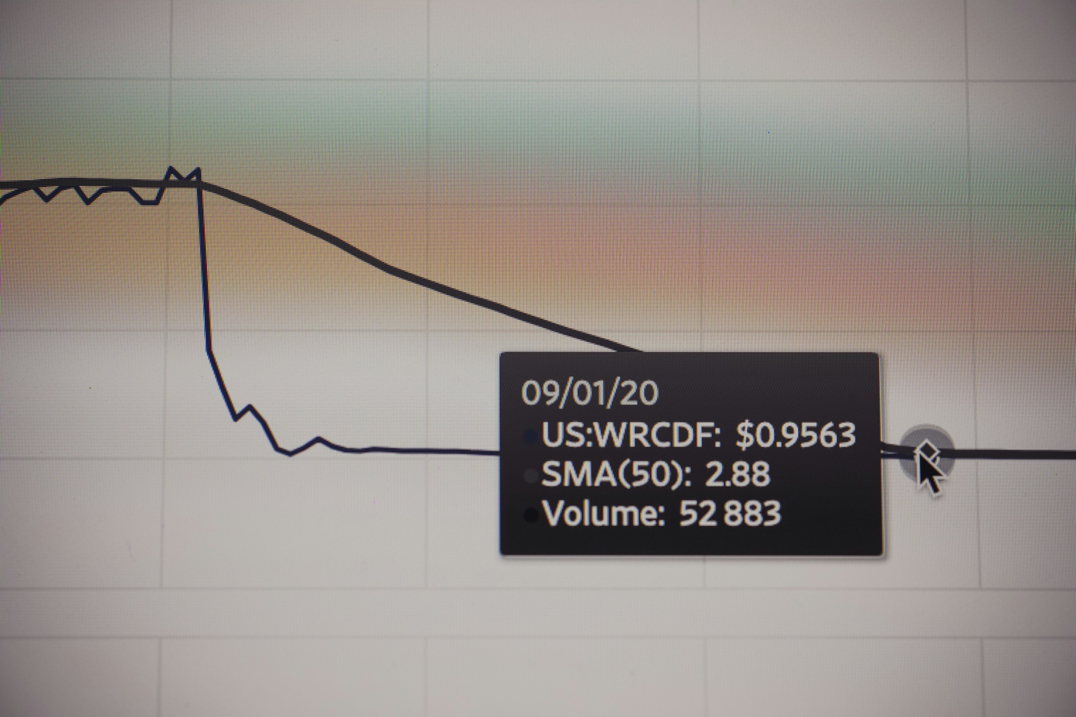
Stock market chart value. Made with analog vintage lens, Leica APO Macro Elmarit-R 2.8 100mm (Year: 1993)
Veins of Scalp, Face, and Neck, CLVIII. Section of Neck at Sixth Cervical Vertebra, CLIX. Diagram of Deep Cervical Fascia CLX. Cervical Plexus, CLXI. Superficial Structures of Neck, CLXII. Superficial Structures of Neck, CLXIII. Diagram of Triangles of Neck, CLXIV. Incisions for Dissection and Lines for Arteries, Veins, and Nerves of Neck, CLXV.

Cervical Plexus In Situ Nerves of the Upper Extremity ...
In this article we will analyze the various aspects of the choroid plexus. Many of us will have heard of cerebrospinal fluid (CSF) and are familiar with its... These vascularized invaginations, are lined by a plexus of specialised cells that produce our CSF. This plexus of cells is called the.The ventricular system The...

Cervical Plexus Block - Hadzic's Peripheral Nerve Blocks ...
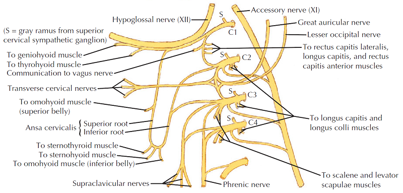
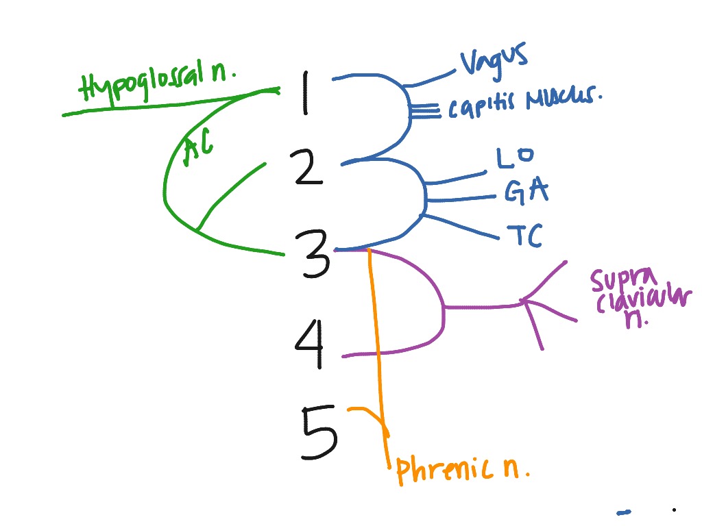
A major loop of nerves in the anterior triangle is part of the ansa cervicalis in the cervical plexus. The superior root from the anterior ramus travels with CN XII and descends the anterior surface of the carotid sheath. The nerve for the thyrohyoid is a branch of the superior root that travels with the hypoglossal to innervate this muscle.

Plexus products, Cervical, Nerve anatomy
Anatomy. Cranial nerves are the 12 nerves of the peripheral nervous system that emerge from the foramina and fissures of the cranium.Their numerical order (1-12) is determined by their skull exit location (rostral to caudal). All cranial nerves originate from nuclei in the brain.Two originate from the forebrain (Olfactory and Optic), one has a nucleus in the spinal cord (Accessory) while the ...

Cervical Plexus Block - Landmarks and Nerve Stimulator ...
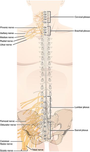
A nerve plexus is a network of nerves that innervate the same region of the body. They are typically named after the regions of the body they innervate also, such as the cervical, brachial, lumbar ...

12.6C: Plexuses - Medicine LibreTexts
Related to cervical: Cervical cancer, Cervical Spondylosis cer·vi·cal (sûr′vĭ-kəl) adj. 1. Of or relating to the uterine cervix. 2. Of or relating to the neck. American Heritage® Dictionary of the English Language, Fifth Edition. Copyright © 2016 by Houghton Mifflin Harcourt Publishing Company. Published by Houghton... nerve cervical plexus cervical polyps cervical range of motion cervical root syndrome cervical smear cervical...
Cervical Plexus Block | Radiology Key
The brachial plexus of ox derives from the ventral branches of the last three cervical and first two thoracic nerves.If you want to get the best guide to learn the anatomy of the brachial plexus of an ox, this article is for you. Here, I will show you the formation of brachial plexus, branches, and their innervation in an ox, sheep, and goat.
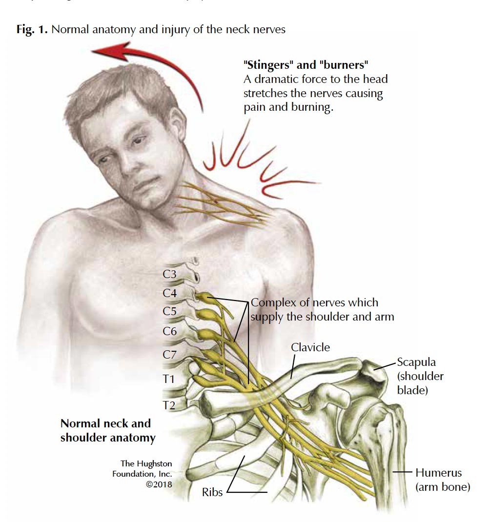
Diagram Of Bones In Neck And Shoulder - Neck Anatomy ...
Diagram of the right brachial plexus of a fetal pig. The ventral cords (shaded) are reflected ventrally. Ax axillary nerve; C5-8 ventral rami of the fifth to eighth cervical nerves; ld latissimus dorsi muscle; R radial nerve; sbs subscapularis muscle; T1 ventral ramus of the first thoracic nerve; tma teres major muscle; 1-4 branches to the ...

Image from page 431 of "The development of the human body : a manual of human embryology" (1914)
The brachial plexus is a network (plexus) of nerves (formed by the anterior rami of the lower four cervical nerves and first thoracic nerve (C5, C6, C7, C8, and T1). This plexus extends from the spinal cord, through the cervicoaxillary canal in the neck, over the first rib, and into the armpit.
ปัà¸à¸žà¸´à¸™à¹‚ดย sensei ใน Han
The cervical plexus is a complex neurologic structure located within the head and neck. The large portion of the cervical plexus is the communication between the anterior divisions of C1 through C4 nerves. While the plexus itself can be complex, it is essential for practitioners to understand the significant motor and sensory functions of the cervical plexus as this information can provide ...

Pin em Physical Therapy
The cervical plexus forms within the muscles of the floor of the posterior triangle. A major branch of this plexus is the phrenic nerve, which arises from the anterior divisions of spinal nerves C3-C5. It descends down the neck, within the prevertebral fascia, to innervate the diaphragm.
Cervical Plexus - Physiopedia, universal access to ...
The brachial plexus is a network (plexus) of nerves (formed by the ventral ramus of the lower four cervical nerves and first thoracic nerve (C5, C6, C7, C8, and T1 ). This plexus extends from the s...

Plexus cervicalis | definition of plexus cervicalis by ...
The brachial plexus is a network of nerves that gives rise to all the motor and sensory nerves of the upper extremity. This plexus arises from the anterior rami ofthat undergo several mergers and splits into trunks and... In addition to terminal branches, the brachial plexus gives rise to several preterminal branches called...

Image from page 339 of "War surgery of the face. A treatise on plastic restoration after facial injury by John B. Roberts ... Prepared at the suggestion of the subsection on plastic and oral surgery connected with the office of the surgeon general. Illust
The cervical plexus is a conglomeration of cervical nerves formed by the anterior/ventral rami of spinal nerves C1-C4 (a.k.a. 1st-4th cervical nerves).These are the roots (limbs) of the cervical plexus. However, most authors include the fifth cervical nerve (i.e. the anterior ramus of spinal nerve C5) to the plexus owing to its contribution to the formation of one of the motor branches of the ...

Deep cervical plexus block. | Download Scientific Diagram
This diagram now includes the main branches and main nerve roots with proper connections. The median nerve is a sensory and motor nerve of the arm (or upper limb) The brachial plexus is a network of nerves that gives rise to all the motor and sensory. Of 3.1) and abnormality in a katz hand diagram.16 although commonly used in .

Instant Anatomy - Head and Neck - Nerves - Somatic nerves ...
Lumbar Plexus Diagram. ... vertebrae are given names corresponding to their locations in the human body. These vertebrae are called the cervical, thoracic, lumbar, and sacral vertebrae, as well as ...

Image from page 90 of "An illustrated encyclopædic medical dictionary. Being a dictionary of the technical terms used by writers on medicine and the collateral sciences, in the Latin, English, French and German languages" (1890)
The great auricular nerve is a cutaneous nerve of the head. It originates from the cervical plexus, with branches of spinal nerves C2 and C3. It provides sensory nerve supply to the skin over the parotid gland and the mastoid process of the temporal bone, and surfaces of the outer ear.

Image from page 416 of "Anatomical technology as applied to the domestic cat; an introduction to human, veterinary, and comparative anatomy" (1882)
Home » Tanpa Label » Cervical Spinal Nerves Diagram - Treating Neurologic Like Symptoms By Addressing Cervical Spine Instability And Disrupted Blood Flow Into The Brain Caring Medical Florida / Circulating within the meninges is a liquid substance known as the cerebrospinal fluid (csf).
Cervical plexus Cervical vertebrae Ansa cervicalis Great ...
One problem with understanding this anatomy is that the traditional wiring diagram for the brachial plexus is unnecessarily complex and intimidating. Fig. 4.1 illustrates that the plexus is formed by the ventral rami of the fifth to eighth cervical nerves and the greater part of the ramus of the first thoracic nerve. In addition, small ...
Cervical Plexus - Anatomy, Function, Injury, Complications ...
Suprascapular Nerve Block Introduction The suprascapular nerve provides sensory innervation to the glenohumeral joint (shoulder). Suprascapular nerve block is indicated for relief of acute shoulder pain e.g., after shoulder surgery and is more effective when combined with blockade of the axillary nerve. It is also useful for... Neck Cervical Plexus Block Upper Limb Upper Limb Axillary Block Infraclavicular Block Lower Limb Lower Limb...

Image from page 54 of "The anatomy of the nervous system, from the standpoint of development and function" (1920)
The brachial plexus is a complex intercommunicating network of nerves formed by spinal nerves C5, C6, C7, C8 and T1. The brachial plexus, frequently appears in examination questions. This guide will cover the brachial plexus and includes a summary diagram. One of the best ways to memorise the brachial plexus is by drawing it.

Bar charts quickstudy nervous system
Cranial nerves vector illustration. Labeled diagram with ...
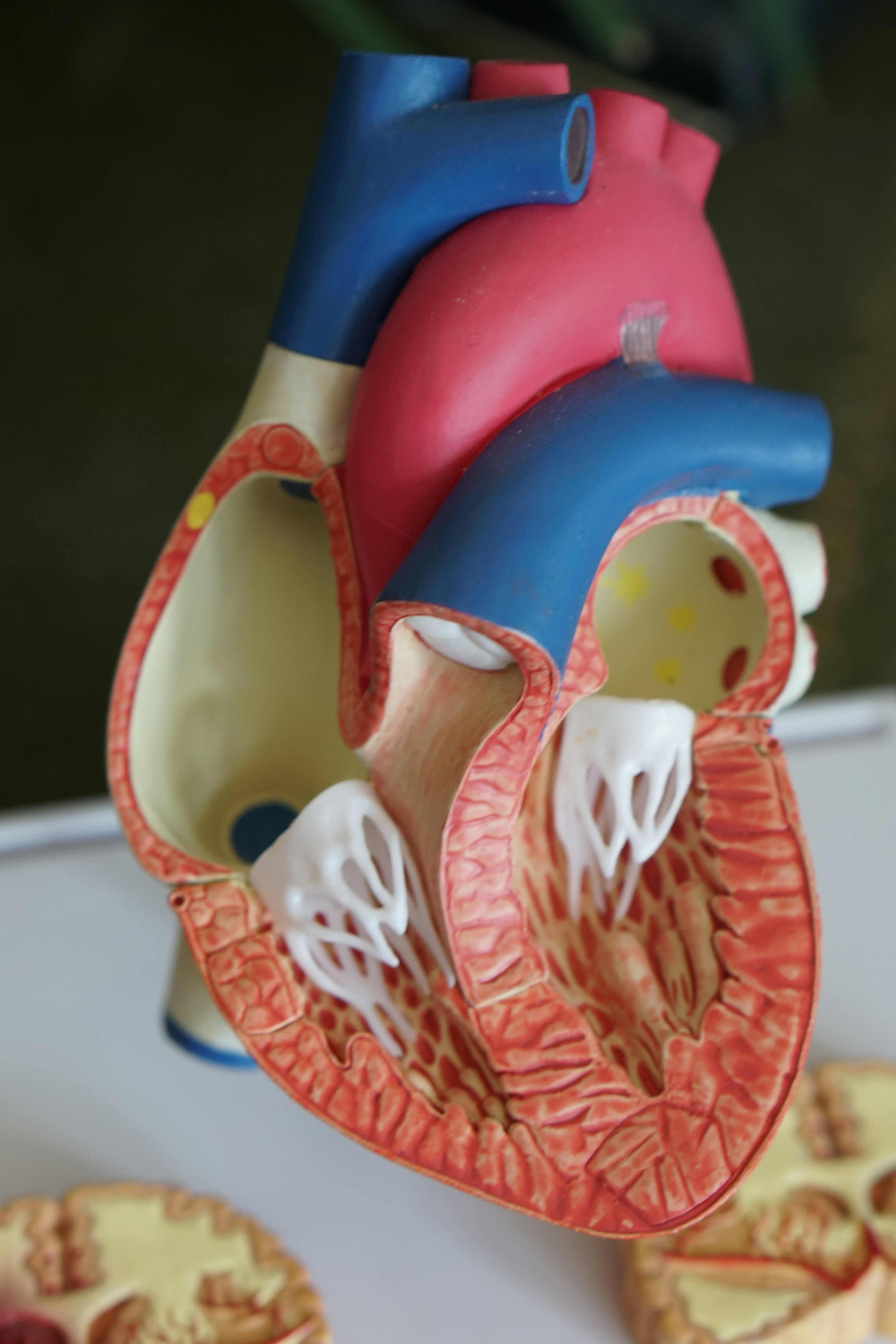
Open heart model

Spinal Nerves: Cervical Plexus (C1-C5)

Image from page 730 of "Biology of the vertebrates : a comparative study of man and his animal allies" (1949)
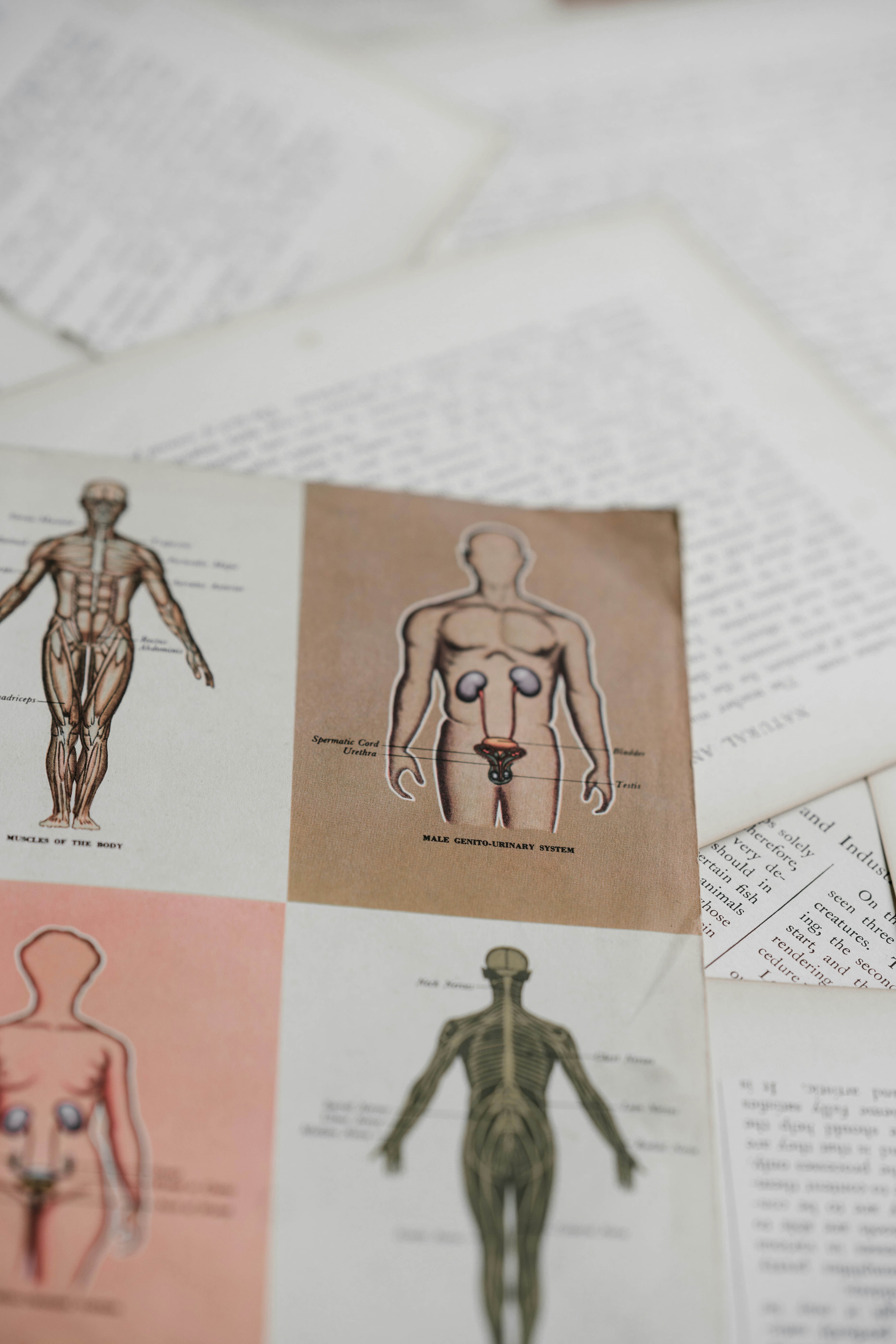
Medical diagram
Cervical plexus. What is the Cervical Plexus? (with ...

Image from page 569 of "The cyclopædia of anatomy and physiology" (1847)

Places To Be , Hamburg
Closeup of skeleton pelvic model
Phrenic Nerve - Physiopedia

Finance investment stock market chart. Made with analog vintage lens, Leica APO Macro Elmarit-R 2.8 100mm (Year: 1993)
Difference Between Cranial and Spinal Nerves | Definition ...

Cervical and Brachial Nerve Plexuses | ClipArt ETC


Comments
Post a Comment