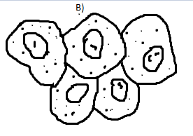
41 cheek cell diagram
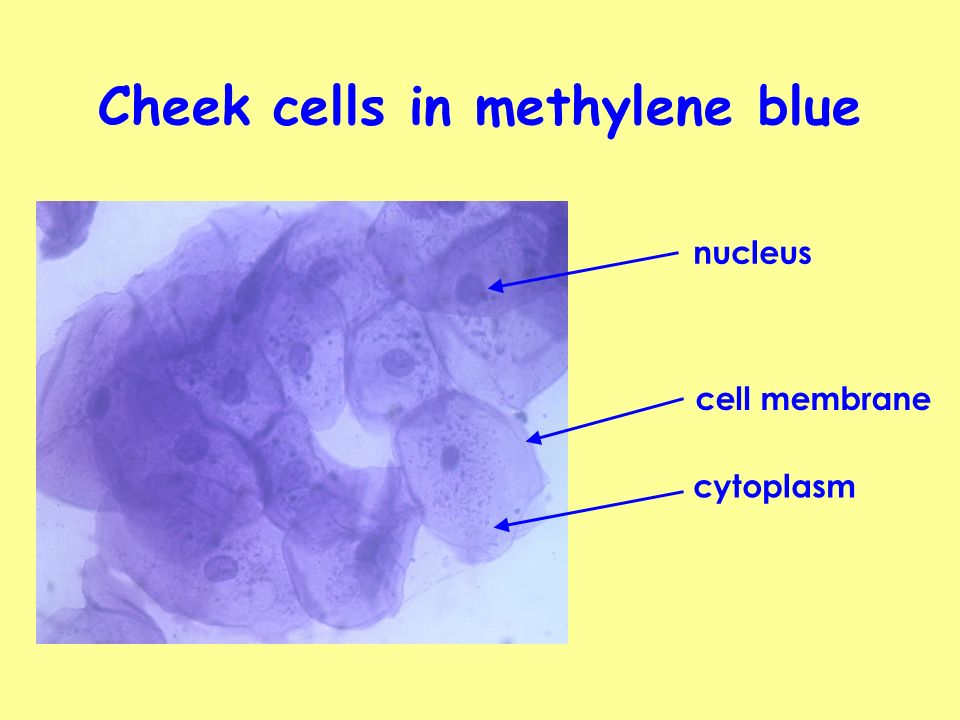
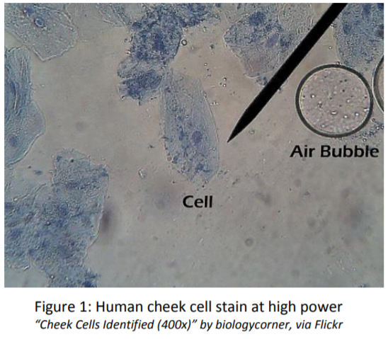
Research Biology Cheek Cell Lab page 1 of 3 Cheek Cell Lab AFTER you have completed the rest of this lab come back to this cover page DRAW & LABEL A CHEEK CELL WITH ALL THE PARTS / ORGANELLES YOU OBSERVE UNDER 40X. Purpose: To observe and identify major animal cell structures and to relate the structure of the cell to its function. In this lab, we took a sample of our own cells by scraping the inside of the cheek with a toothpick. The sample was then stained with methylene blue, so the cells could be viewed at 40x, 100x, and 400x. The following images simulate what would be seen at each magnification. Cheek cells 40x. Cheek Cells, 100x. Cheek cells, 400x
Cheek cells can be easily obtained by gently scraping the inside of the mouth using a clean, sterile cotton swab. Once the cells have been obtained, the following procedure is used for cheek cell wet mount preparation: place a drop of physiological saline on a clean microscopic slide (central part of the slide)

Cheek cell diagram
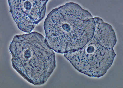
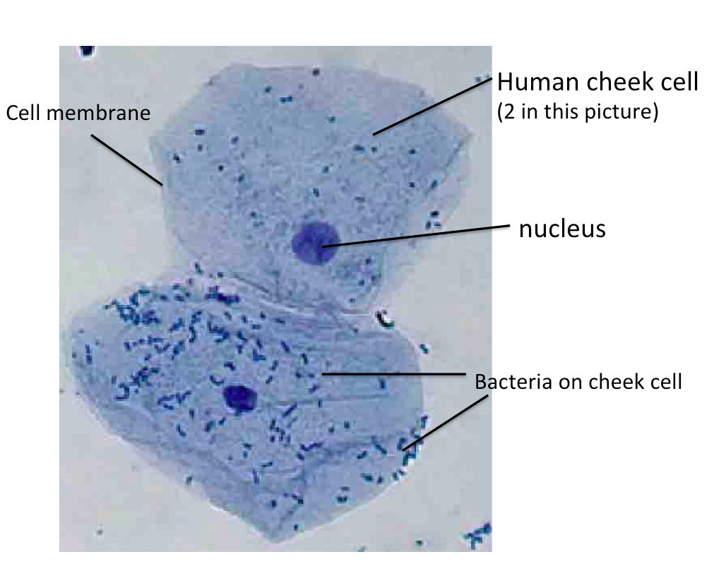
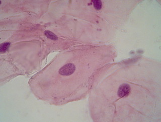
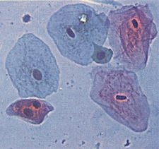
A cell membrane that is semi-permeable surrounds the cytoplasm. The vacuole in an animal cell is smaller in size, or absent. The nucleus is present at the centre of the cytoplasm. The absence of a cell wall and a prominent vacuole are indicators that help identify animal cells, such as cells seen in the human cheek. Drawing Diagrams Diagram 1: Human cheek cells The photo below is of a cluster of human cheek cells that have been stained and magnified 400 times. On your lab sheet draw the cluster of cells and label one cell with the following structures: cell membrane – the cell membrane is the outer edge of the cell Draw a diagram of one cheek cell and label the parts. (You should observe the cell membrane, nucleus, and cytoplasm.) Observation: The following labeled drawings must be completed. Drawings MUST be completed neatly using a pencil/colored pencil. 1. Onion Cell drawing (high power) 2. Cheek cell drawing (any power but preferably high)
Cheek cell diagram. Preparing cheek cell slides to view using a light microscope is described in page 6 of this guide. Cell structure How it is related to its function. Cytoplasm: Cells, The Structural and Functional Units of Life. For Teachers 9th - 12th. Students observe the general structure and organelles of plant and animal cells. Students prepare microscope slides of elodea, onion, check, and cork and identify the cells by size and shape as unicellular, multicellular, plant or animal. Cheek Cells Under a Microscope Requirements, Preparation and Staining. Cell membrane (outer boundary of the cell) Cytoplasm (the fluid within the cell) Nucleus (at the center of the cell and controls cell functions) Organelles (e.g. mitochondria-Organelles are cell structures with specific functions) Cheek cells are generally irregularly shaped and are always flat cells. The cells are made up of many parts including a very thin membrane on the outer part of the cell. Cheek cells are among the most popular cells that are studied in a classroom setting because they can be obtained from a person in a minimally invasive way.
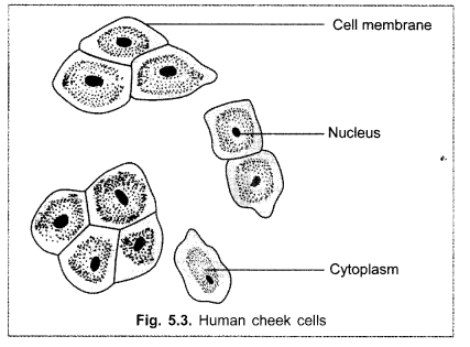
Structure. Cheeks are fleshy in humans, the skin being suspended by the chin and the jaws, and forming the lateral wall of the human mouth, visibly touching the cheekbone below the eye. The inside of the cheek is lined with a mucous membrane (buccal mucosa, part of the oral mucosa).. During mastication (chewing), the cheeks and tongue between them serve to keep the food between the teeth. Sep 18, 2021 · Human Cheek Cell Diagram Labeled. angelo. November 7, 2021. What Is A Cell Animal Cell Project Cells Project Human Cell Diagram. Label The Plant Cell Worksheets Sb11867 Sparklebox Cells Worksheet Plant Cell Plant Cells Worksheet. Diagram Of Animal Cell Anatomy Premium Vector Free Vector Freepik Vector Freebackground Freemedical Animal Cell ... Title: how to draw human cheek cell | how to draw human cheek cell easily | how to draw human cheek cellHello Friends in this video I tell you about how to d... 1.The cell has an irregular shape. 2.Cells are flat with dense cytoplasm. 3.The ectoplasm is also clearly visible. 4.The boundary of a cell has a thin cell membrane. 5.A centrally located dark stained nucleus is seen. PRECAUTIONS. 1.Gently scrap the cheek or it may cut the cheek. 2. Don’t over stain the material.
Q1.Figure 1 shows a human cheek cell viewed under a light microscope. ... The cell shown in the diagram is usually found with similar cells. Draw a ring around the correct answer to complete the sentence. ... light purple liquid in the cell. The sample was then stained with methylene blue so the cells could be viewed at 40x 100x and 400x. Draw A Neat Diagram Of Human Cheek Cell And Label Its Three Parts Brainly In Chapter 1 page 7 histologyolm. Human cheek cell labeled diagram. Cheek cells are eukaryotic cells cells that contain a nucleus and other A cheek cell, an epithelial cell found in the tissue on the inside lining of the mouth, continually secretes mucus to maintains a moist environment in the mouth. Together with salivary glands that secrete saliva, the cheek cells supply enough moisture in the mouth for enzymes to thrive. This moisture softens food, assists in swallowing and starts digestion. Remove any excess solution by allowing a paper towel to touch one side of the coverslip. Place the slide on the microscope, with 4 x or 10 x objective in position and find a cell. Then view at higher magnification. Methylene blue stains negatively charged molecules in the cell, including DNA and RNA. This dye is toxic when ingested and it ...
Cheek cell image using brightfield and darkfield microscopy.
To prepare a temporary mount of human cheek epithelial cells, and to study its characteristics. Theory The body of all animals including humans is composed of cells. Unlike plant cells, animal cells do not have cell wall. The outermost covering of an animal cell is a cell membrane. The cytoplasm, nucleus and other cell organelles are enclosed ...

Cheek Cells | Cell, Things under a microscope, Cheek
A cell membrane that is semi-permeable surrounds the cytoplasm. The vacuole in an animal cell is smaller in size, or absent. The nucleus is present at the centre of the cytoplasm. The absence of a cell wall and a prominent vacuole are indicators that help identify animal cells, such as cells seen in the human cheek.

Cells, Tissues, Organs, and Systems | T h e P e l i c a n P ...
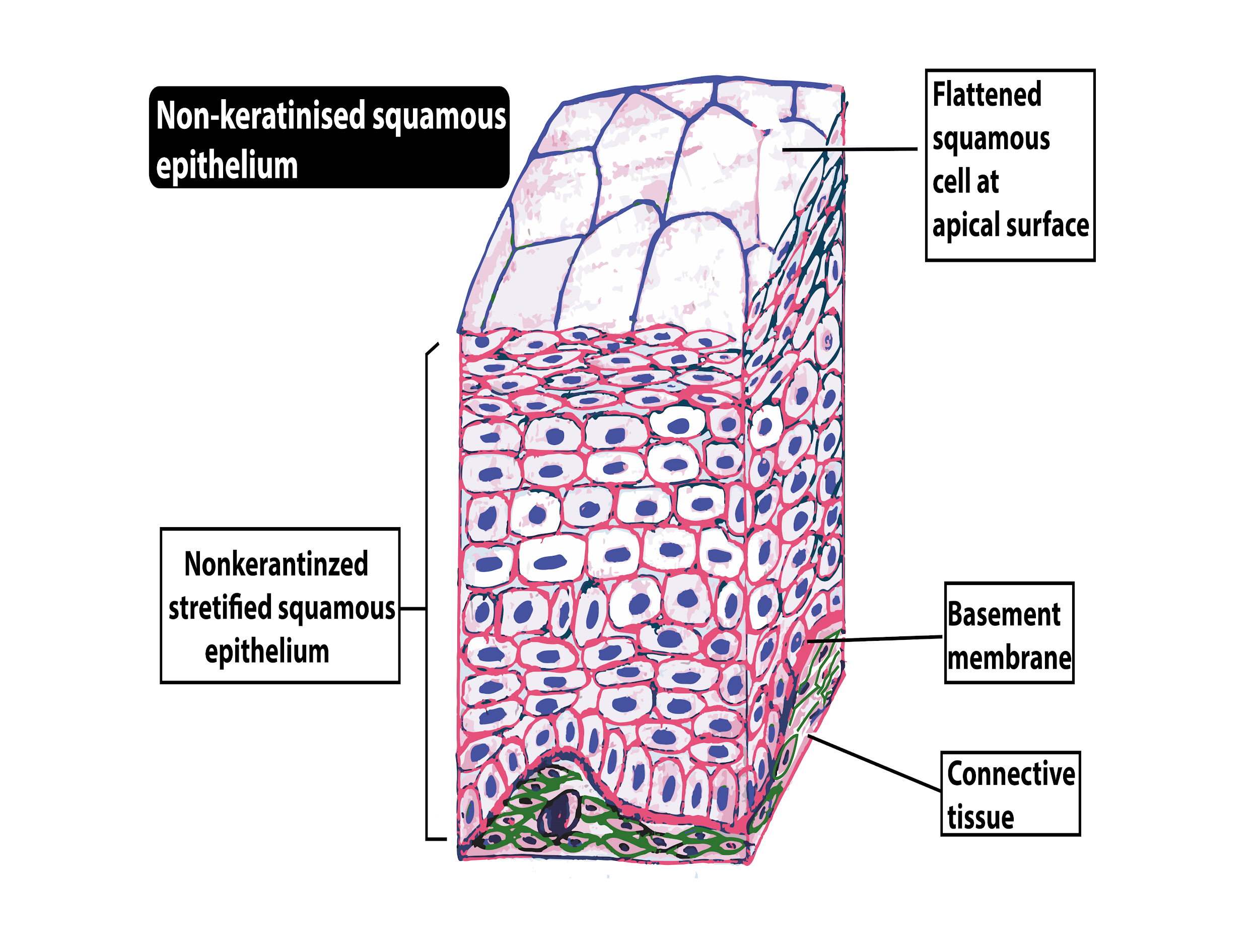
Human Cheek Epithelial Cells. The tissue that lines the inside of the mouth is known as the basal mucosa and is composed of squamous epithelial cells. These structures, commonly thought of as cheek cells, divide approximately every 24 hours and are constantly shed from the body.
Cheek cells are made up of aMuscle cells bEpithelial class 11 ...
Onion cell and human cheek cell are two types of epithelial cells observes under the light microscope. Both cells are easy to obtain. Both are in a single layer. Both are transparent. Both have a cell membrane, cytoplasm, nucleus, mitochondria, Golgi apparatus, ER, ribosomes, lysosomes, and plastids. Both cells do not have chloroplasts.
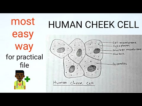
How to draw #human cheek cell || Most easy way || Step by step
Before exploring the details of cell structure, let's understand the differences in the structure of an onion cell and a human cheek cell. Onion Cell. An onion is a multicellular (consisting of many cells) plant organism.As in all plant cells, the cell of an onion peel consists of a cell wall, cell membrane, cytoplasm, nucleus and a large vacuole.
Easy diagram for HUMAN CHEEK CELL.....by TEJBIR MAND... - YouTube
Cheek cells are epithelial cells that line the interior surface of our mouths. All epithelial cells share the same characteristics. ... The base layer of cells in an epithelial structure are not actually cells, but a sticky layer on which the cells anchor. The other surface of the epithelial cell touches the outside world (like skin) or an open ...

Draw a well labelled diagram for the cell observed in onion ...
Human Cell Diagram, Parts, Pictures, Structure and Functions The cell is the basic functional in a human meaning that it is a self-contained and fully operational living entity. Humans are multicellular organisms with various different types of cells that work together to sustain life.

Microscopic views of human buccal cells. The cells were ...
Cheek Cell Lab - observe cheek cells under the microscope Cheek Cell Virtual Lab - virtual microscope view of cells. Plant Cell Lab - microscope observation of onion and elodea Plant Cell Lab Makeup - can be done at home or at the library Plant Cell Virtual Lab - use a virtual microscope to view plant cells.. Comparing Plant and Animal Cells - looks at cheek and onion cells
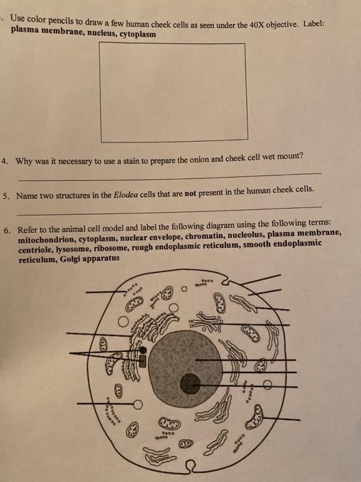
Solved -. Use color pencils to draw a few human cheek cells ...
Download scientific diagram | DIC image of a cheek cell from publication: Quantitative phase restoration in differential interference contrast (DIC) microscopy | Phase contrast imaging is a ...

drawing of nucleus in cheek cells ​ - Brainly.in
2. Cell Structure Practical Exercises: ( 3 Parts in one report). ( 2-3 pages) a. Onion Cells b. Human Cheek Cells c. Fungal Cells. Biology 2. Cell Structure Practical Exercises. Introduction. In this practicalexercise you will look at three different categories of cell: Fungal, Animal and Plant.

Cheek Epithelial Cell Practical Experiment
The diagrams show a cheek cell from a human and a leaf cell from a plant. (a) The two cells have a number of parts in common. (i) On the cheek cell, label three of these parts which both cells have. (3) (ii) In the table, write the names of the three parts you have labelled above and describe the main function of each part.

Lab: Observing Cells – Michael Eng – Michael's Blog
Human cheek cells are made of simple squamous epithelial cells, which are flat cells with a round visible nucleus that cover the inside lining of the cheek.C...

CELL LAB HOW DID YOU DO??? I love cheek cells!!. Your animal ...
Human cheek cells are made of simple squamous epithelial cells, which are flat cells with a round visible nucleus that cover the inside lining of the cheek. Cheek cells are easy to obtain and easy to see under a microscope. As such it is a favorite in biology classrooms to show what a typical animal cell looks like.
Buccal Epithelial Cells | Nikon's MicroscopyU
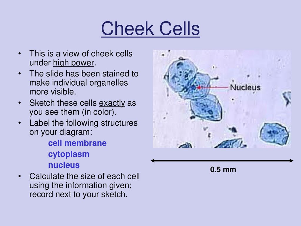
Draw a diagram of one cheek cell and label the parts. (You should observe the cell membrane, nucleus, and cytoplasm.) Observation: The following labeled drawings must be completed. Drawings MUST be completed neatly using a pencil/colored pencil. 1. Onion Cell drawing (high power) 2. Cheek cell drawing (any power but preferably high)

Were there more structures visible in the Elodea sample or in ...
Drawing Diagrams Diagram 1: Human cheek cells The photo below is of a cluster of human cheek cells that have been stained and magnified 400 times. On your lab sheet draw the cluster of cells and label one cell with the following structures: cell membrane – the cell membrane is the outer edge of the cell

Increased Human Buccal Cell Autofluorescence Is a Candidate ...
A cell membrane that is semi-permeable surrounds the cytoplasm. The vacuole in an animal cell is smaller in size, or absent. The nucleus is present at the centre of the cytoplasm. The absence of a cell wall and a prominent vacuole are indicators that help identify animal cells, such as cells seen in the human cheek.
DNA Isolation from wheat germ – work in pairs!

15. The following diagram showing cells of onion peel, label ...
Can you expect to see mitochondria while using a light ...
CBSE Class 9 Science Practical Skills – Slide of Onion Peel ...
Lab 5: Cells - Biology LibreTexts
The following diagram shows cells of onion peel label class ...
Solved Using this table from the Size Estimation module ...
What type of cells are cheek cells? - eNotes.com

Cheek Cell - an overview | ScienceDirect Topics

Barr body in Cheek cells – Microcosmos

drawing of nucleus in cheek cells ​ - Brainly.in
Virtual Microscope – Cheek Cells - ppt download
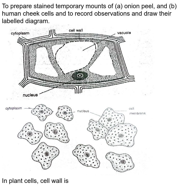
To prepare stained temporary mounts of (a) onion peel, and (b ...

Cell organelle present in both prokaryotic and eukaryotic ...

Unit 1: Cheek Cell Experiment Diagram | Quizlet
Tabulate differences between plant and animal cell with ...

Lab: Observing Cells – Michael Eng – Michael's Blog

Human cheek cells, light micrograph - Stock Image - C020/8175 ...
Cell Nucleus | Contexo.Info

Cell theory - 1 Human cheek cell Diagram | Quizlet
investigating human cheek cell ss

Cheek Cell Lab

HUMAN CHEEK CELL ( Class : 8 Lesson No : 8 )
1. Identify the organelles labelled A - E in the following ...
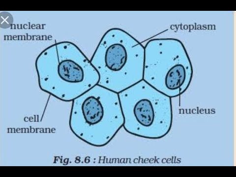
![Draw a labelled diagram of human cheek cells. [3 MARKS]](https://search-static.byjusweb.com/question-images/byjus/tnl-content-images/moodle-migration/8464_f2de758c56c0cce78b2222ae53ba00c93da2a1fc_a.png)
Draw a labelled diagram of human cheek cells. [3 MARKS]


Comments
Post a Comment