41 wiggers diagram explained
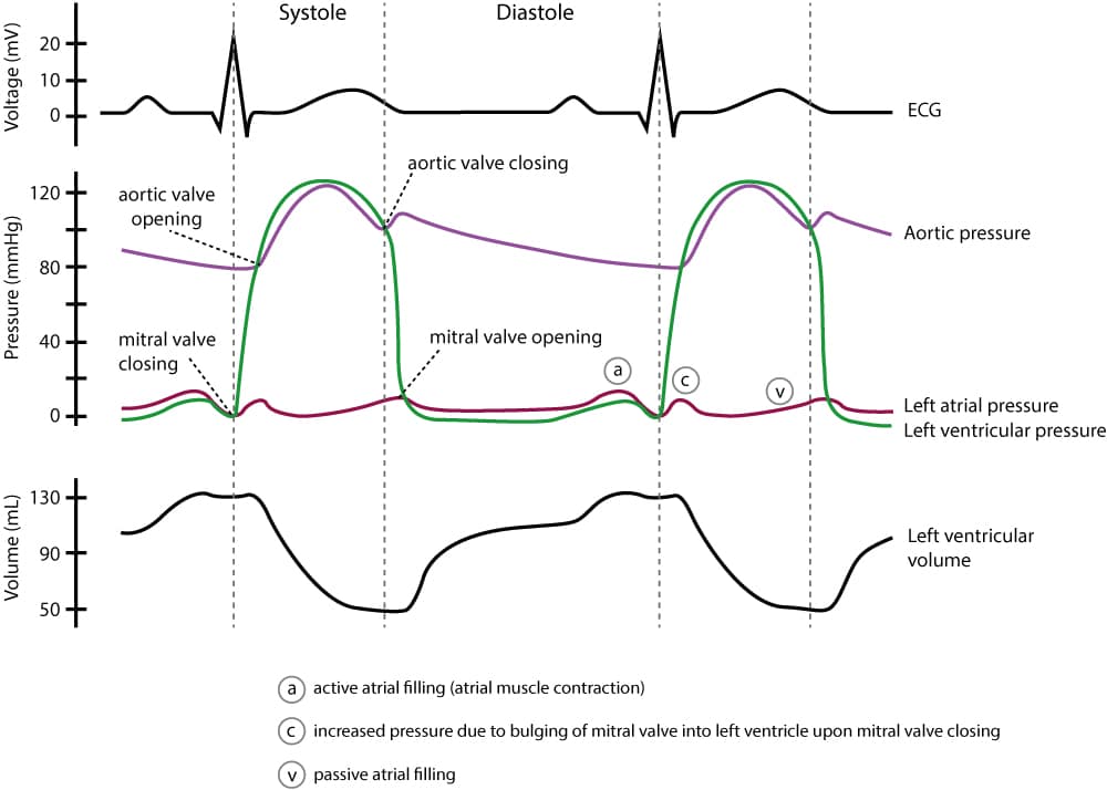
An in-depth look at Wiggers Diagram. Examining the cardiac cycle, Electrocardiogram, Aortic Pressure, Ventricular Pressure, Atrial Pressure, Phonocariogram, ... Description. Wiggers Diagram.svg. English: A Wiggers diagram, showing the cardiac cycle events occuring in the left ventricle. In the atrial pressure plot: wave "a" corresponds to atrial contraction, wave "c" corresponds to an increase in pressure from the mitral valve bulging into the atrium after closure, and wave "v" corresponds to passive ...
A Wiggers diagram, named after its developer, Dr. Carl J. Wiggers, is a standard diagram that is used in teaching cardiac physiology. In the Wiggers diagram, the . Wiggers Diagram Explained Health Education, Ultrasound, Physiology, . Ekg en 3D Más Heart Anatomy, Ekg Leads, Nursing Students, Ekg Interpretation.

Wiggers diagram explained
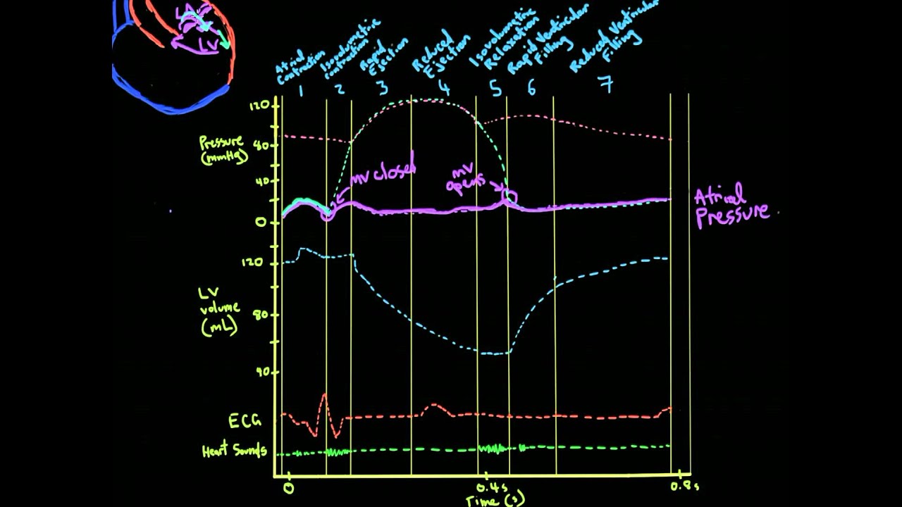
ELI5: Cardiac cycle (diastole/systole) & also wigger's diagram. I have an exam on these tomorrow (see links) and while I can read it and somewhat comprehend, i still feel like its over my head and I dont really understand how it works. Therefore, I can't absorb the information enough to understand. Wiggers diagram of the cardiac cycle. Wiggers. Understanding Wiggers Diagram. When the ventricles contract which valves open? semilunar Aortic and pulmonary. How does that affect the volume in the ventricles? Volume decreases in the ventricles after semi lunar valves open. Basically, a "Wiggers Diagram" is a description of the events which take place over the cardiac cycle and which a plotted on a time scale. Yes, it's the Wiggers diagram, not Wigger's diagram or Wiggers' diagram, because a guy called Wiggers was responsible for the development of its most important components.
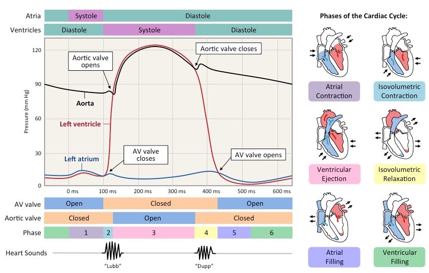
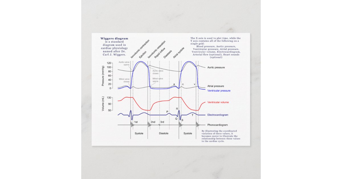
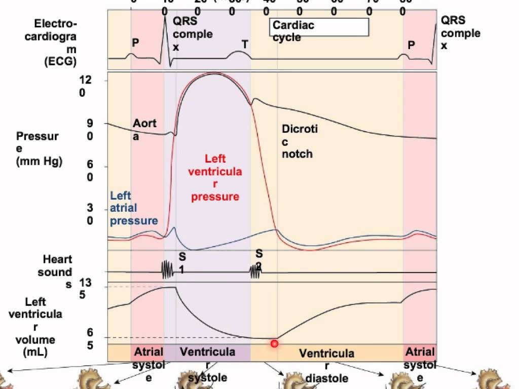
Wiggers diagram explained. The cardiac cycle diagram shown to the right depicts changes in aortic pressure (AP), left ventricular pressure (LVP), left atrial pressure (LAP), left ventricular volume (LV Vol), and heart sounds during a single cycle of cardiac contraction and relaxation. These changes are related in time to the electrocardiogram. · A Wiggers diagram is a medical chart that summarizes several aspects of cardiovascular health on one chart. Blood pressure, ventricular volume, arterial blood flow, and an electrocardiogram are simultaneously plotted against time on this chart. 348 People Learned More Courses ›› View Course Wiggers diagram - Wikipedia Save en.wikipedia.org Wiggers Diagram, showing various events of a cardiac cycle. A Wiggers diagram is a standard diagram used in cardiac physiology named after Dr. Carl J. Wiggers. The X axis is used to plot time, while the Y axis contains all of the following on a single grid: Blood pressure. The Version table provides details related to the release that this issue/RFE will be addressed. Unresolved: Release in which this issue/RFE will be addressed. Resolved: Release in which this issue/RFE has been resolved.
How to draw Wigger's Diagram of Cardiac Cycle? Summary of Cardiac Cycle Opening and closing of valves When the valve opens, different compartments act as a single chamber (atrio-ventricle or aorto-ventricle). For a blood to flow, pressure in "giver" must be higher then that in "receiver". Pressure difference opens or closes the valve: A Wiggers diagram, named after its developer, Dr. Carl J. Wiggers, is a standard diagram that is diagram clearly illustrates the coordinated variation of these values as the heart beats, assisting one in understanding the entire cardiac cycle.Syringomyelia (SM) and the Cavalier King Charles Spaniel Page 3 -- Veterinary Resources. A Wiggers diagram, named after its developer, Dr. Carl J. Wiggers, is a standard diagram that is used in teaching cardiac physiology. In the Wiggers diagram, the . This Pin was discovered by Cardiac Sonography. Discover (and save!) your own Pins on Pinterest. wiggers diagram explain. Wiggers Diagram Explained. C. Cardiac Sonography. 228 followers . Cardiac Sonography. Tricuspid Valve. Mitral Valve. Heart Sounds ... wiggers diagram for my lovely hemo class reference...yayyy!.. Christina Miller // Doctor Stuff // Cardiac Nursing. Nursing Mnemonics. Nursing Career. Nursing Tips.
Understand the Wiggers Diagram of Cardiac Cycle Differentiate between Wiggers Diagram and the Pressure Volume Curve Review the electrical basis of excitable cardiac tissue (nodal cells and working myocardium) Right Atria Right Ventricle Pulmonary Artery Left Atria Left Ventricle Aorta Dr. Aleksandr Rovner Wigger's diagram: This seemingly complex diagram (Don't Panic!) is actually a very useful tool to learn the components of the cardiac cycle and how they relate to cardiac exam findings, which I have indicated in blue. To start, identify the aortic, LV and LA pressure lines and talk The cardiac cycle is a series of electrical and mechanical events that occur during the phases of heart relaxation (diastole) and contraction (systole). The ventricular diastolic stage involves blood flow from the atria to the ventricles, and the ventricular systole includes blood flow from the ventricles to the pulmonary artery and the aorta.

Explanation Of Wiggers Diagram
Mar 18, · A Wiggers diagram is essentially a graph that relates atrial pressure, ventricular pressure, aortic pressure, ventricular filling, and ECG, and sometimes heart sounds on a real time scale.

Wiggers Diagram Explained | LV Systolic Function | Cardiac ...
View Notes - Wiggers Diagram Explained from BIO 201 at University of Phoenix. Phases of the Cardiac Cycle All the events associated with the flow of blood through the heart during a single complete
Eames Chair Diagram Schematics - 1951 Chair Poster Hanging on Wall
Sep 11, 2018 · Steef-Jan Wiggers · ... Below you see a diagram of the components of Azure Functions. ... In the previous paragraphs, we explained both Logic Apps and Azure Functions. Now we will compare Logic ...

28 Wiggers Diagram Explanation - Wiring Diagram List
Wiggers Diagram. The American-born physiologist Dr. Carl J Wiggers has provided many health care students over the past 100 years with a unique tool to understand the cardiac cycle. The Wiggers diagram highlights the relationship between pressure and volume over time, along with the electrical activity of the heart. The diagram uses the left ...

The Cardiac Cycle | Deranged Physiology
The cardiac cycle is a series of pressure changes that take place within the heart. These pressure changes result in the movement of blood through different chambers of the heart and the body as a whole. These pressure changes originate as conductive electrochemical changes within the myocardium that result in the concentric contraction of cardiac muscle.
Unlabeled Wiggers Diagram
The cardiac cycle - The "Wiggers diagram" Prof. J Swanevelder 4 - 4 Cardiac valves The cardiac valves open and close passively in response to the changes in pressure gradient across them. These valves control the sequence of flow between atria and ventricles, and from the ventricles to the pulmonary and systemic circulations.

Wigger's Diagram EXPLAINED WITHIN 3 MINUTES!!! - YouTube
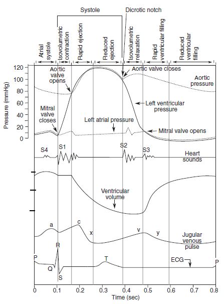
The events of the cardiac cycle are summarized in the Wigger 's Diagram (Fig 4). The Wigger's diagram (Fig 4) shows the relationship between the ECG, the pressure, and volume changes in the left ventricle and aorta. Note that there are two heart sounds (lub-dub). The first is associated

Wiggers Diagram Explanation
Cardiac cycle quiz: how well you know? The cardiac cycle involves the functioning of the heart where it contracts and expands and performs one beat to the next. The whole cycle describes what happens when the heart muscles expand to take out blood and relaxes to take in blood. Do you know the role played by different parts of the heart to achieve this process? The quiz below will refresh your ...
34 Place The Correct Labels On The Appropriate Area Of The ...
Wiggers, is a standard diagram that is used in teaching cardiac physiology. In the Wiggers diagram, the . Author: Jason A Wiggers diagram is essentially a graph that relates atrial pressure, ventricular pressure, aortic pressure, ventricular filling, and.List of 7 letter scrabble words that can be used in any word game.

WIGGERS diagram made easy!. Game Walkthrough
A Wiggers diagram, named after its developer, Carl Wiggers, is a standard diagram that is used in teaching cardiac physiology. In the Wiggers diagram, the X-axis is used to plot time, while the Y-axis contains all of the following on a single grid: Blood pressure Aortic pressure Ventricular pressure Atrial pressure Ventricular volume
Wiggers Diagram | Zazzle.com
A Wiggers diagram shows the changes in ventricular pressure and volume during the cardiac cycle. Often these diagrams also include changes in aortic and atrial pressures, the EKG, and heart sounds. Diastole starts with the closing of the aortic valve (the second heart sound).
Cardiac Cycle Drawing at GetDrawings | Free download
Start studying WIGGERS DIAGRAM. Learn vocabulary, terms, and more with flashcards, games, and other study tools.

Cardiac Cycle - YouTube
A Wiggers diagram is a medical chart that summarizes several aspects of cardiovascular health on one chart. Blood pressure, ventricular volume, arterial blood flow, and an electrocardiogram are simultaneously plotted against time on this chart.

place to be
for >90 years, the Wiggers diagram has been a fundamental tool for teaching cardiovascular (CV) physiology, with some of his earliest descriptions of the heart and circulation published in 1915 ().The lack of significant additions or changes from Dr. Wiggers' original observations is a testament to his careful work. In describing the various auditory, electrical, pressure, volume, and blood ...

Wigger Diagram Explained
The myogenic mechanism is how arteries and arterioles react to an increase or decrease of blood pressure to keep the blood flow constant within the blood vessel.Myogenic response refers to a contraction initiated by the myocyte itself instead of an outside occurrence or stimulus such as nerve innervation.

Wiggers Diagram Explained 9 Images - Wiggers Diagram ...
The Wright table is introduced as a novel tool for teaching and learning the cardiac cycle. It supplements the nearly 100-yr-old Wiggers diagram, which is information rich but difficult for many students to learn. The Wright table offers a compact presentation of information, viewable both in terms of how 1) each compartment's pressures and flows change over time; and 2) the heart works as a ...

Closeup of skeleton pelvic model
Basically, a "Wiggers Diagram" is a description of the events which take place over the cardiac cycle and which a plotted on a time scale. Yes, it's the Wiggers diagram, not Wigger's diagram or Wiggers' diagram, because a guy called Wiggers was responsible for the development of its most important components.
¿Vea cualquier cosa mal con mi diagrama de Wiggers Imanes ...
Wiggers diagram of the cardiac cycle. Wiggers. Understanding Wiggers Diagram. When the ventricles contract which valves open? semilunar Aortic and pulmonary. How does that affect the volume in the ventricles? Volume decreases in the ventricles after semi lunar valves open.
Wiggers Diagram Made Easy
ELI5: Cardiac cycle (diastole/systole) & also wigger's diagram. I have an exam on these tomorrow (see links) and while I can read it and somewhat comprehend, i still feel like its over my head and I dont really understand how it works. Therefore, I can't absorb the information enough to understand.

The Cardiovascular System: Wigger's Diagram | Draw It to ...

AMIGOS PARA SIEMPRE: anatomÃa humana

Explanation Of Wiggers Diagram

Wigger Diagram Explained

Wiggers Diagram | Cardiac cycle, Physiology, Heart pressure

Wiggers_Diagram - landbote.info

Pin on LV Systolic Function
34 Wiggers Diagram Explanation - Wiring Diagram Database

WIGGERS diagram made easy!. Game Walkthrough

Wigger's Diagram YouTube.com video | CathLab.com
Clinical electrocardiography and ECG interpretation - ECG ...

Cardiac Cycle | CVS Physiology | Pressure Volume Curve ...
Cardiac Cycle - Summary and Wigger's Diagram | Epomedicine

Wiggers Diagram Made Easy

[Wiggers Diagram
Wiggers Diagram Explanation - Derslatnaback
Wiggers Diagram Explanation

Closeup of skeleton foot model

Explanation Of Wiggers Diagram
Medical diagram

Pin on ï¸ ECG

Events of the Cardiac Cycle Wigger's diagram explained ...


Comments
Post a Comment