43 ap psychology eye diagram
AP Psychology Name: _____ Eye and Ear Anatomy Learning Target: Describe the sensory process for vision and audition including the specific nature of energy transduction, relevant anatomical structures, and the specialized pathway to the brain. Dec 26, 2011 - This site was designed for students of anatomy and physiology. It contains textbook resources, such as chapter review guides, homework sets, tutorials, and printable images. Each chapter has a practice quiz and study tips for learning the topic.
Jeff Milner's backmasking site is a great demonstration of top-down v. bottom-up processing. Instructions: 1. Click on a song on the right-hand side 2. Listen to the music forward, then backwards. 3. When listening backwards, try to make out what is being said.

Ap psychology eye diagram
Start studying A&P2 Lab 13 HW, A&P2 Lab 12 HW, A&P2 Lab 11 HW, A&P2 Lab 10 HW, Lab 9 HW, Lab 8 HW, A&P2 Lab 1 HW, A&P2 Lab 2 HW, A&P2 Lab 3 HW, A&P2 Lab 4 HW, A&P 2 Lab 5 HW, A&P2 Lab 6 HW, A&P2 Lab 7 HW. Learn vocabulary, terms, and more with flashcards, games, and other study tools. 18.01.2022 · Happy New Year, Readers. It’s been so long since I posted to you, I think my chin hairs have turned into a beard. (Shhh, forget I said that; … Eye Diagram Handout Author: National Eye Health Education Program of the National Eye Institute, National Institutes of Health Subject: Handout illustrating parts of the eye Keywords: parts of the eye, eye diagram, vitreous gel, iris, cornea, pupil, lens, optic nerve, macula, retina Created Date: 12/16/2011 12:39:09 PM
Ap psychology eye diagram. Module 9 Flip It Video - Action Potential. Module 10 Flip It Video - The Reflex Arc. Module 10 Flip It Video - Structure of the Nervous System. Module 11 Flip It Video - Limbic System. Module 12 Flip It Video - Structure and Function of the Cortex. Module 13 Flip It Video - Split-Brain Research. AP Psychology Study Guide ... take to ring a bell) nerve leaves the eye oMedulla - vital organs (HR, BP) o Pons - sleep/arousal (Ponzzzzzz) • Midbrain o Reticular formation: attention (if you can't pay attention, You R F'd) • Forebrain: higher thought processes Best AP Psychology Review Book for Low-Scoring Students: Cracking the AP Psychology Exam, 2020 Edition. Cost: $18 for print, $13 for digital Written by The Princeton Review, this is by far the best book for learning test-taking strategies for the AP Psychology test.The content is high quality as well, but it's not as easy to study from if you don't have much time on your hands. Psychology. 8. View this sample Book/movie review. READING ASSIGNMENT. Undergrad. (yrs 3-4) Religious studies. 4. View this sample Research paper. Barriers to Entry. Undergrad. (yrs 3-4) Logistics. 2. View this sample Research paper. Reflecting on Adult Education/Training . Master's. Education. 6. View this sample Essay (any type) Cultural Activity. Undergrad. (yrs 3 …
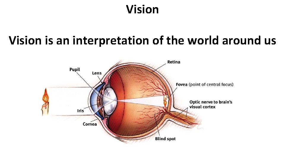
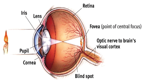
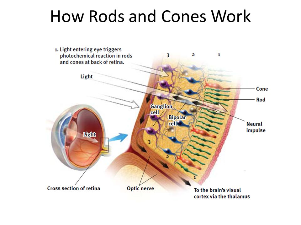
AP PSYCHOLOGY: Home Course Description Homework > > Projects Extra Help AP Exam wednesday 10/9. FRQ Practice HW= Module 16. thursday 10/10. DN: What is selective attention? an you give me an example? ... -Introduce Eye Diagram - use as lecture - Watch Videos and discuss Eye HW= Module 18 (Due Tuesday 10/15) Seeing. AP Psychology is a completely manageable subject if you prepare for the exam responsibly. Memorizing key terms, applying confusing psychology concepts to your everyday life, using common sense to solve practice questions, and staying focused on the free-response section will help you be successful, both in class and on the AP test. Full-page diagrams of the eye and ear that has students label each part. I've used this worksheet as both a note guide during AP Psychology lectures and as a take-home worksheet. They go with the Meyers' textbook PowerPoint slides if you use those! Step Three: Transduction Psychology Definition. Ok, so how does our eye turn the light into neural impulses so that our brain can understand. Most of the process occurs on the back of the eye called the retina. The retina is the most important part of our eye (it is often referred to as the brain of the eye). First, it is important to know that ...
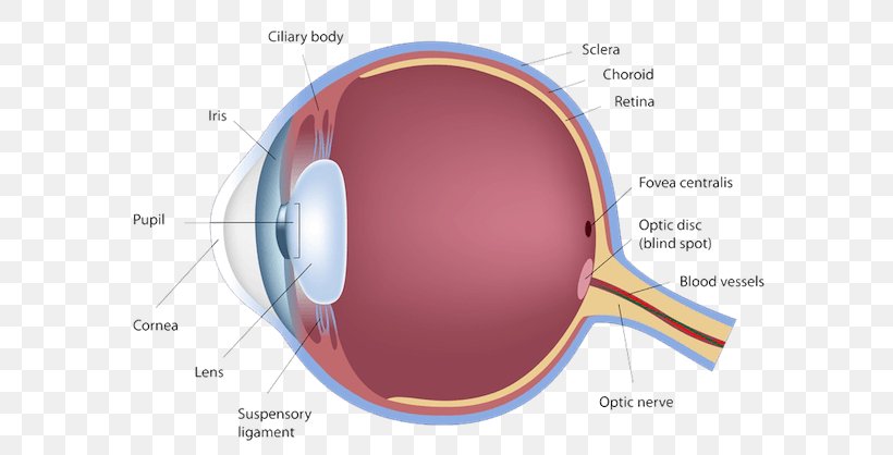
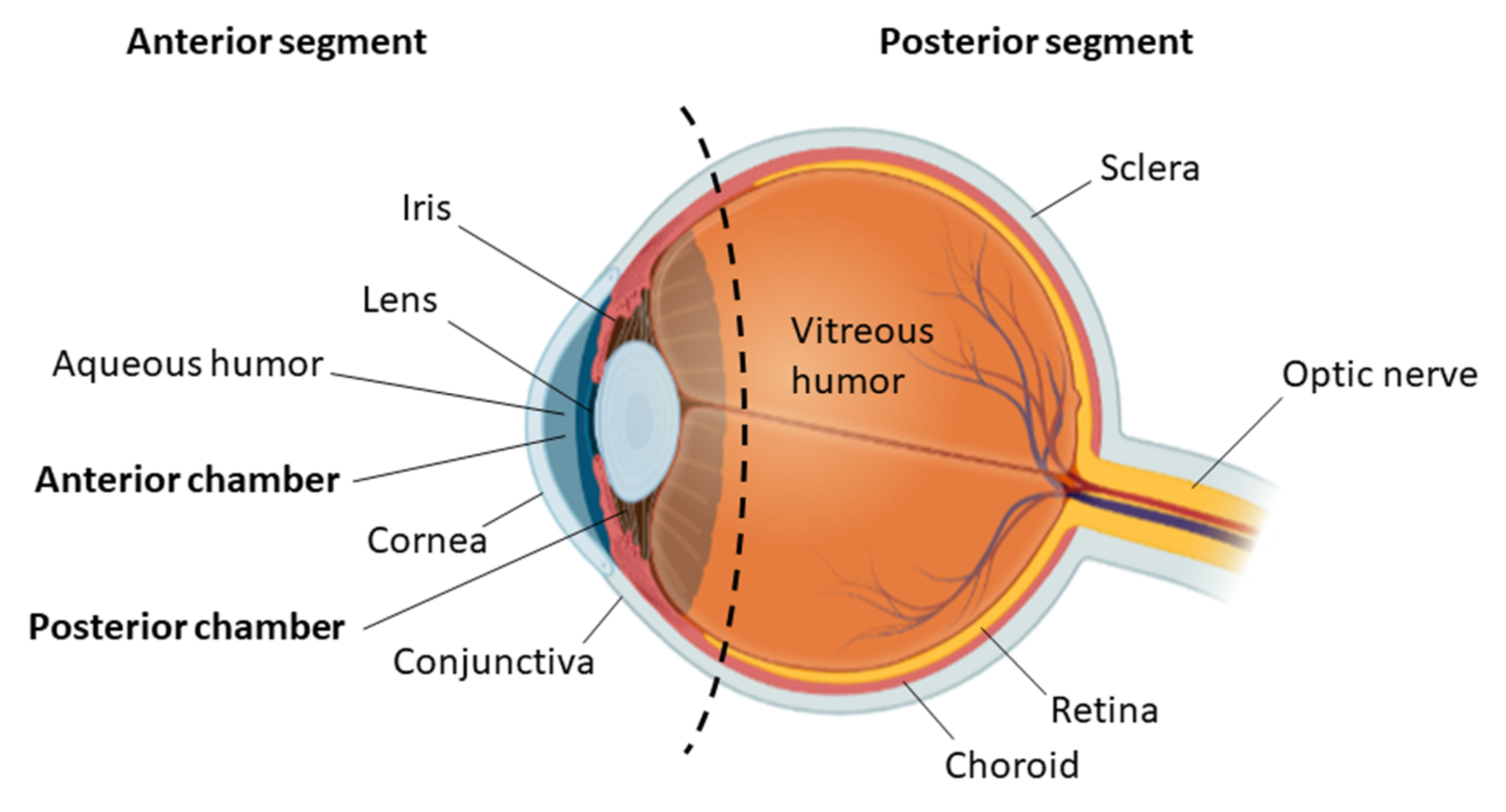
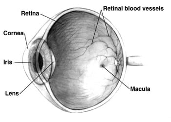
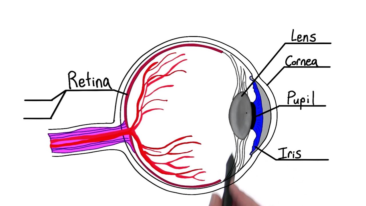
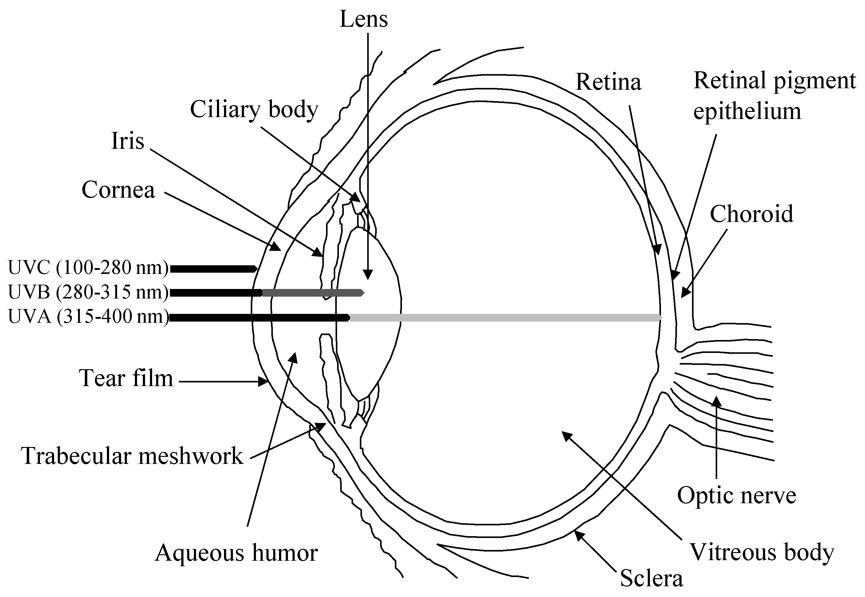
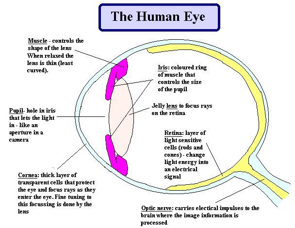
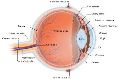
BI 335 - Advanced Human Anatomy and Physiology Western Oregon University Figure 4: Mid-sagittal section of brain showing diencephalon (includes corpus callosum, fornix, and anterior commissure) Marieb & Hoehn (Human Anatomy and Physiology, 9th ed.) - Figure 12.10 Exercise 2: Utilize the model of the human brain to locate the following structures / landmarks for the Free Body Diagram Questions and Answers. Get help with your Free body diagram homework. Access the answers to hundreds of Free body diagram questions that are explained in a way that's easy for ... AP Psychology Please check back here for more information, assignments, notes, etcetera. Sign up for REMIND by texting the code @acpsych to the number 81010. of light entering the eye. Lens: The lens is a clear part of the eye behind the iris that helps to focus light, or an image, on the retina. Macula: The macula is the small, sensitive area of the retina that gives central vision. It is located in the center of the retina. Optic nerve: The optic nerve is the largest sensory nerve of the eye.
Occipital Lobe - Hook AP Psychology 3B
Ultimate Study Guide for AP Psychology ... • how light waves travel through eye and get transduced • major eye anatomy • (including rods and cones - see diagrams ) • feature detectors in occipital lobe in brain • why we have a blind spot • trichromatic color theory • opponent-process color theory ...

20 Questions Tuesday: 419 - Vision — 20 Questions Tuesday
along the back of the eye and it contains the rods, cones, bipolar and ganglion cells. Use "red tin" as your mnemonic and imagine that the back of your eye is covered with red tin. Fovea: is a spot in the eye that is directly behind the lens. There is a very high concentration of cones in this area which means that images t
Eye Anatomy Handout
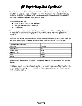
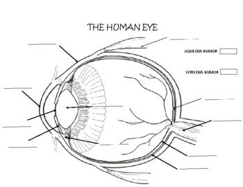
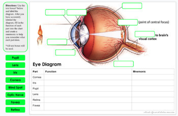
AP Psychology - Unit 4 Assignment ... Draw a diagram of the eye. Label and explain the function of the iris, lens, pupil, cornea, retina, fovea, blind spot, and optic nerve. 5. What is transduction and how does this process occur in the photoreceptors of the eye and cochlea of the ear? 6. Draw a diagram of the ear.

AP Psychology - Sensation & Perception - Part 2 - Vision
AP Psychology Eye diagram. The light-sensitive inner surface of the eye, containing the receptor rods and cones plus layers of neurons that begin the processing of visual information. The central focal point in the retina, around which the eye's cones cluster. Nice work!
Human Eye Diagram Retina Anatomy, PNG, 600x418px, Watercolor ...
06.02.2014 · Thinking Outside the Box: A Misguided Idea The truth behind the universal, but flawed, catchphrase for creativity. Posted February 6, 2014

Vision - Coach Wise's AFNORTH LIONS Social Sciences
An Error Occurred. Services for this domain name have been disabled.
Sensation and Perception A P Psychology Assignment 13
Where the optic nerve leaves the eye is a blind spot, as a result of the absence of receptor cells there. Image Courtesy of Myers' AP Psychology Textbook - 2nd Edition As mentioned before, feature detectors were discovered by Hubel and Wiesel in the visual cortex.
Medical laser safety
Diagram showing the lobes of the brain. Image Source: Wikimedia Commons. The parietal lobe is located at the top of the brain, between the frontal and occipital lobe. It consists of the somatosensory cortex and is responsible for integrating sensory information from different parts of the body, especially visual information related to navigation and spatial orientation.

Anatomy of the human eye and the retina with its specialized ...
Information for Mrs. Carlton's AP Psychology students. Our Mission Statement at West Broward High: Promote integrity, respect and dignity by creating lifelong learners in a safe and trusting environment. Our Vision is building a spirit of collaboration and pride. To grow a community of self-reliant young adults who will explore and challenge their individual talents for future success.
AP Psychology chapter 4: Sensation
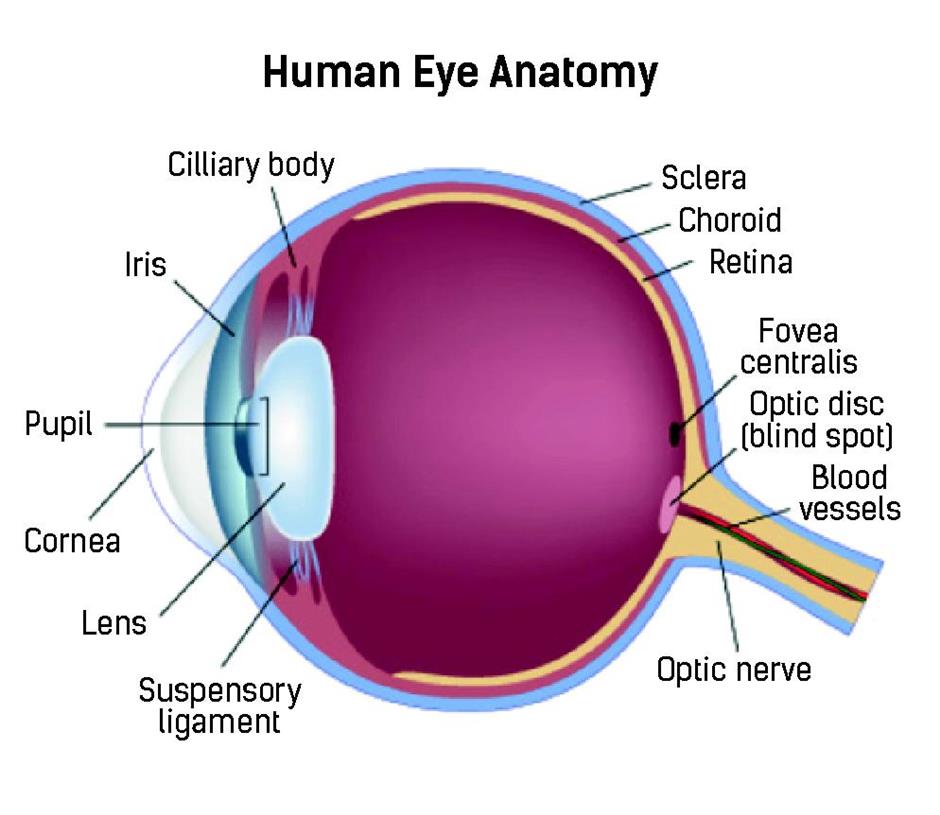
Human Eye Diagram: Contrary to popular belief, the eyes are not perfectly spherical; instead, it is made up of two separate segments fused together. Explore: Facts About The Eye To understand more in detail about our eye and how our eye functions, we need to look into the structure of the human eye.

What is the Pupil of the Eye? - Definition & Function - Video ...
Virtual Learning Week #3 AssignmentsMonday, April 6 - Thursday, April 9 (No School Friday April 10, School Holiday)All work assigned on Monday at 8am and due by Thursday at 4pm. 1- Complete the Unit 1 Progress Check on AP Classroom. 2- Complete the Unit 2 Progress Check on AP Classroom.
Bioengineering | Free Full-Text | Biofabrication of ...
30.09.2021 · Mental Health and Psychopathology Defined. Mental health is a positive mental status, with an individual capable of coping with normal life …
AP Psychology on Twitter: "Eye diagram. #APPsych https://t.co ...
A human eye is roughly 2.3 cm in diameter and is almost a spherical ball filled with some fluid. It consists of the following parts: Sclera: It is the outer covering, a protective tough white layer called the sclera (white part of the eye). Cornea: The front transparent part of the sclera is called cornea. Light enters the eye through the cornea.
AP Psychology Eye Diagram Project by Jennifer Kasyan | TpT
(Brain Diagrams - LEFT flip-up (Body & Behavior Lecture (printed for you) - right-left match (hemisphere diagram too) ... The product is eye-catching and original, creating an impact on the reader. The student has given . careful attention. to . ... AP Psychology Notebook ...
Eyes (Anatomy): Overview, Parts and Functions | Biology ...
AP Psych Eye Diagram STUDY PLAY Cornea Clear, curved bulge of tissue on the front of the eye that bends light rays to begin focusing them and protects the eye (lots of nerve endings) Iris Ring of muscle tissue that forms the colored portion of the eye; surrounds the pupil and controls its size Pupil
Parts of the eye - Intro to Psychology
AP Psychology > > > > AP Seminar Modern World Blog Voices Unit 3: Sensation and Perception ... Eye Diagram Retina to Brain Diagram: F. Describe the vision process, including the specific nature of energy transduction, relevant anatomical structures, and specialized pathways in the brain for each of the senses. ... Ear Diagram I. Describe the ...

AP Psychology: Sensation - Diagram #1 (The Eye) Diagram | Quizlet
Parts of the Neuron. Neurons are our body's nerve cells which make up the nervous system. For a neuron to fire, or communicate with another neuron, information must first be gathered in by the dendrites of the receiving neuron. From there, the information passes through the cell body to the axon. Part of the Neuron.

Optical Express - Did you know, patients who have waited ...
15.01.2022 · Flow diagram depicting criteria from the PRISMA guidelines for systematic reviews and meta-analyses as well as inclusion and exclusion criteria which were applied in the course of search and inclusion of studies for these meta-analyses. Additional file 1: Table S1 contains a comprehensive list of all studies included in our meta-analyses. Full size image. Inclusion and …

Eye and Ear Diagrams
January 27, 2022. AP Psychology 3.03.pdf. . The human ear is an essential organ that provides humans with the sense of hearing. AP Psychology CDoerrer. 11/1/2018 Test: AP Psycholo

The Locations & Functions of Parts in the Eye - AP Psychology ...
Eye Diagram Handout Author: National Eye Health Education Program of the National Eye Institute, National Institutes of Health Subject: Handout illustrating parts of the eye Keywords: parts of the eye, eye diagram, vitreous gel, iris, cornea, pupil, lens, optic nerve, macula, retina Created Date: 12/16/2011 12:39:09 PM

AP Psychology Review on Twitter: "Anatomy and Function of the ...
18.01.2022 · Happy New Year, Readers. It’s been so long since I posted to you, I think my chin hairs have turned into a beard. (Shhh, forget I said that; …

AP Psychology Review 3.1, 3.3 Sensation and Visual Anatomy ...
Start studying A&P2 Lab 13 HW, A&P2 Lab 12 HW, A&P2 Lab 11 HW, A&P2 Lab 10 HW, Lab 9 HW, Lab 8 HW, A&P2 Lab 1 HW, A&P2 Lab 2 HW, A&P2 Lab 3 HW, A&P2 Lab 4 HW, A&P 2 Lab 5 HW, A&P2 Lab 6 HW, A&P2 Lab 7 HW. Learn vocabulary, terms, and more with flashcards, games, and other study tools.
Antioxidants | Free Full-Text | Antioxidant Defenses in the ...
Vision AP Psych Transduction – converting one form of energy ...

Psych Sensation and Perception Myers 20-25 - Quizizz
AP Psychology chapter 4: Sensation

Human Eye Ball Anatomy & Physiology Diagram
The Human Eye Diagram Worksheets & Teaching Resources | TpT
AP Psychology on Twitter: "Take a LOOK st this. Parts of the ...

Sight - Hook AP Psychology 2A
Sunglasses to hide behind may also prevent melanoma of the ...
Eye Diagram Worksheets & Teaching Resources | Teachers Pay ...
Human Eye Iris Diagram Mammalian Eye - Parts Of The Eye ...

Anatomy of the human eye and the retina with its specialized ...

Myers' PSYCHOLOGY (6th Ed) Chapter 5 Sensation. The spectrum ...
AP Psychology Ch. 3 Flashcards | Memorang

Lutein and zeaxanthin are... - Energlo Lutein and Zeaxanthin ...

Psychology Parts of the Eye Diagram | Quizlet
Phacoemulsification the gold standard of cataract surgery ...
Ms. Scott's A.P. Psychology Summer Assignment due on the ...

Eyes - AP Psychology
Sensation & Perception
Sensation & Perception


Comments
Post a Comment