43 reticular formation diagram
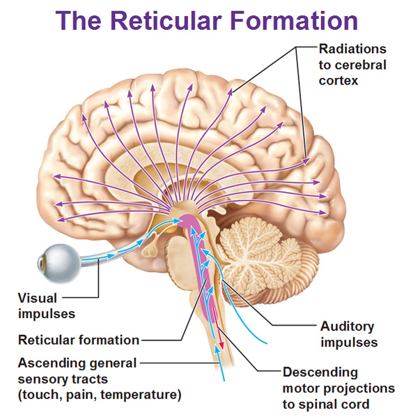
Reticular Formation Diagram. angelo on December 3, 2021. Understanding The Brain A Work In Progress Professor Keith Kendrick Reticular Activating System Nervous System Parts System. Reticular Formation Reticular Formation Reticular Activating System Medicine. Ascending reticular activating system: Reticular formation sends efferent impulses to almost all areas of cerebral cortex through ARAS. In the diagram (Fig. 9.45) descending tracts numbered in black color are reticulospinal tracts which would descend down into spinal cord.
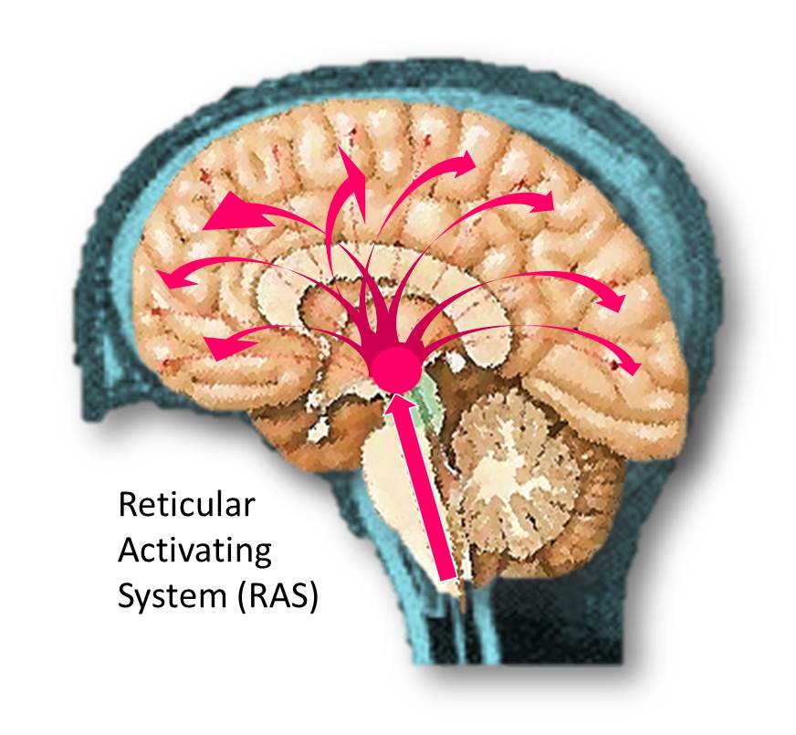
The Reticular Formation Descending Reticular Formation Sleep and ArousalNeuronal Basis of Changes in the EEG Sleep Disorders The Ascending Reticular Activating System The brainstem contains many small neural networks that regulate essential functions, including the arousal system, cardiovascular and respiratory control, and the control of ...

Reticular formation diagram
Reticular tissue is a special type of connective tissue that predominates in various locations that have a high cellular content. It has a branched and mesh-like pattern, often called reticulum, due to the arrangement of reticular fibers (reticulin).These fibers are actually type III collagen fibrils.In comparison to the predominant type I collagen, type III fibrils are narrower, do not form ... Brain diagram reticular formation. It is also the origin of the descending analgesic pathways. The reticular formation cranial extension is upto the dienceph-alon subthalamus hypothalamus and. All the functions are carried out without a single glitch and before you. Peer Reviewed Journal High visibility Open Access. The reticular formation is a cluster of nerves within the brainstem that relay sensory and motor signals to and from the spinal cord and the brain. This diagram labels the cranial nerves, including olfactory, oculomotor, trochlear, abducens . Describe the functions of the reticular formation region of the pons.
Reticular formation diagram. The above diagram illustrates the reticular nuclei in the brainstem in a tiered fashion The Medial Reticular formation Surrounding the previously discussed ridge of serotonergic cells, the medial reticular formation has many roles and functions. The medial reticular formation is filled with a mixture of large and small neurons. The reticular formation of the pons and medulla gives rise to reticulospinal fibres. Axons arising from the pontine reticular formation descend ipsilaterally as themedial (orpontine) reticulospinal tract. Axons from the medulla descend bilaterally in thelateral (ormedullary)reticulospinal tracts. Both tracts are located in the ventral funiculus. The Reticular Formation. Is a network of nerve pathway situated in the brainstem. This area connects the spinal cord, cerebellum and cerebrum. It mediates conscious activity and uses sensory and other impulses from the brain stem. It is important in cortex activation, muscle tone (specifically those affected by gravity), regulation of heartbeat ... The brainstem gives us our most basic functions like consciousness 💭 (reticular formation), breathing and heartbeat ️ (medulla), and coordination of movement 🏃 (pons). The thalamus sits on top of the brainstem and receives and sorts all sensory input (except smell) to other parts of the brain. Lastly, the cerebellum sits at the rear of ...
The reticular formation is a nerve network of nuclei clusters found in the human brain stem. The dorsal tegmental nuclei are in the midbrain, the central tegmental nuclei are in the pons, and the... Reticular Activating System. The ARAS is part of the reticular formation, which consists of a network of neurons in the central portion or core of the brainstem from the medulla through the pons and midbrain and into the diencephalon. From: de Lahunta's Veterinary Neuroanatomy and Clinical Neurology (Fifth Edition), 2021. Related terms: Thalamus The reticular formation has the ascending reticular activating system, containing nuclei that release neurotransmitters, and the descending reticulospinal tract, containing pontine and medullary ... Download scientific diagram | a Post-traumatic aspect of the reticular formation in a Fiesta sequence equally weighing T1/T2 ratios. This Figure comes from a 34 years-old women reporting the onset ...
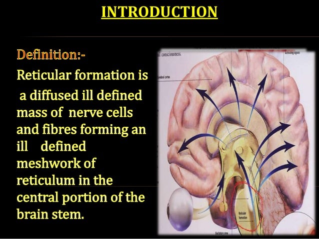
The reticular formation resembles a net made up of nerve fibers and nerve cells. It is a deeply placed diffuse network of fibers and nuclei. This network can be diffusely divided into three longitudinal columns; median column, medial column, and the lateral column. The median column occupies the median plane. It Throughout the life of the vertebrates, the core of the central nervous system, sometimes called the reticular formation, has retained the power to commit the whole animal to one mode of ... has retained the power to commit the whole animal to one mode of behavior rather than another. Its anatomy, or wiring diagram, is fairly well known, but to ... The reticular formation is a set of interconnected nuclei that are located throughout the brainstem.It is not anatomically well defined, because it includes neurons located in different parts of the brain.The neurons of the reticular formation make up a complex set of networks in the core of the brainstem that extend from the upper part of the midbrain to the lower part of the medulla oblongata. Reticular interstitial pattern is one of the patterns of linear opacification in the lung. It can either mean a plain film or HRCT/CT feature. Pathology Causes Reticulation can be subdivided by the size of the intervening pulmonary lucency in...
Reticular Formation and Limbic System - Textbook of ...
Brain diagram labeled reticular formation. The reticular formation is a cluster of nerves within the brainstem that relay sensory and motor signals to and from the spinal cord and the brain. The skull consists of 22 bones 14 of which form the facial bones and the remaining. Activity in the brain stem is important for.

Image from page 153 of "The anatomy of the nervous system, from the standpoint of development and function" (1920)
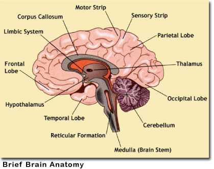
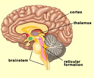
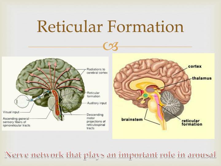
The central core consists of the thalamus, pons, cerebellum, reticular formation and medulla. These five regions are the central areas that regulate breathing, pulse, arousal, balance, sleep and early stages of processing sensory information. The thalamus interprets the sensory information and helps determine what is good and bad.

reticular formation critical for attention arousal and ...
The reticular formation within the pons is partly responsible for postural control functions. The cortico-reticulospinal tract is involved in proximal stability and regulating postural tone. The premotor cortex is able to identify appropriate axial musculature to enable distal movement.
The Limbic System and the Reticular Formation
Our interactive diagram helps you explore the anatomy of the human brain and learn The reticular formation controls muscle tone in the body and acts as the. According to MedlinePlus Dictionary, the reticular activating system is "a part of the reticular formation that extends from the brain stem to the midbrain and.
Schematic diagram of the reticular activating system ...
The reticular (from the Latin reticulum, meaning net) formation is a far-reaching network of neurons extending from the spinal cord to the thalamus, with connections to the medulla oblongata, midbrain (mesencephalon), pons, and diencephalon.

Image from page 67 of "The cell in development and inheritance" (1896)
The reticular formation is located in the brainstem but extends into the spinal cord and thalamus; it passes through the medulla, pons, midbrain, and diencephalon. The RF does not completely fill the brainstem but is loosely split into three columns of nuclei (groups of nerve cells with their own set of functions) that run along its length.
Cerebrum anatomy
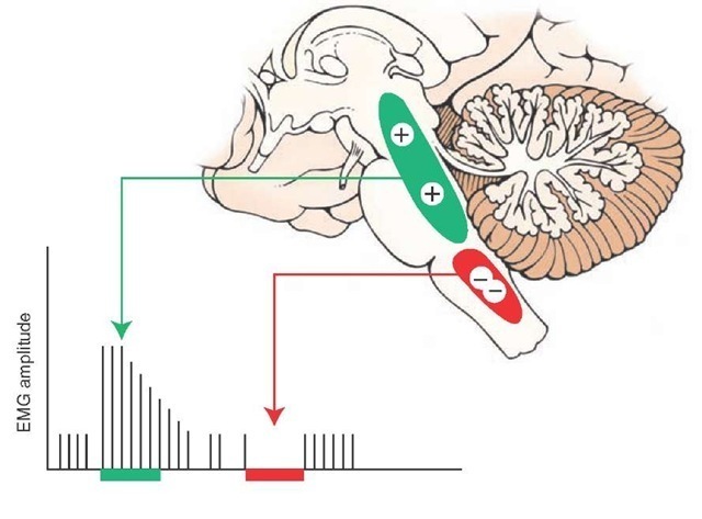
the reticular formation of the brainstem the periaqueductal grey matter of the midbrain a region of the diencephalon known as A11 the Cingulate Gyrus of the cerebral cortex (part of the Limbic System) The diagram also shows the importance of aminesin influencing nociceptive processing in the dorsal horn.

U.S. H-type BSE (B14842): reticular formation. (A and B ...
Nervous Cell Diagram. Here are a number of highest rated Nervous Cell Diagram pictures upon internet. We identified it from obedient source. Its submitted by management in the best field. We say yes this kind of Nervous Cell Diagram graphic could possibly be the most trending topic afterward we allowance it in google benefit or facebook.
Ophthalmology | Neupsy Key
reticular formation and the disorders of the reticular formation. Historical reviews The term ''reticular formation'' was coined in the late 19th century, coinciding with Cajal (1909) who commented on the extensive multiple branching of the reticular formation neurons as the fibers ascended and descended through the middle of the brain stem. Papez
Reticular formation
The reticular formation gets habituated to excluding meaningless and repetitive signals, that are deemed to be consistently unimportant. That explains why people can sleep through any kind of noise, once they have become habituated to it, while waking up in a startled state, to an auditory signal like a gunshot. ...

Schematic illustration of the reconstruction of the ...
The reticular formation is a cluster of nerves within the brainstem that relay sensory and motor signals to and from the spinal cord and the brain. This diagram labels the cranial nerves, including olfactory, oculomotor, trochlear, abducens . Describe the functions of the reticular formation region of the pons.

Everything about the reticular formation | Reticular ...
Brain diagram reticular formation. It is also the origin of the descending analgesic pathways. The reticular formation cranial extension is upto the dienceph-alon subthalamus hypothalamus and. All the functions are carried out without a single glitch and before you. Peer Reviewed Journal High visibility Open Access.

lecture 16 neuroanatomy [ reticular formation - internal ...
Reticular tissue is a special type of connective tissue that predominates in various locations that have a high cellular content. It has a branched and mesh-like pattern, often called reticulum, due to the arrangement of reticular fibers (reticulin).These fibers are actually type III collagen fibrils.In comparison to the predominant type I collagen, type III fibrils are narrower, do not form ...

BIOL 214 Midbrain & Reticular Formation (Ch 14 Part 5 ...
THE BRAIN FROM TOP TO BOTTOM
Closeup of skeleton pelvic model
Cns 14

Light photomicrograph showing reticular fibers (Rf) extend ...
Structures of the Brain Notes (Chapter 3)

Places To Be , Hamburg

Image from page 108 of "The brain from ape to man; a contribution to the study of the evolution and development of the human brain" (1928)
Pinned map of the United States of America
:fill(FFFFFF,true):format(jpeg)/images/container/connective-tissue/Connective_Tissue_1.png)
Connective tissue (Histology) - Study Guide | Kenhub
Physiology of Vomiting

Image from page 110 of "The brain from ape to man; a contribution to the study of the evolution and development of the human brain" (1928)

Image from page 82 of "The brain from ape to man; a contribution to the study of the evolution and development of the human brain" (1928)

Brainstem | Neupsy Key | Limbic system, Brain facts ...
Motivation and emotion/Book/2014/Reticular formation ...
Closeup of skeleton pelvic model
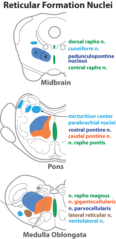
Reticular Formation Nuclei

Image from page 268 of "The brain from ape to man; a contribution to the study of the evolution and development of the human brain" (1928)

Figure 1 from Reticular formation: A Review | Semantic Scholar

The Reticular Formation in: Journal of Neurosurgery Volume ...

Closeup of skeleton hand model
place to be

Reticular Formation Pons Nucleus Raphe Nuclei Brainstem ...

Position of reticular formation-right sagittal section of ...
Success Tips: Use a Goal Board to create the Motivation ...
PPT - Anatomy and Functions of the Brain PowerPoint ...

Reticular formation and ascending reticular system (ARAS ...

Image from page 337 of "A reference handbook of the medical sciences, embracing the entire range of scientific and practical medicine and allied science" (1913)

Mirages II
The Reticular Formation (Integrative Systems) Part 3


Comments
Post a Comment