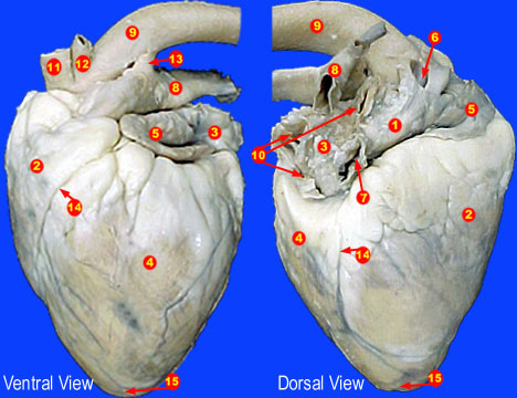
43 sheep heart diagram labeled
Take this practice quiz that covers information related to the sheep heart & the heart model. It is intended for use as a supplemental study aid. As is the case in the lab practical, each correct answer counts. So, make sure you learn from the feedback. Questions and Answers. 1. Today's Demonstration: Sheep Heart DissectionLearn about the parts of the heart as you observe a sheep heart dissection.#arizonasciencecenter #athomelearning...
About this Quiz. This is an online quiz called Sheep Heart Labeling. This quiz has tags. Click on the tags below to find other quizzes on the same subject. Comparative Anatomy.

Sheep heart diagram labeled
The mammalian heart is the central organ of the circulatory or cardiovascular system. It pumps blood to the body's organs and tissues delivering oxygen and nutrients, while transporting wastes away. Dissection of a preserved sheep or pig heart offers students an excellent opportunity to learn about mammalian heart anatomy. This page contains photos of the sheep heart dissection. All the major vessels are represented, many are labeled with colored pencils so that you can see ... Diagram Of The Cow Basic Internal Ans And Beef Cuts Chart. Circulatory Systems In Animals Transport. Pig Heart At Paintingvalley Explore Collection Of. Vector Heart Medical Diagram Clip Art Ox Anatomy. Left Side Of Heart Diagram A Cow Clipart Full Size. Lab Human Anatomy At Colby. Cat Sheep Heart Dissections.
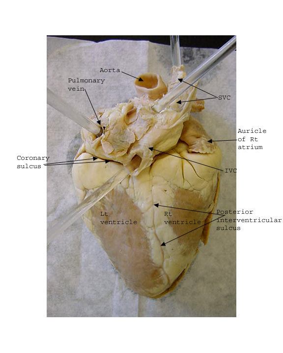
Sheep heart diagram labeled. Human Heart: Diagram and Anatomy of the Heart internal anatomy of the heart Heart Diagram: Right/left Atria, Right/left Ventricles, Pulmonary Trunk, Aorta, ... (a) Anterior view of the external heart C' 2019 Pearson Education. Aort'c arch Ligamentum arteriosum Left pulmonary artery Left pulmonary ve ns Auricle of left atrium Circumflex artery Left coronary artery (in atrioventricular sulcus) Great cardiac vein Left ventricle Anterior interventricular artery (in anterior interventricular sulcus) Apex Sheep Heart Unlabeled. Sheep Heart Leader-lined. Sheep Heart Labeled. San Diego Mesa College 7250 Mesa College Drive San Diego, CA 92111-4998 Student Support San Diego Community College District San Diego City College San Diego Mesa College San Diego Miramar College San Diego Continuing Education. Sheep Heart Dissection Procedure (Day 2) - you will be cutting the heart open today! a. Review the outer part of the heart and make sure you know where these structures are: left and right ventricle, left and right atrium, pulmonary artery, pulmonary veins, aorta, coronary artery, apex, superior and inferior vena cava. b. Locate the pulmonary ...
Sheep heart dissection Lab Anatomy & Physiology. Purpose of this lab: To review the structural characteristics of the human heart and to examine the major features of a mammalian heart. Procedure A—The Human Heart. 1. Using your textbook and/or your notes and heart diagram worksheet, label the diagram of the human heart on your lab report ... Heart diagram - Label and sketch the internal and external heart and all mentioned parts Clean up Put the dissected heart, and all associated organic matter, in the plastic bag of the appropriately labeled container. Place organically contaminated materials, gloves and towels, in the appropriate marked container. 4. Compare the structures of the sheep heart with those of the human heart. 5. Know the path of blood through and out of the heart Materials Preserved sheep heart Dissecting tray and instruments Vinyl dissecting aprons Disposable gloves Anatomy & Physiology / Revealed, Version 2.0 CD-ROM Human heart model 2 atria, 2 ventricles, completely closed circulatory system with no mixing of blood. Base. the wide upper or anterior end of the heart, where the aorta and vena cava are located. apex. somewhat blunt tip of the heart, composed entirely of the left ventricle. musculi pectinati. parallel ridges in the walls of the atria of the heart.
1. Using your notes and wonderful memory, label the diagram of the human heart on your lab report sheet. 2. Read through the analysis questions on your lab report as you should be answering them as a groups as you dissect . the sheep heart. Procedure B—Dissection of a Sheep Heart. 1. Obtain a preserved sheep heart. 14+ Heart Arteries Diagram Labeled. Labeled heart diagram showing the heart from anterior. Inner body parts with their names. CoronaryArteriesComplete from faculty.etsu.edu (taken from johnson, weipz and savage lab book). This is an excellent human heart diagram which uses different colors to show different parts and also labels a number… Recognition of the chambers and valves of the heart and the blood vessels connected to it in dissected hearts or in diagrams of heart structure. Labeled Sheep Heart Picture #4 Done. 36,092 views. 0 faves
On holiday in Florence (Firenze), Tuscany, Italy, where there was a lot of street art and graffiti, however most of it was extremely good, including this simple one that, to me, tells a powerful and strong story.
Thank you for your help! I am just frightened that it would be considered improper to source an annotated diagram.

Label the parts of the dissected sheep heart. | Study.com
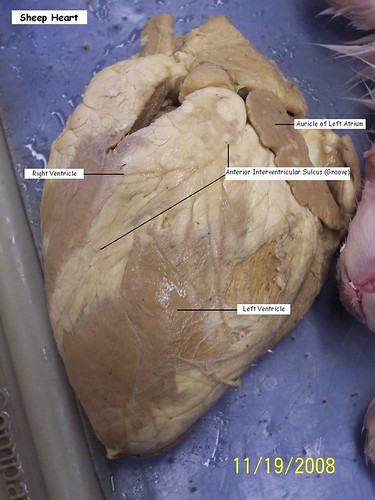
Sheep Heart Dissection Sheep heart (anterior view) This image shows an external view of the anterior side of a preserved sheep heart. Note the pointed apex of the heart and the wide superior end of the heart which is termed the base. The large blood vessels (i.e., the great vessels of the heart) which carry blood to and from the heart are ...

Smith, K / Anatomy and Physiology Activities
sheep heart. Observation: External Anatomy Most heart diagrams show the left atrium and ventricle on the right side of the diagram. Imagine the heart in the body of a person facing you. The left side of their heart is on their left, but since you are facing them, it is on your right. 1. Identify the right and left sides of the heart. Look closely and on

Multiple good visuals for pig heart dissection | Medical ...
Sheep Heart Dissection External Anatomy 1. Identify the right and left sides of the heart. Look closely and on one side you will see a diagonal line of blood vessels that divide the heart, this line is called the interventricular sulcus. The half that includes all of the apex(pointed end) of the heart is the left side. 2.

SHEEP HEART - Biology Forums Gallery
27 S H E E P H E A R T D I S S E C T ION SHEEP HEART DISSECTION LOGISTICS • Big Idea: Anatomy: Form and Function of the Heart • Type of Activity: Sheep Heart Dissection • Length of Activity: 5 minutes - 1 hour • Group Size: 1-5 per heart • Space Needed: One table per group of fi ve The smell of the preservative bothers some people. Make sure they have somewhere else to

Heart Models | Heart anatomy, Medical anatomy, Anatomy and ...
Can someone please help me label this sheep heart diagram? Thank you so much! Or at least . This is what I got so far: Right ventricle - 9. left ventricle - 11. auricle of right atrium - 16. chord teninaea - 7. aortic semilunar valve - 8. right atrium- 1. left atrium- 4. right cornoary- 20. aorta - 18
a moment of connection between the two lambs in the foreground
ANATOMY- Sheep HEART DISSECTION Sheep Heart Dissection Grace Boshart and Anja Stichter. Lab Report. 1. Purpose: To get a better understanding of the mammalian heart. We were able to make connections between what we had learned about the structure and function of the heart with what we could observe on a real heart. 2.

cow heart diagram printable - Google Search | Heart ...
This page contains photos of the sheep heart dissection. All the major vessels are represented, many are labeled with colored pencils so that you can see. In this lab guide, students are given instruction on how to remove the dura mater, and locate the main structures of the external and internal brain.

19 Best Cow Eye Dissection Labeled
Sheep Heart Dissection from Science Interactive on Vimeo
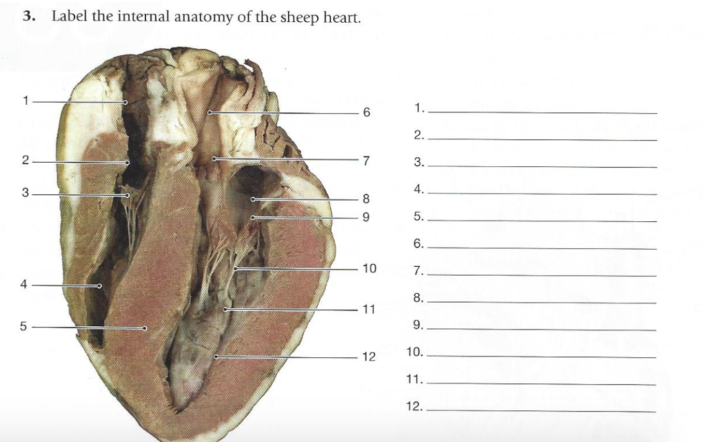
Solved: 3. Label The Internal Anatomy Of The Sheep Heart ...
Lab guide outlining the procedure for dissecting the sheep's heart. It includes photos to diagram where major vessels are and where incisions should be made to view internal structures, such as the mitral valve and papillary muscles. ... Human Heart: Diagram and Anatomy of the Heart internal anatomy of the heart Heart Diagram: Right/left Atria ...

Sheep Heart Images
Sheep Heart Dissection. Sheep have a four-chambered heart, just like humans. By studying the sheep's anatomy, you can learn how your own heart pumps blood through your body, thereby keeping you alive!. Use this sheep heart dissection guide in a lab for high school students.

Girls Rock Bio: Sheep Heart Dissection
Identify the chambers of the heart and the major blood vessels that lead into and out of those chambers Trace the path that blood takes through the heart Pre-lab: 1. Label the heart diagrams (dorsal and ventral sheep heart views) on the last page of this lab. (Parrot pp. 948-951, Dragonfly pp. 943-950). 2.
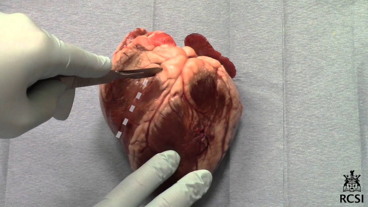
Sheep's Heart Dissection - YouTube
To view details of the aortic arch, ductus arteriosus, and pulmonary artery, it will be helpful to remove the left lung. With the left lung removed, the heart can be pushed to the right side to reveal the aorta and other blood vessels shown in the diagram below. Figure 30. Thoracic cavity. Figure 31. Thoracic cavity. Figure 32. Major vessels ...
anatomy of heart and sheep
Pics of : Anterior View Of Sheep Heart Labeled. Anterior View Of Sheep Heart Diagram Quizlet. Anterior View Of Sheep Heart Diagram Quizlet. On The Cutting Edge Sheep Heart Dissection Carolina Com. See also Jeep Cherokee Trailhawk Tire Size. My Pbl Project Stemsos2017 Heart And Dissection. Lab 04 Heart Anatomy.

Sheep Heart Dissection | | A&P | Pinterest | Heart ...
Mar 06, 2021 · Start studying Sheep heart: labeled. Learn vocabulary, terms, and more with flashcards, games, and other study tools.
WithLuke.com / Instagram: @WithLuke / Email for business enquiries: info@withluke.com
Diagram Of The Cow Basic Internal Ans And Beef Cuts Chart. Circulatory Systems In Animals Transport. Pig Heart At Paintingvalley Explore Collection Of. Vector Heart Medical Diagram Clip Art Ox Anatomy. Left Side Of Heart Diagram A Cow Clipart Full Size. Lab Human Anatomy At Colby. Cat Sheep Heart Dissections.

Sheep Heart Images
This page contains photos of the sheep heart dissection. All the major vessels are represented, many are labeled with colored pencils so that you can see ...
Young Sheep
The mammalian heart is the central organ of the circulatory or cardiovascular system. It pumps blood to the body's organs and tissues delivering oxygen and nutrients, while transporting wastes away. Dissection of a preserved sheep or pig heart offers students an excellent opportunity to learn about mammalian heart anatomy.

Sheep Heart Anatomy flashcards | Quizlet

Collection of Heart Dissection Lab Worksheet - Bluegreenish

Saddleback Sciences: HAVE A HEART...

Sheep Heart Specimen | Dissection, Sheep, High school science

Labeled Sheep Heart Picture #4 - a photo on Flickriver
Sheep heart
How would you label the structures (both external and ...

Sheep river waterfall at sunset with sun flare. Kananaskis Country in the Canadian Rocky Mountain landscape.

Heart Structures Flashcards | Easy Notecards

Diagram Of Cow Heart Anatomy - All About Cow Photos

sheep heart anatomy - YouTube
![New Page 1 [classroom.sdmesa.edu]](http://classroom.sdmesa.edu/anatomy/IMAGES/Sheep_heart_labeled/Heart_medial_labeled.jpg)
New Page 1 [classroom.sdmesa.edu]

Pin on School Days

Anatomy and Physiology : The Heart Dissection

Pin on Teaching: Human Body - Circulatory System

8 best Anatomy (Heart) images on Pinterest | Anatomy ...

Sheep Heart Dissection | Anatomy Corner | Cardiac anatomy ...

Pin on Cardiovascular System
Index of /anatomy/images/Sheep_heart_labeled

Heart Lake in the Adirondacks

Pig Heart: Pulmonary Trunk, Aorta, Atria, Pulmonary Veins ...

Sheep and lamb on hill

Sheep Heart Images

Bio 151

Sheep Heart Dissection Diagram Labeled - Diagram Media


Comments
Post a Comment