38 human eye diagram with labels
Students learn about the anatomical structure of the human eye and how humans see light, as well as some causes of color blindness. They conduct experiments as an example of research to gather information. During their investigations, they test other students' vision, gathering data and measurements about when objects appear blurry. These topics help students prepare to design solutions to an ... 57 Free Flowchart Templates for Word, PowerPoint, Excel, and Google Docs. Featured Bonus Content: Download 57 Flow Chart Templates for FREE! Click Here To Download It. To improve efficiency in your organization, all team members and employees must be on the same page regarding your company's procedures and processes.
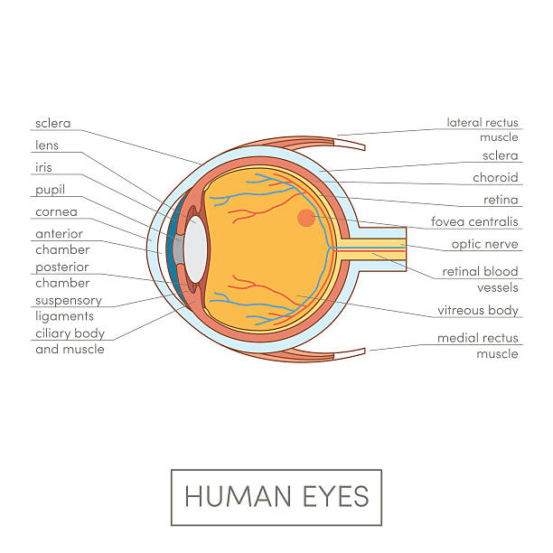
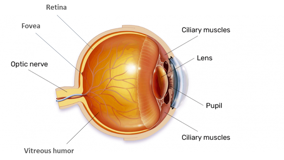
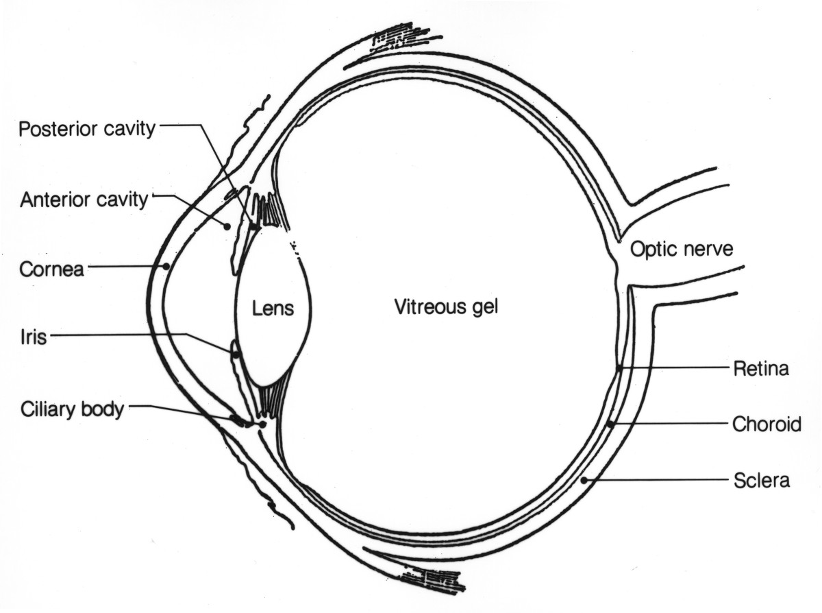
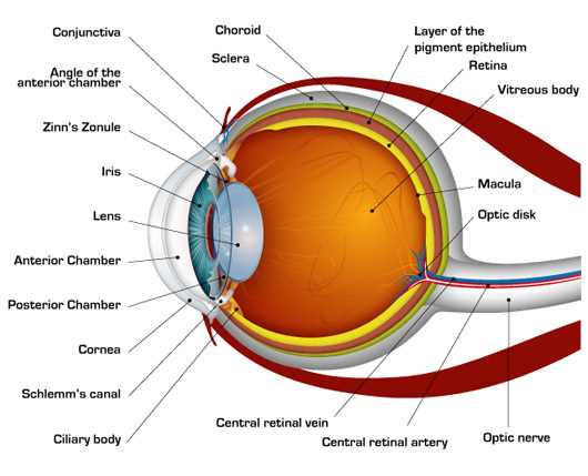
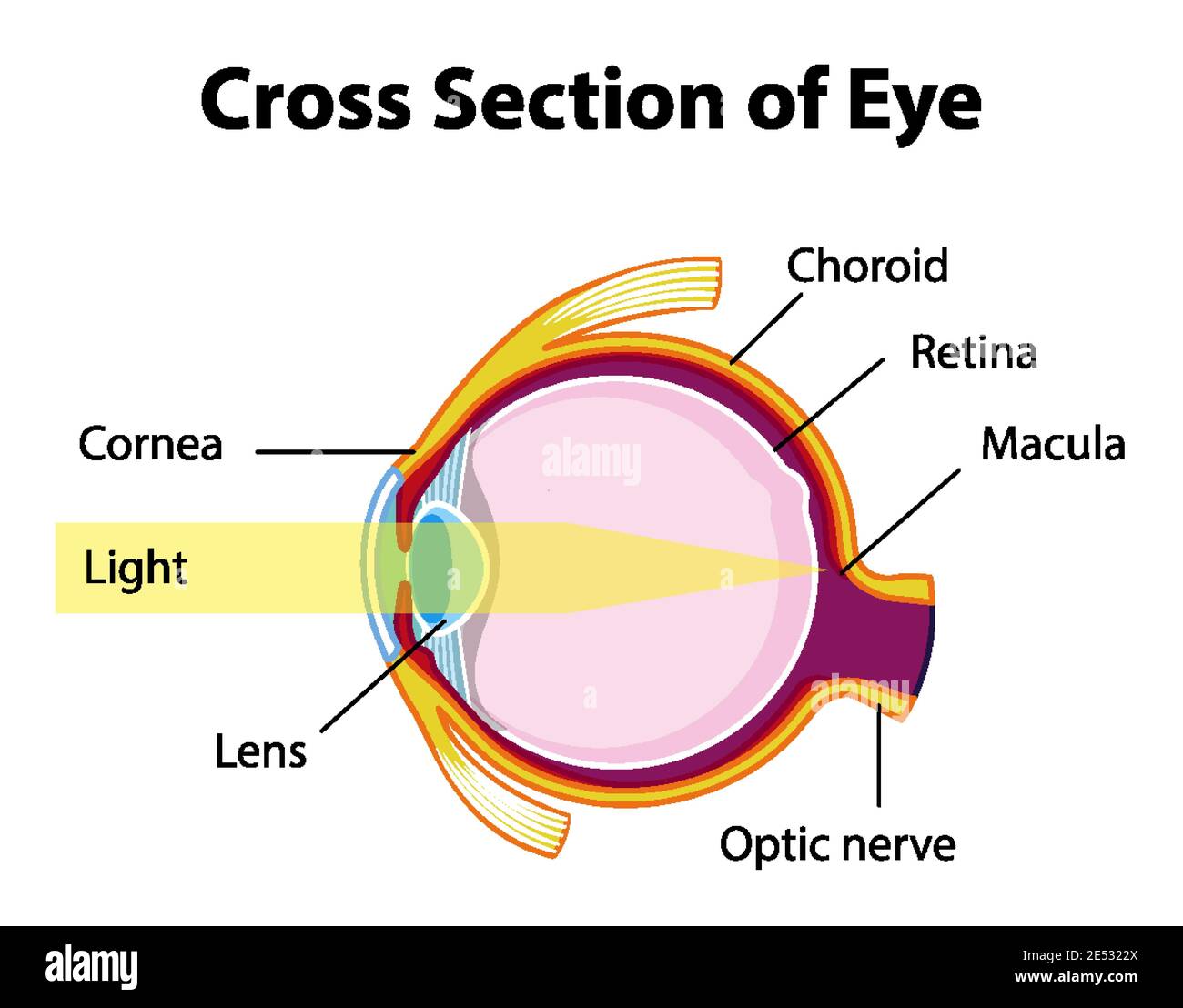
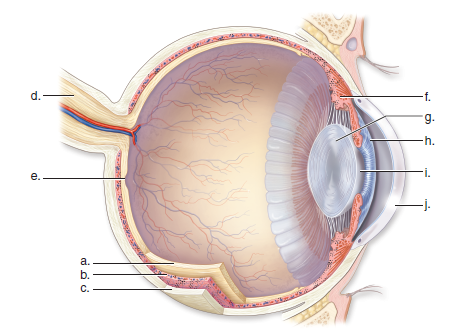
6. Fills posterior cavity of eye 7. Area where optic nerve exits the eye 8. Smooth muscle that controls the pupil size and light entering the eye 9. Fills anterior and posterior chambers of the anterior cavity of the eye 10. Contains photoreceptor cells called rods and cones 11. Connects lens to ciliary body 12. Cause lens to change shape

Human eye diagram with labels
Revolving Nosepiece or Turret: Turret is the part of the microscope that holds two or multiple objective lenses and helps to rotate objective lenses and also helps to easily change power. Objective Lenses: Three are 3 or 4 objective lenses on a microscope. The objective lenses almost always consist of 4x, 10x, 40x and 100x powers. The most common eyepiece lens is 10x and when it coupled with ... the cochlea is lined with sensitive hairs which trigger the generation of nerve signals that are sent to the brain. Read the definitions below, then label the ear anatomy diagram.- (also called the incus) a tiny bone... from the stirrup to the cochlea. This is the smallest bone in the human body (it is 0.25 to 0.33 cm long). Suppliers and employers must use and follow the WHMIS 2015 requirements for labels and safety data sheets (SDSs) for hazardous products sold, distributed, or imported into Canada. Please refer to the following other OSH Answers documents for more information: WHMIS 2015 - General. WHMIS 2015 - Pictograms.
Human eye diagram with labels. Atoms are really small. So small, in fact, that it's impossible to see one with the naked eye, even with the most powerful of microscopes. At least, that used to be true. Now, we have a photograph ... Here are a number of highest rated Human Spine Diagram With Labels pictures upon internet. We identified it from obedient source. Its submitted by paperwork in the best field. We consent this nice of Human Spine Diagram With Labels graphic could possibly be the most trending topic once we allocation it in google gain or facebook. This diagram depicts Picture Of Female Reproductive System Diagram 10241204 with parts and labels. Image Result For Eye Structure And Function Eye Anatomy Eye Anatomy Diagram Basic Anatomy And Physiology. Anatomy Of The Body. Drawing isnt for everybody. Posted on June 7 2016 by admin. The following diagram below is the human body muscle diagram. ©| The museum of science, art and human perception
The eye is linked together with the nervous system which allows the brain to take in information from the eyes and make the appropriate decisions on how to act upon this information. Label Parts of the Human Eye. The eye is the organ responsible for our sense of sight. Drawing an Eye with only one Pencil. Suppliers and employers must use and follow the WHMIS 2015 requirements for labels and safety data sheets (SDSs) for hazardous products sold, distributed, or imported into Canada. Please refer to the following OSH Answers documents for information about WHMIS 2015: WHMIS 2015 - General. WHMIS 2015 - Labels. lateral eye movement. While we had 500ms saccades with the left-aligned labels, with right-aligned labels, the saccade times between the labels and the input... —For a brief introduction to how human eyes work, take a look at my article “.” Moreover, we measured shorter saccade times of just 50ms in cases where users did... Heart Diagram - Diagram of a heart - Human Heart - Human Heart Anatomy - The human heart consists of the following parts aorta, left atrium, right atrium, left ventricle, right ventricle, veins, arter
As part of our unit on the human body we learned about our amazing skeletal system through a variety of hands on projects and this informational, free printable skeletal system worksheet pack.These free printable skeletal system worksheets are handy to use with students from kindergarten, first grade, 2nd grade, 3rd grade, 4th grade, Skeletal system worksheet grade 5 pdf, and 6th grade students. Mammary Glands Function in Male and Female with Labelled Diagram Which is the only organ of your body not fully developed at the time of birth? Do males also... Isaac combines his vast experience with a keen and critical eye to create practical and inherently engaging content on the human body. His background as a researcher... Body Organs Body Systems Human... Human Body What Are Ovaries? Facts, Structure and Location in Human... Ear Diagrams For Kids Ear Diagram Human Ear Diagram Ear Anatomy . Mar 31 2016 image of the ear is colored according to the directions where structures such as the tympanum malleus incus stapes and cochlea are indicated. Inside ear diagram for kids. This excellent ear diagram labels all the important parts of the human ear system the labeled ... Internal organs diagram 502. Download human body diagram with name png free browse more than 1358 png and clipart related with human body diagram with name. The following human organ diagram shows you the front and back view of the human body diagram. Lungs liver kidneys joints tooth brain heart colon eye ear in shape of stomach.

Eye Diagram - Cliparts.co
Zip file with 6 EU energy labels of this lamp. The lamp's performance in the lumen-Watt field, with the energy efficacy fields indicated. Eulumdat light diagram. This light diagram below comes from the program Qlumedit, that extracts these diagrams from an Eulumdat file. The light diagram giving the radiation pattern.

Eye Anatomy 1 Illustration photo | Eye anatomy, Eye anatomy ...
Function: Secretion of a lubricating fluid. 6. Skene's glands: These are also known as the lesser vestibular glands and are located on either side of the urethra.These glands secrete fluid to lubricate the urethra opening and the vulva during sexual arousal. Function: Secretion of a lubricating fluid. Below are the internal parts of the female reproductive system and their functions.
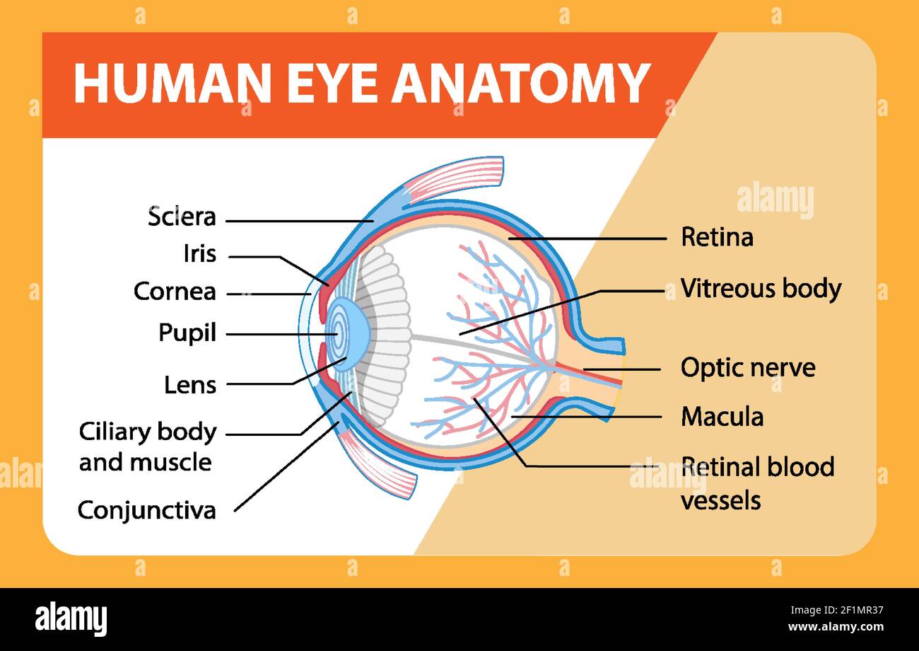
Diagram of human eye anatomy with label illustration Stock ...
Anatomy of the human female pelvis: how to use the anatomical labels On "Anatomical parts", the user can choose the type of anatomical labels he wishes to display on the exam: Genital organs: uterus (with uterine zonal anatomy: endometrium, myometrium, junctional zone), cervix (central zone, cervical stroma), parametria, vagina, ovaries and ...
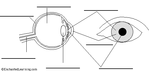
Label Eye Printout - EnchantedLearning.com
Award Winning Human Anatomy and Physiology Home Study Course - For Practitioners, Students, Medical Professionals,Paramedics & Academia
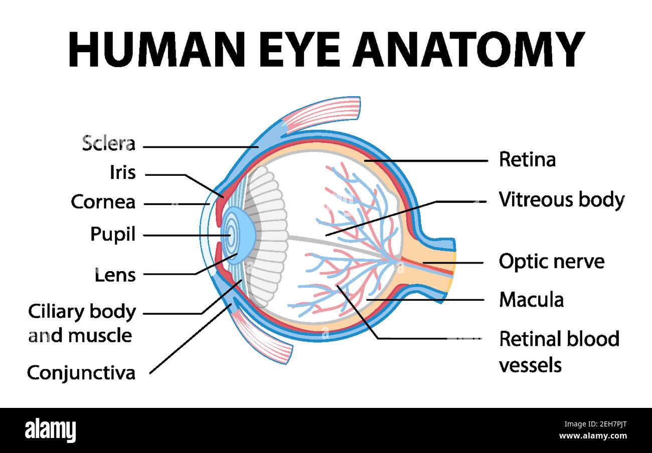
Diagram of human eye anatomy with label illustration Stock ...
of Human Eye Label Diagram of Earth's Magnetism Label the Scientific Method Diagram Label the Diagram of Simple Machines Label the Diagram of Human Ear Label the Diagram of States of Matter Identify and Label the Types of Forces Label the Periodic Table Groups Label the Diagram of a Comet Label the Structure of the Earth Label...
Eye diagram with easy steps | How to draw human eye.
The bones of the face and neck were labeled using different colors to facilitate comprehension. The bone structures are rather more difficult to view on a weighted MRI T2 than on a CT-Scan: for more details on the bones of the face, please refer to the e-Anatomy module "Face-CT-Scan". The teeth were numbered using the FDI World Dental ...

draw a labelled diagram of human eye and give functions of ...
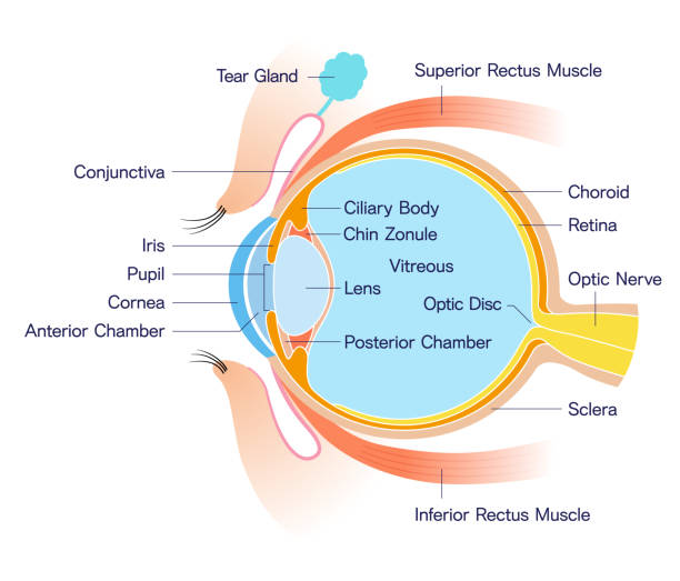
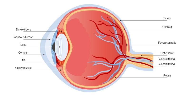
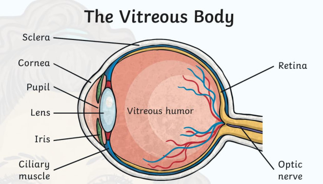
Structure of the Human Eye illustrated and explained using a diagram of the human eye and definitions of the parts of the human eye.

Labelled Diagram Of Human Eye , Png Download - Label A Human ...
What is Osmosis? By definition, osmosis is the movement of any solvent through a selectively permeable membrane into an area of higher solute concentration, the result of which will be an equalizing of solute concentration on either side of the membrane.. This equilibrium is important for the efficient and optimized function of cells; as mentioned before, balance is the preferred state in a ...

a Draw a labelled diagram of the human eye. Label the ...
A fully supervised approach for this study would need one label per detected cell, this means more than 3.7 million labels done by human experts, which is infeasible.

Human eye diagram Images, Stock Photos & Vectors | Shutterstock
Optical Illusions Book HOME > Chapter 2, The Eye > Anatomy of the Human Eye 1 2 Chapter 2 - The Eye: How the Eye Influences Optical Illusions Chapter 2 - The Eye... It is interesting to compare the eye with the camera. In the case of the camera and the photographic process, we have (1) an inverted light-image, a facsimile of... The same is true with other refractive errors, like... the Human Eye VisualIllusion .net Site Map | Terms of...

the human eye Diagram | Quizlet
There are three major parts of the ear, the outer, middle and inner ear. Each contains several parts that are essential to the overall function of the ear. The outer ear is the portion of the ear that sits atop the skull, which is made of flesh and cartilage. It is the visible part which serves to protect the eardrum.
1,883 Eye Cross Section Illustrations & Clip Art - iStock
1 크레딧 Essentials 컬렉션 이 이미지: $12 1개월 정액제 이용 시 $4 ($40에 Essentials 이미지 10개 다운로드) 구매 계속 진행 요금제 및 가격 보기 이 일러스트 편집 최대 사이즈:벡터(EPS) – 어떤 크기로든지 확장 가능 스톡 일러스트 ID:611104910 업로드 날짜:2016년 10월 03일 (월) 자주 묻는 질문 로열티 프리 라이선스는 무엇인가요? 로열티 프리 라이선스는 한 번 결제로... human body with...
72,789 Human Eye Illustrations & Clip Art - iStock
Furthermore, contrary to popular belief, the eye is not perfectly spherical; Ebm was equally as an outgrowth of both punk and industrial music … Binocs will explain the diffe. It is made up of several muscles and … It combines sequenced repetitive basslines, programmed dance music rhythms, and mostly undistorted vocals and commandlike ...

Human Eye Anatomy Diagram stock vector. Illustration of ...
Read Hatchet Chapter 15 and complete summary, t-charts, and plot diagram by Monday of next week. Wednesday, December 4, 2019 Reading Diagnostic Testing. Hatchet is about a boy who gets on a plane to go to his dads house. The boys name is Brian Robson. While he is in the plane the pilot has a heart attack and dies.

Cross-section of the human eye with labels - Stock ...
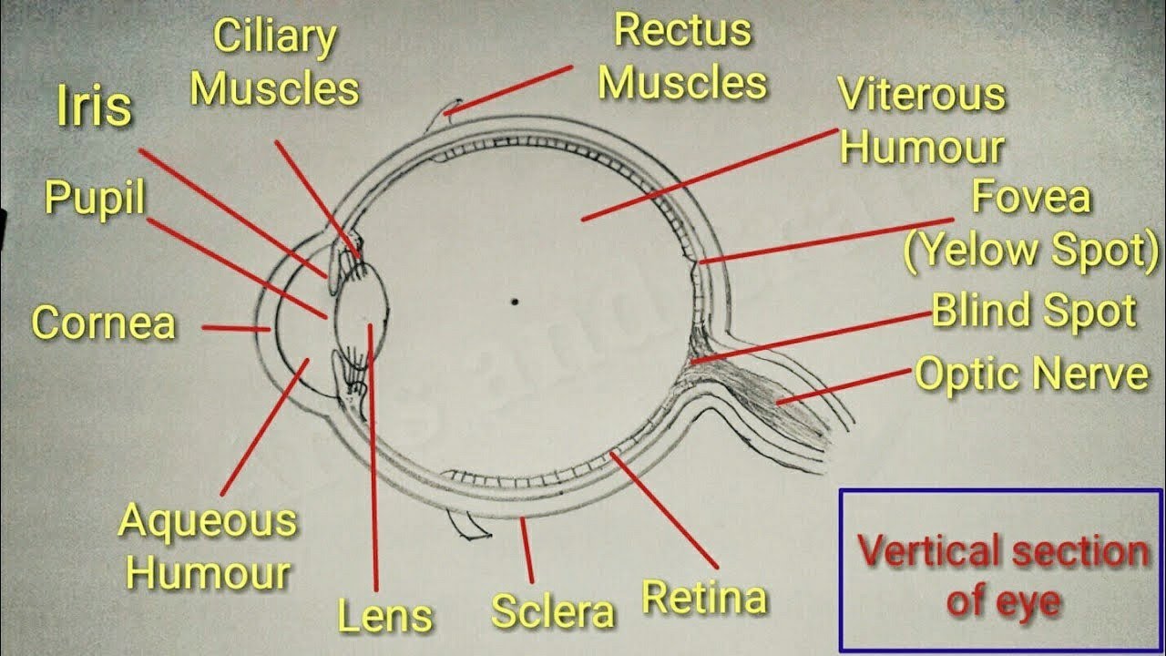
Name the ernica 20 the STRUCTURE OF THE HUMAN EYE Label the parts of the human eye on the diagram below. a aqueous humor b. cornea c. pupi d.lons e. iris f. ciliary body b Ovitreous humor hretina optic nerve J. choroid coat k. sclera 1. suspensory...
The Anatomy of Human Eye with Diagram | EdrawMax Online
Label the part of the human body. Label the parts of the body in French. Label the parts of the body in German. Label the parts of the body in Italian. Label the parts of the body in Spanish. Label the lateral section brain anatomy diagram. Label the digestive system. Label the digestive system. Label the ear anatomy diagram....

Anatomy of the Eye Diagrams for Coloring/Labeling, with ...
The world at the start of the 21st Century is also the result of what we call the Great Acceleration - the most rapid transformation of the human relationship with the natural world in history. 'Many human activities reached take-off points sometime in the mid-20th Century and sharply accelerated towards the end of the century....

Human Eye Anatomy - Parts of the Eye Explained | Eye anatomy ...
Circulatory System Diagram. The circulatory system is the most vital systems of your body that is required for the optimal distribution of oxygenated blood to all the body organs and tissues. A fully functional circulatory system aims to maintain adequate concentration of oxygen in the biological tissues to ensure longevity and health.

Human Eye Ball Anatomy & Physiology Diagram
Here are a number of highest rated Anatomical Diagram Of The Human Eye pictures upon internet. We identified it from reliable source. Its submitted by running in the best field. We agree to this nice of Anatomical Diagram Of The Human Eye graphic could possibly be the most trending topic later we ration it in google benefit or facebook.
Human Eye Information – Learning and Teaching Resource
To further delineate the location of opticin protein in the human eye, we performed immunohistochemical analysis on eye sections from a human male donor (Fig. ). Several components of the eye were labeled with the anti-opticin antibody, with the most intense staining in the vitreous base and cortex regions (Fig.H). The basal...

Human Eye Credits - Draw And Label A Human Eye PNG Image ...
Read the definitions, then label the skin anatomy diagram below.- Tubes that carry blood as it circulates. Arteries bring oxygenated blood from the heart and lungs; veins return oxygen-depleted blood back to the heart and lungs. - (also called the cutis) the layer of the skin just beneath the epidermis. - the outer layer of the...

human eye Diagram | Quizlet
Read on to explore the human brain structure, diagram, parts of the human brain and the body functions controlled by the human brain. Find out how some people live with just half a brain. On average, an adult brain... It also controls the reflex movements of the head, eye and neck muscles. It provides a passage for the...

Labeled Eye Diagram | Human eye diagram, Eye anatomy, Diagram ...
Blank Eye Diagram To Label. Here are a number of highest rated Blank Eye Diagram To Label pictures upon internet. We identified it from obedient source. Its submitted by paperwork in the best field. We admit this nice of Blank Eye Diagram To Label graphic could possibly be the most trending subject with we portion it in google gain or facebook.

Diagram human eye anatomy with label Royalty Free Vector
Four Chambers of the Heart and Blood Circulation. The shape of the human heart is like an upside-down pear, weighing between 7-15 ounces, and is little larger than the size of the fist. It is located between the lungs, in the middle of the chest, behind and slightly to the left of the breast bone. The heart, one of the most significant organs ...
Draw a well labelled diagram of the human eye and write class ...
For each label type, 504 labels are created. The colored vessel segmentation labels are used to execute the deep-learning-optimized method to segment the vessels from the original retinal image, which is taken from the RVM dataset. The images and their labels, for type-1 and type-2, have a size of 2002 \(\times\) 2000 pixels. Each original ...

Draw the well labelled diagram of V.S. human eye.
Suppliers and employers must use and follow the WHMIS 2015 requirements for labels and safety data sheets (SDSs) for hazardous products sold, distributed, or imported into Canada. Please refer to the following other OSH Answers documents for more information: WHMIS 2015 - General. WHMIS 2015 - Pictograms.

Diagram Of The Human Eye With Parts Labeled Stock ...
the cochlea is lined with sensitive hairs which trigger the generation of nerve signals that are sent to the brain. Read the definitions below, then label the ear anatomy diagram.- (also called the incus) a tiny bone... from the stirrup to the cochlea. This is the smallest bone in the human body (it is 0.25 to 0.33 cm long).

Schematic drawing of the human eye. Adapted from ...
Revolving Nosepiece or Turret: Turret is the part of the microscope that holds two or multiple objective lenses and helps to rotate objective lenses and also helps to easily change power. Objective Lenses: Three are 3 or 4 objective lenses on a microscope. The objective lenses almost always consist of 4x, 10x, 40x and 100x powers. The most common eyepiece lens is 10x and when it coupled with ...
/GettyImages-695204442-b9320f82932c49bcac765167b95f4af6.jpg)
Structure and Function of the Human Eye

Diagram human eye anatomy with label Royalty Free Vector
Anatomy of the Eye: Human Eye Anatomy - Owlcation

human eye | Definition, Anatomy, Diagram, Function, & Facts ...

Diagram human eye anatomy with label Royalty Free Vector
How the Human Eye Works | Cornea Layers/Role | Light Rays
Human eye anatomy with cross section of eye diagram ...

Structure Of Human Eye Without Label Transparent PNG ...
Solved: Label this diagram of the human eye. State a function ...

Diagram showing cross section human eye Royalty Free Vector


Comments
Post a Comment