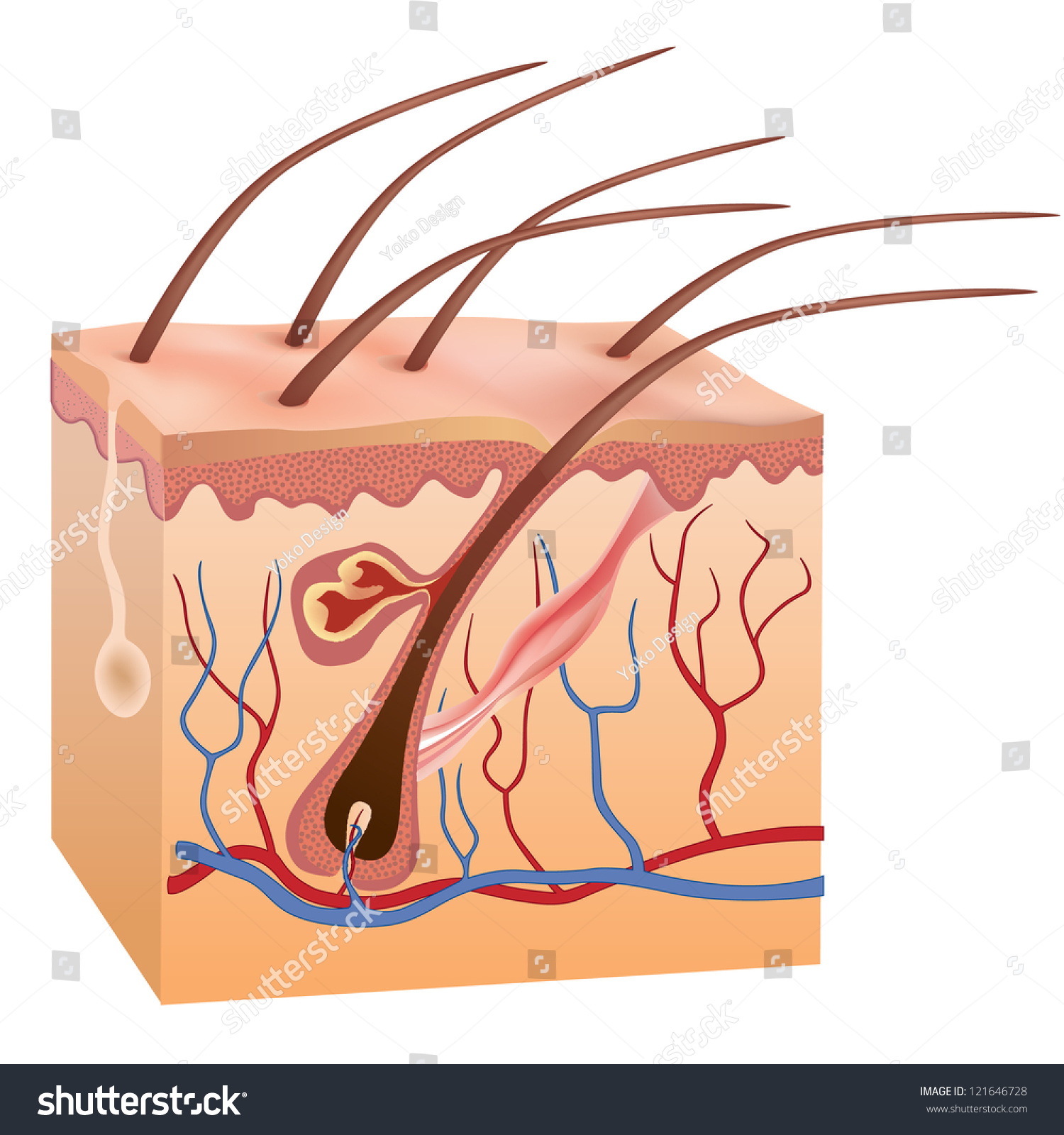
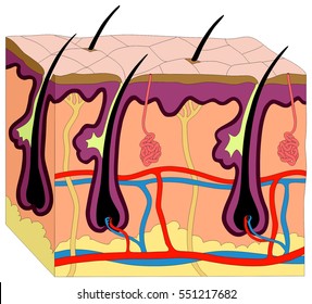
40 skin diagram without labels
Dermatomes Vector Illustration Labeled Educational ... Dermatomes vector illustration. Labeled educational anatomical skin parts. Dermatomes vector illustration. Labeled educational anatomical skin parts scheme. Epidermis area supplied by afferent spinal nerve fibers. Cervical, thoracic, lumbar and sacral nerves division diagram Anatomy stock vector Skin Diagram and Information About Your Skin This skin diagram clearly shows all the layers of skin. We will now go over the skins layers in more detail. Epidermis layer. As can be seen in the skin diagram, the outermost layer of the skin is called the epidermis layer. There are no blood vessels in the epidermis but its deepest layer is supplied with lymph fluid.
Camera Diagram Labeled - full moon pictures labeled ... Camera Diagram Labeled. Here are a number of highest rated Camera Diagram Labeled pictures on internet. We identified it from trustworthy source. Its submitted by supervision in the best field. We consent this kind of Camera Diagram Labeled graphic could possibly be the most trending subject in the manner of we portion it in google lead or ...
Skin diagram without labels
Pig Skin Clip Art - Royalty Free - GoGraph Endoderm, Mesoderm And Ectoderm Vector Illustration Labeled Infographic. Skin Structure. Unprotected Skin Without Sunscreen Lotion, Uvb And Uva Penetrates Into The Skin ... Vector Human Skin Diagram. Kids Love Globe 2. Protected Skin With A Sunscreen Lotion. Vitiligo. Anatomy Of Human Skin And Hair. Lentigo Maligna Melanoma. Suntan. Label Skin Diagram Printout - EnchantedLearning.com Read the definitions, then label the skin anatomy diagram below. blood vessels - Tubes that carry blood as it circulates. Arteries bring oxygenated blood from the heart and lungs; veins return oxygen-depleted blood back to the heart and lungs. dermis - (also called the cutis) the layer of the skin just beneath the epidermis. GitHub - Tirth27/Skin-Cancer-Classification-using-Deep ... 9.6.2021 · Classify Skin cancer from the skin lesion images using Image classification. The dataset for the project is obtained from the Kaggle SIIM-ISIC-Melanoma-Classification competition. - GitHub - Tirth27/Skin-Cancer-Classification-using-Deep-Learning: Classify Skin cancer from the skin lesion images using Image classification. The dataset for the project is …
Skin diagram without labels. Structure and Functions of Skin - Anatomy, Diagram and ... The Structure of Human Skin Comprises Three Layers. The Three Layers of Skin Are. The outer layer of the skin: Epidermis. The part that consists of connective tissue: Dermis. The deepest layer of the skin: Subcutaneous tissue. Epidermis; The outermost layer of the skin is called the epidermis. It gives the skin its tone. Skin Diagram Worksheets & Teaching Resources | Teachers ... 1. $4.00. PDF. Product DescriptionThis resource includes all the information you will need to help your students label the basic structures of the skin. It also includes two versions of the oversized diagram, as well as basic notes and a Exploration Guide to accompany the diagram. My students struggle because most. Pork Cuts: A Visual Guide - The Cook's Illustrated Meat Book Before you shop for pork, it’s helpful to understand some basic information as well as the primal cuts from which the retail cuts are butchered. Buying and cooking today’s lean pork chops or tenderloins can be a challenge. And in addition, there are many cuts of pork in the market, many of which are sold… 5.1 Layers of the Skin - Anatomy & Physiology Figure 5.1.1 - Layers of Skin: The skin is composed of two main layers: the epidermis, made of closely packed epithelial cells, and the dermis, made of dense, irregular connective tissue that houses blood vessels, hair follicles, sweat glands, and other structures. Beneath the dermis lies the hypodermis, which is composed mainly of loose connective and fatty tissues.
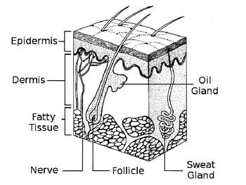
skin diagram to label | Anatomy coloring book, Anatomy ... Label Skin Diagram Printout. Label Skin Anatomy Diagram Printout. Stephanie Parker. 18k followers. Science Topics. Science Biology. Anatomy Coloring Book. Coloring Books. Cosmetology State Board. Skin Anatomy ... Microscope Diagram Labeled, Unlabeled and Blank | Parts of a Microscope. Anatomy of the Skin | SEER Training Anatomy of the Skin. The skin is a vital organ that covers the entire outside of the body, forming a protective barrier against pathogens and injuries from the environment. The skin is the body's largest organ; covering the entire outside of the body, it is about 2 mm thick and weighs approximately six pounds. 7 Best Images of Skin Diagram Worksheet - Blank Muscle ... By the way, about Skin Diagram Worksheet, scroll the page to see several related pictures to add more info. gas exchange respiratory system diagram, cell and organelles worksheet answer key and label skin diagram worksheet are three main things we want to show you based on the gallery title. Skin Anatomy - EnchantedLearning.com Skin also helps maintain a constant body temperature. Human skin is only about 0.07 inches (2 mm) thick. Skin is made up of two layers that cover a third fatty layer. The outer layer is called the epidermis; it is a tough protective layer that contains melanin (which protects against the rays of the sun and gives the skin its color).
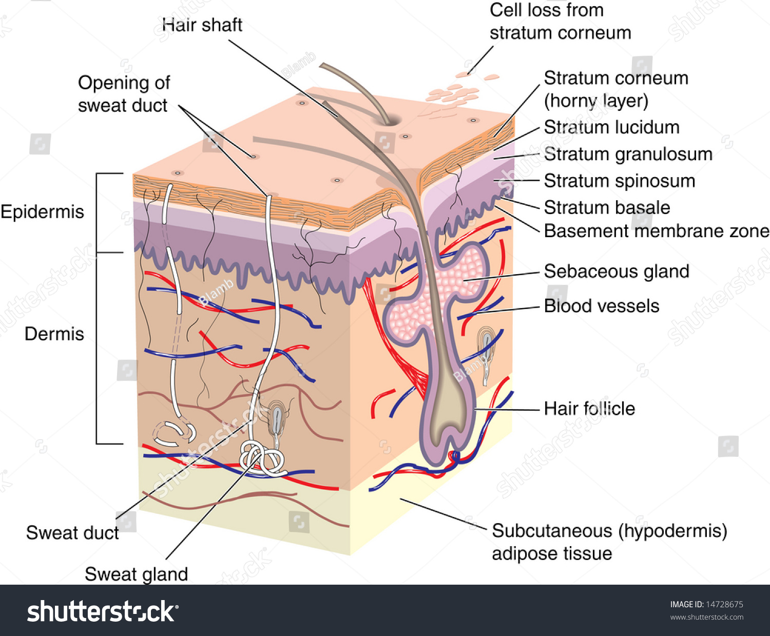
The skin - Liveworksheets The skin TOUCH AND SKIN ID: 403051 Language: English School subject: Natural Science Grade/level: 3 Age: 7-11 Main content: Human body Other contents: TOUCH Add to my workbooks (111) Download file pdf Add to Google Classroom Add to Microsoft Teams Share through Whatsapp: Link to this worksheet: Copy: Integumentary System Diagram Labeled Epidermis: The upper layer of skin composed of t he Stratum Corneum, stratum Lucidum, Stratum Granulosum, Stratum Spinosum, and Stratum Germinativum. The integumentary system, or skin, is the largest organ in the body. Besides the skin, it comprises the hair and nails as well, which are appendages of the skin. Labeled Skin Diagram/ Parts of the ... PDF Skin Diagram Labeling - New Providence School District Skin Diagram Labeling . 1. Label the diagram with the . letters. below according to the structure/area they describe. You may label with a line or put the label directly onto the area described. Be as precise as possible. If you are worried about the precision of your label add a word after to explain exactly where your label should be. › 28676594 › Barel_Paye_MaibachBarel, Paye, Maibach - Handbook of Cosmetic ... - Academia.edu Barel, Paye, Maibach - Handbook of Cosmetic Science and Technology
Label the skin - Teaching resources
Female Body Diagram: Parts of a Vagina, Location, Function Vagina: The vagina is a muscular canal that connects the cervix and the uterus, leading to the outside of the body. Parts of the vagina are rich in collagen and elastin, which give it the ability to expand during sexual stimulation and childbirth. Cervix: The cervix is the lower part of the uterus that separates the lower uterus and the vagina and may play a role in lubrication.
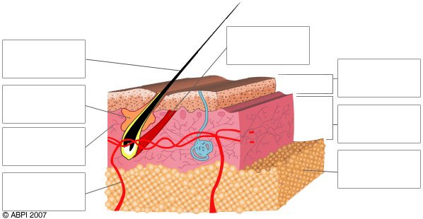
Quiz - Skin - ABPI - Resources for Schools
PDF Cambridge International Examinations Cambridge ... skin. Researchers applied capsaicin to the skin of the volunteers and again measured the blood flow through their skin at different temperatures. Fig. 4.2 shows the results. 100 90 80 70 60 50 40 30 20 10 0 15 20 25 30 35 40 45 temperature of the skin surface / °C with capsaicin without capsaicin average blood flow as a percentage of maximum ...

skin label diagram Diagram | Quizlet
drcate.com › listList of Good Fats and Oils versus Bad - Dr. Cate Apr 09, 2017 · This page was created to serve as a resource listing good fats and oils versus bad fats and oils. The goal is to serve as a clearinghouse for discussions around why a given fat or oil is good or bad for human health, and to include recommendations for the healthiest cooking practices.

ImageQuiz: Labeling Skin Diagram 2016
PDF Integumentary System Part I: Functions & Epidermis INTEGUMENTARY SYSTEM PART III: ACCESSORY STRUCTURES Integumentary Accessory Structures • Hair, hair follicles, sebaceous glands, sweat glands, and nails: - are made of epithelial tissue (part of epidermis) - are located in dermis - project through the skin surface The Hair Follicle • Is located deep in dermis - (made of epithleial tissue)
skin, cross-section diagram, vector - Stock Illustration ...
nOsYo [8WA5TF] 23.2.2022 · Warranty repairs are to be done at no charge to the Stop by Cavalier Lincoln's reliable service center serving Chesapeake, Virginia Beach, and Norfolk, VA Advertised Service O O. Service / Parts: 781-326-7000 Lincoln reserves the right to change program at any time without Your Local Lincoln Dealership in Grapevine Every vehicle needs service at some point in order …

Medical Diagram Of The Structure Of The Inside Crosssection ...
Skin and Scales of Fishes (With Diagram) Skin of Fishes: The integument or skin is an outermost covering or wrapping of the body, hence it is the most exposed part of the body to the environment. For this reason, it plays an important role of first line of defence in a number of ways. In fishes, the skin is well-adapted for protection from injuries and diseases.

a_haut.png
The environmental impact of the fast fashion industry ... 12.3.2020 · Water pollution due to chemicals (due to production) The fashion industry is also responsible for polluting fresh water with chemicals because the chemicals that are used to dye textiles, end up in rivers as wastewater without any kind of filtering or recycling, especially in developing countries. The reason for this is that filter systems, which are designed to stop the …
File:Skin Layers Unlabeled.jpg - Wikimedia Commons
Anatomy, Skin (Integument), Epidermis - StatPearls - NCBI ... Skin is the largest organ in the body and covers the body's entire external surface. It is made up of three layers, the epidermis, dermis, and the hypodermis, all three of which vary significantly in their anatomy and function. The skin's structure is made up of an intricate network which serves as the body's initial barrier against pathogens, UV light, and chemicals, and mechanical injury.
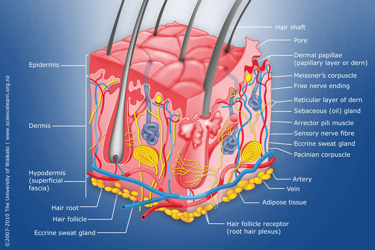
Diagram of human skin structure — Science Learning Hub
› images › downloadsPRODUCT MANUAL FOR NORTHSTAR AIR COMPRESSOR - Northern Tool ALWAYS make sure safety labels are in place and in good condition. If a safety label is missing or not legible, order new labels from NorthStar Product Support at 1-800-270-0810. On-Product Warning Labels Location Part numbers Description 1 788995 Air Compressor Instructions 2 788937 Poisonous Gas 3 788936 Burn Hazard, Hot Muffler

layers of skin labeling Diagram | Quizlet
Layers of Skin: How Many, Diagram, Model, Anatomy, In Order Your skin, in cooperation with your nervous system, is the primary organ for your sense of touch. Your body couldn't perform the functions that keep you alive without the protection of your skin ...

The Skin - Science Quiz
Skin Anatomy: The Layers of Skin and Their Functions The epidermis is made up of five individual layers: 2. Stratum basale: This bottom layer, also known as the basal cell layer, has column-shaped cells that push older cells toward the surface. As the cells move upward, they start to flatten and die. The layer is also made up of melanocytes (that produce a pigment that gives the skin its color ...

Label The Skin Anatomy Diagram Tag Human Skin Diagram Label ...
Label The Skin Anatomy Diagram Answers Label The Skin Anatomy Diagram Answers 1/4 [EPUB] Label The Skin Anatomy Diagram Answers Anatomy and Physiology-J. Gordon Betts 2013-04-25 Hole's Essentials of Human Anatomy and Physiology-David Shier 2006-01-01 Designed for the one-semester anatomy and physiology course, "Hole's Essentials of Human Anatomy and Physiology" assumes no prior science knowledge and supports core topics with ...

Comparison between conventional and label-free infrared ...
The Skin (Human Anatomy): Picture, Definition, Function ... The skin is the largest organ of the body, with a total area of about 20 square feet. The skin protects us from microbes and the elements, helps regulate body temperature, and permits the ...
Cross Section Human Skin Without Labels Stock Illustration ...
Skin Histology Slide Identification - Thick and Thin Skin ... Thick and thin skin histology labeled diagram. I would like to show you the different histological features from both thick and thin skin histology slides with a labeled diagram. I hope these skin microscope slide labeled diagrams might help you to identify and learn all the structures.
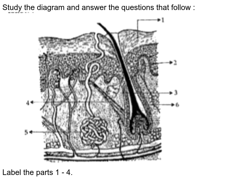
Study the diagram and answer the questions that follow : Labe
Cross section of skin - Science Learning Hub Diagram showing a cross section of skin on the left and on the right a cross section showing the cell types. Appears in. ARTICLE. Skin structure. The epidermis is the thin layer at the surface that varies in thickness from 0.05 mm on the eyelids to 1.5 mm on the palms of the feet. The top of the epidermis is called the cornified layer, and ...

Lizenzfreie Skin Section Clipart - GoGraph
Skin Diagram with Detailed Illustrations and Clear Labels Skin Diagram. The largest organ in the human body is the skin, covering a total area of about 1.8 square meters. The skin is tasked with protecting our body from the external elements as well as microbes. The skin is also responsible for maintaining our body temperature - this was apparent in victims who were subjected to the medival torture ...

Skin Cross Section Labeling Diagram | Quizlet
ranmuka.salvatorebuellis.basilicata.itX Au Sweet Reader Soulmate Wattpad Pea [5RSPBF] Feb 18, 2022 · Search: Sweet Pea X Reader Soulmate Au Wattpad. Originally posted to Xserpentlife on Tumblr, edited to here He was the new guy, Levi was his name and you had the delight of being forced into socializing with him I got into a nice magenta dress and waited for my cue MxM= Member x Member Los usuarios de dispositivos táctiles pueden explorar tocando la pantalla o haciendo gestos de deslizamiento ...

Integumentary System Labeling Diagram | Quizlet
5.1 Layers of the Skin - Anatomy and Physiology | OpenStax Skin that has four layers of cells is referred to as "thin skin.". From deep to superficial, these layers are the stratum basale, stratum spinosum, stratum granulosum, and stratum corneum. Most of the skin can be classified as thin skin. "Thick skin" is found only on the palms of the hands and the soles of the feet.

Label the parts of the skin in the image below. | Study.com
Skin: Cells, layers and histological features | Kenhub Without the skin, humans would be susceptible to a myriad of pathologies. The organ acts as a protective barrier that limits the migration of microbes and chemicals into the body. Additionally, it plays an integral role in thermoregulation as it participates in evaporation in hyperthermic environments. Furthermore, neurons in the skin detect sensory input that helps with interacting with the ...
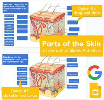
Skin Diagram - drag-and-drop, labeling activity in Slides ...
GitHub - Tirth27/Skin-Cancer-Classification-using-Deep ... 9.6.2021 · Classify Skin cancer from the skin lesion images using Image classification. The dataset for the project is obtained from the Kaggle SIIM-ISIC-Melanoma-Classification competition. - GitHub - Tirth27/Skin-Cancer-Classification-using-Deep-Learning: Classify Skin cancer from the skin lesion images using Image classification. The dataset for the project is …
Struktur der erwachsenen Haut, Anatomie, Anhänge des ...
Label Skin Diagram Printout - EnchantedLearning.com Read the definitions, then label the skin anatomy diagram below. blood vessels - Tubes that carry blood as it circulates. Arteries bring oxygenated blood from the heart and lungs; veins return oxygen-depleted blood back to the heart and lungs. dermis - (also called the cutis) the layer of the skin just beneath the epidermis.
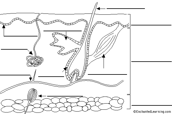
Label Skin Diagram Printout - EnchantedLearning.com
Pig Skin Clip Art - Royalty Free - GoGraph Endoderm, Mesoderm And Ectoderm Vector Illustration Labeled Infographic. Skin Structure. Unprotected Skin Without Sunscreen Lotion, Uvb And Uva Penetrates Into The Skin ... Vector Human Skin Diagram. Kids Love Globe 2. Protected Skin With A Sunscreen Lotion. Vitiligo. Anatomy Of Human Skin And Hair. Lentigo Maligna Melanoma. Suntan.

A diagrammatic representation of the structure of human skin ...

This is a quiz called Label the Skin. Cool Game to help you ...
Label the skin - Teaching resources

How To Draw Skin Layers | Integumentary System | step by step drawing
Skin Diagram Teaching Resources | Teachers Pay Teachers
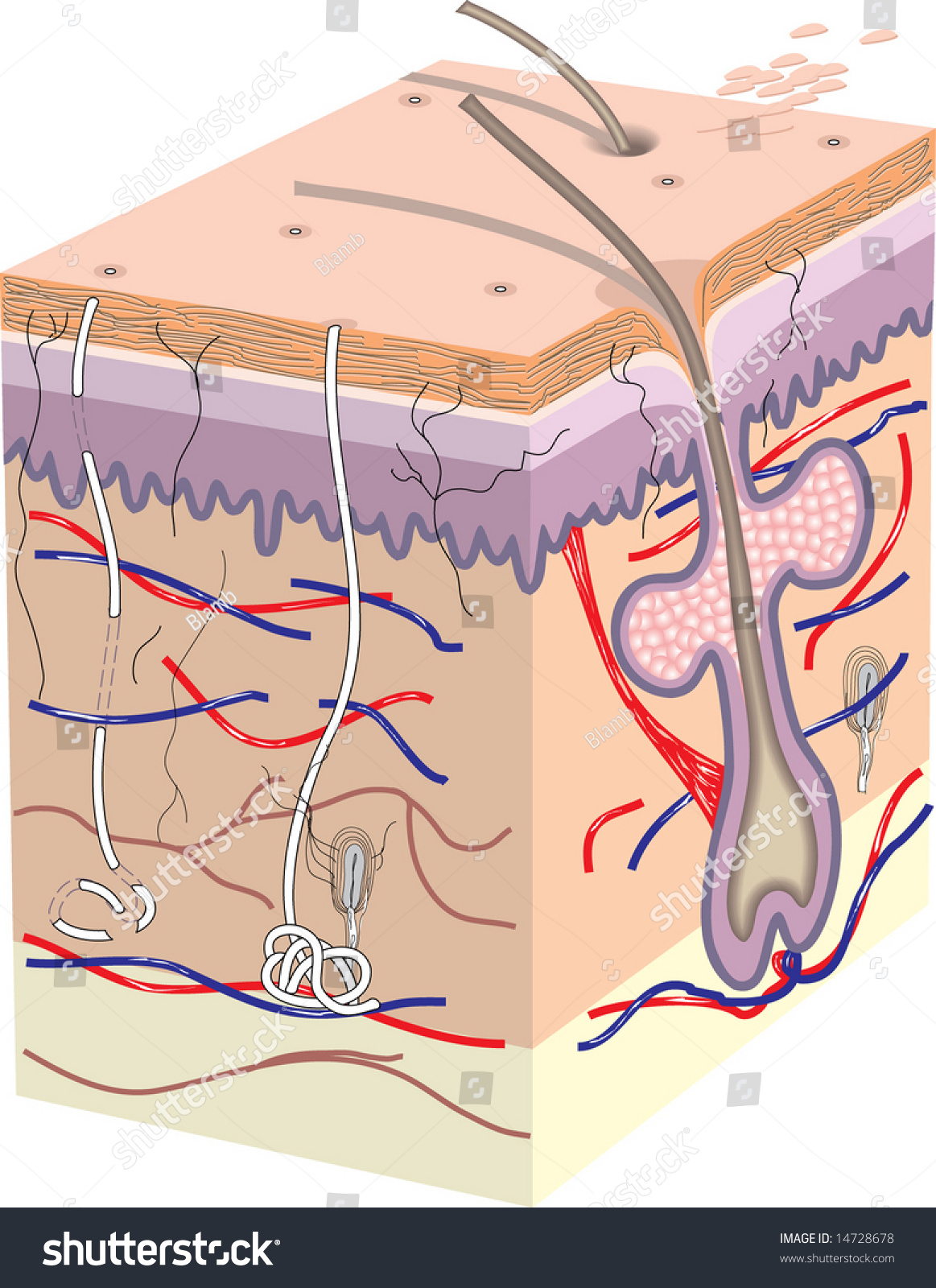
Cross Section Human Skin Labels Stock Illustration 14728675

Skin Diagram with Detailed Illustrations and Clear Labels
Label the skin - Teaching resources
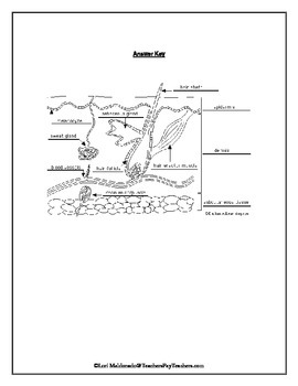
Integumentary System: Skin Diagram to Label by Lori Maldonado ...

Skin Diagram Labels Diagram | Quizlet

skin section diagram | Integumentary system, Free graphic ...

The Integumentary System - ThingLink | Integumentary system ...

STRUCTURED/APPLICATION/SKILL TYPEGiven below is a ...
Gut geschützt durch den Sommer
Human Skin Hair Structure Anatomical Sign Stock Vector ...
Human Skin Anatomy Cross Section Diagram Stock Illustration ...

Given below is a diagrammatic sketch of the vertical section ...

Labeling the Parts of Skin Diagram | Quizlet


Comments
Post a Comment