42 microscope lens diagram
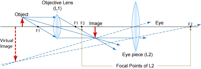
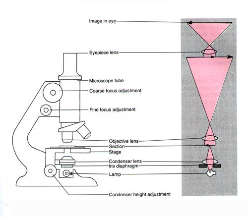
Microscope, Microscope Parts, Labeled Diagram, and Functions Eyepiece Lens: the lens at the top that you look through, usually 10x or 15x power. Tube: Connects the eyepiece to the objective lenses. Illuminator: Illuminator is the most important microscope parts and it serve as light source for a microscope during slide specimen visualization.It is a continuous source of light (110 volts) used in place of a mirror. PDF Chapter 4 Optics - UNC School of Medicine A ray diagram like this can be interpreted in both directions. If you put a point light source one focal length from a converging lens, parallel light ... focal plane of the microscope's objective lens is on the side that faces the specimen. The front focal plane of the condenser is the side that faces the field iris and the illuminator. The ...
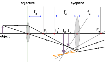
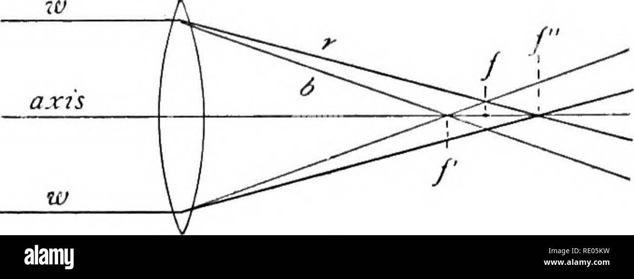
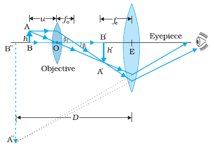
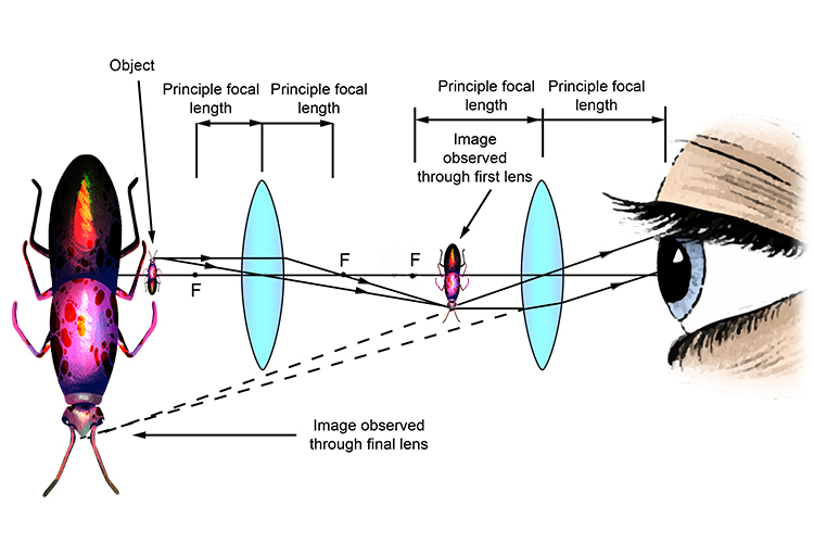
r/AskPhysics - Microscope lenses confusion Microscope lenses confusion. We just looked a ray diagram with lenses of microscope in class. We saw that rays of small object go through objective lens and form larger image on other side of lens at focal length. Then, these rays go through another lens and because this lens is focal length of this lens away, all the rays will go parallel to ...
Microscope lens diagram
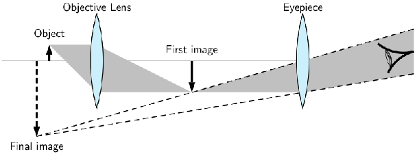
Understanding Microscopes and Objectives - Edmund Optics With a microscope, a relay lens system replaces the single lens; an objective and an eyepiece work in tandem to project the image of the object onto the eye, or a sensor - depending upon the application. There are two parts to a microscope that increase the overall system magnification: the objective and the eyepiece. Microscope Lab - csun.edu There is a diagram link below. Have the students study this diagram so that they know the parts, which objective to use. It is very important that the students don't focus to close to the slide with the high power objective, as this can permanently damage the lens. 16 Parts of a Compound Microscope: Diagrams and Video ... Aperture. Illuminator. Condenser. Diaphragm. Parts of a Compound Microscope. Video: Parts of a compound Microscope with Diagram Explained. As a side note, the microscope used in this post is a great entry level or beginner microscope if you are trying to get someone interested in microscopes, microbiology, or science in general.
Microscope lens diagram. Microscope Lenses Diagram - Micropedia Mar 28, 2020 · Total Internal Reflection And Lenses. Microscope Magnification Calculator Microscopy Pengonversi Unit. Class 12 Compound Microscope Ray Diagram. Assignment 1 Part 3 Building Your Transillumination Microscope. Show By Ray Diagram The Formation Of Image In Case Of A Compound. Notes On Microscope Grade 11 Physics Optical Instruments. Geometrical Construction of Ray Diagrams Microscope Optical Components Interactive Tutorials Geometrical Construction of Ray Diagrams. A popular method of representing a train of propagating light waves involves the application of geometrical optics to determine the size and location of images formed by a lens or multi-lens system. Amazon.com: Microscope Lenses - Lenses / Microscope ... Microscope Lens,40X 185 Biological Microscope Achromatic Objectives Lens 160/0.17 5×5×5cm Eliminate Unwanted Reflections 4.7 out of 5 stars 5 $18.69 $ 18 . 69 Compound Microscope - Diagram (Parts labelled), Principle ... A compound microscope: Is used to view samples that are not visible to the naked eye. Uses two types of lenses - Objective and ocular lenses. Has a higher level of magnification - Typically up to 2000x. Is used in hospitals and forensic labs by scientists, biologists and researchers to study microorganisms.
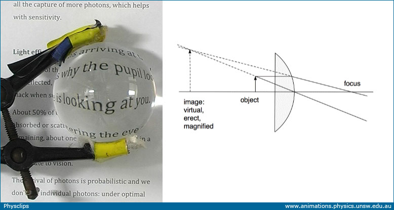
A Study of the Microscope and its Functions With a Labeled ... The microscope is an important instrument in the world of biological science. Diagrams have always been of great help in understanding both the structural and functional aspects of entities. These labeled microscope diagrams and the functions of its various parts, attempt to simplify the microscope for you. Parts of Microscope | Function | Labeled Diagram ... Parts of Microscope with its Function Head. The Head is a part of a microscope that is at the upper side of the microscope and carries an optical lens.. Base. The base is the overall support of the microscope & carries microscope illuminators, brightness adjustment switch, and light switch. Microscope Types (with labeled diagrams) and Functions Simple microscope is a magnification apparatus that uses a combination of double convex lens to form an enlarged, erect image of a specimen. The working principle of a simple microscope is that when a lens is held close to the eye, a virtual, magnified and erect image of a specimen is formed at the least possible distance from which a human eye ... Microscope Parts and Functions With Labeled Diagram and ... Microscope Parts and Functions With Labeled Diagram and Functions How does a Compound Microscope Work?. Before exploring microscope parts and functions, you should probably understand that the compound light microscope is more complicated than just a microscope with more than one lens.. First, the purpose of a microscope is to magnify a small object or to magnify the fine details of a larger ...
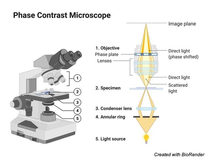
Important Types of Microscopes used in Biology (With Diagram) In simple microscope, convex lens of short focal length is used to see magnified image of a small object. The object is placed between the optical centre and the focus of a convex lens, its image is virtual, erect and magnified and on the same side as the object. ... (With Diagram) electron microscope is around 20,000 times. 4. PhaseContrast ... Microscope diagram Flashcards | Quizlet Microscope diagram. STUDY. Flashcards. Learn. Write. Spell. Test. PLAY. Match. Gravity. Created by. ErinnFergison. Anatomy and Physiology. Terms in this set (14) #1. Body tube #2. Revolving nosepiece #3. Red lens (4 power) #4. Yellow lens (10 power) #5. Blue lens (40 power) #6. Stage clips #7. Diaphram #8. Light #9. Ocular lens #10. arm #11 ... Types of Microscopes: Definition, Working Principle ... F is the focal length of the convex lens; Simple Microscope Diagram. Principle of Simple Microscope. The working principle of a simple microscope is that when a sample is placed within the focus of the microscope, a virtual, erect and magnified image is obtained at the least distance of distinct vision from the eye that is held at the lens. Compound Microscope Parts, Function, & Diagram | What is a ... Nov 04, 2021 · The compound microscope, also called compound light microscope, is an upright microscope that utilizes two lenses to magnify objects. It gets its name because it uses two lenses added to each ...
Microscope Objective, Tube, and Scan Lens Tutorials
Compound Microscope Parts - Labeled Diagram and their ... Always lift a microscope by holding both the arm and base with two hands. There are two major optical lens parts of a microscope: Eyepiece (10x) and Objective lenses (4x, 10x, 40x, 100x). Total magnification power is calculated by multiplying the magnification of the eyepiece and objective lens. The illuminator provides a source of light.

i) Observe the following diagram and answer the questions ...
The Microscope Optical Train | Nikon's MicroscopyU The microscope optical train typically consists of an illuminator (including the light source and collector lens), a substage condenser, specimen, objective, eyepiece, and detector, which is either some form of camera or the observer's eye ( Table 1 ). Research-level microscopes also contain one of several light-conditioning devices that are ...

Schematic diagram of the of the WFLPCF microscope. | Download ...
Parts of a microscope with functions and labeled diagram Dec 24, 2021 · Figure: Diagram of parts of a microscope. There are three structural parts of the microscope i.e. head, base, and arm. Head – This is also known as the body, it carries the optical parts in the upper part of the microscope. Base – It acts as microscopes support. It also carries microscopic illuminators.
MAGNIFICATION IN MICROSCOPE | cell in life
Simple Microscope - Definition, Types, Working Principle ... A simple microscope consists of a convex lens of a short focal length. The below figure shows the ray diagram which subsequently forms the image of an object (or we can say a source of light). (Image will be Updated soon) F is the focal length of the lens. An object is placed between the focal length and the centre of the curvature.

Fill in the blank space.In case of compound microscope, the ...
Microscope: Types of Microscope, Parts, Uses, Diagram - Embibe Feb 02, 2022 · The difference between a compound and a simple microscope is that a simple microscope uses only one lens, while the compound microscope uses more than one lens. Stereo Microscope The stereo microscope, also called a dissecting microscope, provides magnification of up to \(300\) times.
Electricity - detailed contents
Microscope Diagram Diagram | Quizlet Start studying Microscope Diagram. Learn vocabulary, terms, and more with flashcards, games, and other study tools.

A diagram of an inverted microscope with an infinity ...
Microscope- Definition, Parts, Functions, Types, Diagram, Uses A simple microscope is a type of microscope that uses a single lens for magnification. It uses a single convex lens of a small focal length for magnification. In general, its magnification is about 10X. Its magnifying power (m) is given by; m=1+ D/F . where, D = least distance of distinct vision. F = focal length of the lens of a microscope

Systematic design of microscope objectives. Part II: Lens ...
PDF The Microscope Parts and Use - Plainview the year 1590. The compound microscope uses lenses and light to enlarge the image and is also called an optical or light microscope (vs./ an electron microscope) . The simplest optical microscope is the magnifying glass and is good to about ten times (10X) magnification. The

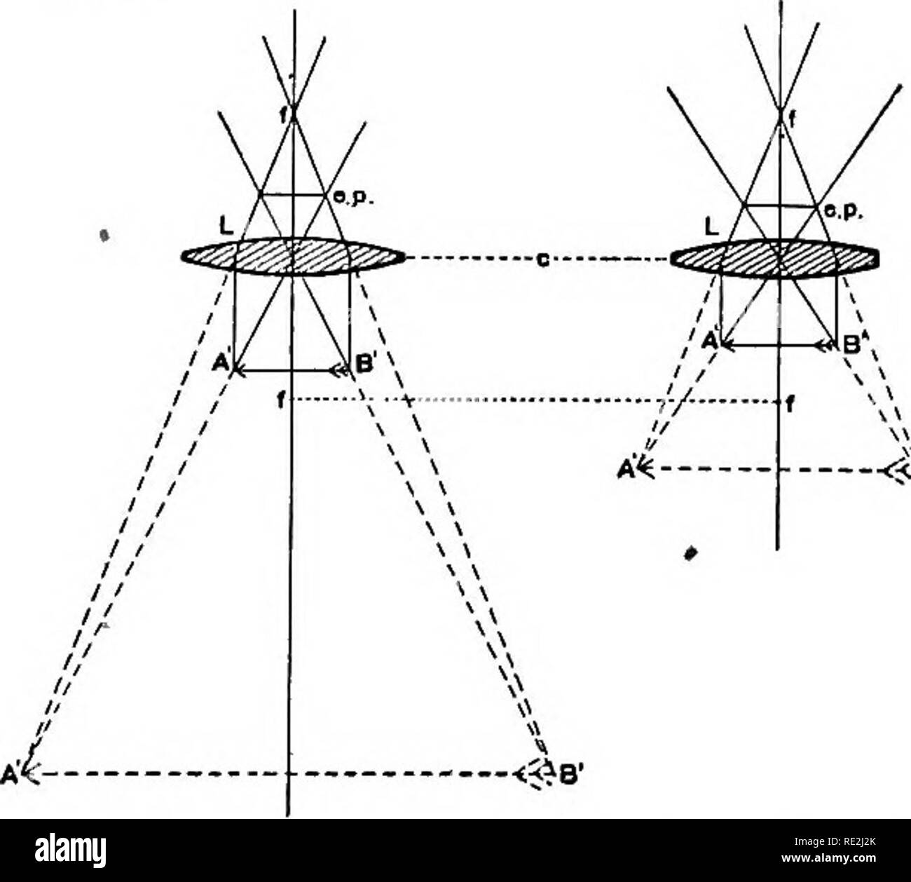
Optical ray diagram with an optical objective lens showing ...
Light Microscope- Definition, Principle, Types, Parts ... A light microscope is a biology laboratory instrument or tool, that uses visible light to detect and magnify very small objects and enlarge them. They use lenses to focus light on the specimen, magnifying it thus producing an image. The specimen is normally placed close to the microscopic lens.

Principles of imaging with an optical microscope: (a) ray ...
Parts of Stereo Microscope (Dissecting microscope ... Labeled part diagram of a stereo microscope Major structural parts of a stereo microscope. There are three major structural parts of a stereo microscope. The viewing Head includes the upper part of the microscope, which houses the most critical optical components, including the eyepiece, objective lens, and light source of the microscope.

Optical Microscopes – Some Basics | Science Lab | Leica ...
Diagram of a Compound Microscope - Biology Discussion 1. It is noted first that which objective lens is in use on the microscope. 2. Stage micrometer is positioned in such a way that it is in the field of view. 3. The eyepiece is rotated so that the two scales, the eyepiece or ocular scale and the stage micrometer scale, are parallel. 4.

Microscope Diagram Vector Illustration. Labeled Zoom ...
Compound Microscope: Definition, Diagram, Parts, Uses ... Compound microscope is a type of optical microscope that is used for obtaining a high-resolution image. There are more than two lenses in a compound microscope. Learn about the working principle, parts and uses of a compound microscope along with a labeled diagram here.

Schematic drawing of microscope objective with pupil fixation ...
16 Parts of a Compound Microscope: Diagrams and Video ... Aperture. Illuminator. Condenser. Diaphragm. Parts of a Compound Microscope. Video: Parts of a compound Microscope with Diagram Explained. As a side note, the microscope used in this post is a great entry level or beginner microscope if you are trying to get someone interested in microscopes, microbiology, or science in general.
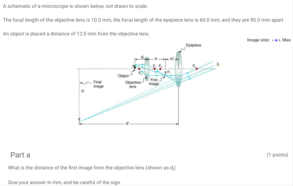
Solved A schematic of a microscope is shown below, not drawn ...
Microscope Lab - csun.edu There is a diagram link below. Have the students study this diagram so that they know the parts, which objective to use. It is very important that the students don't focus to close to the slide with the high power objective, as this can permanently damage the lens.
Which ray diagram is correct for a Compound microscope ...
Understanding Microscopes and Objectives - Edmund Optics With a microscope, a relay lens system replaces the single lens; an objective and an eyepiece work in tandem to project the image of the object onto the eye, or a sensor - depending upon the application. There are two parts to a microscope that increase the overall system magnification: the objective and the eyepiece.

Optical Instrument - Compound Microscope
The microscope : an introduction to microscopic methods and ...
Microscopes and magnifiers: Physclips - Light

Optical microscope

a Draw a ray diagram for the formation of image by a compound ...
Exercises, Telescopes and microscopes, By OpenStax (Page 2/2 ...

The schematic diagram of a compound microscope is shown in ...
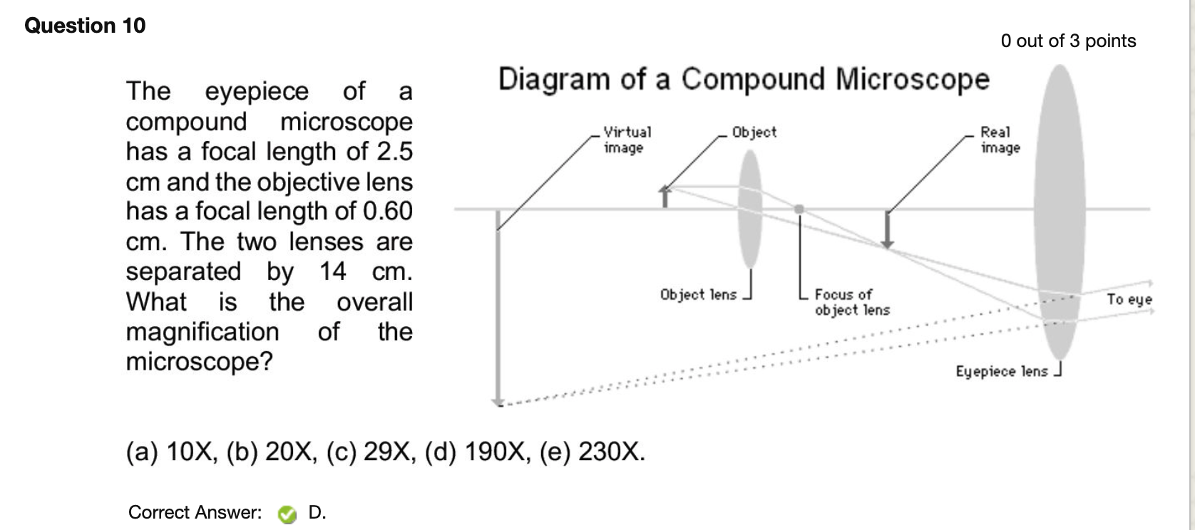
Solved Question 10 O out of 3 points Diagram of a Compound ...

Light Microscope vs Electron Microscope - Accelerating Microscopy
Why does the objective lense in a compound microscope have ...

With the help of ray diagram, describe the construction ...

Describe the construction and working of a compound ...
The microscope; an introduction to microscopic methods and to ...

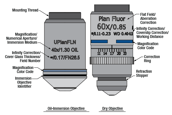
Understanding Microscopes and Objectives | Edmund Optics
What is Histology: The Histology Guide

Draw a labelled diagram of an image formed by a compound ...

Understanding Microscopes and Objectives | Edmund Optics
draw a labelled ray diagram showing imagr formation in ...
Microscope, Microscope Parts, Labeled Diagram, and Functions
Which ray diagram is correct for a Compound microscope ...

Optical Microscopes – Some Basics | Science Lab | Leica ...

Draw the ray diagram of image formation in case of compound ...
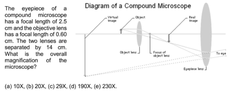
Solved Diagram of a Compound Microscope Virtual image Object ...

File:Microscope simple diagram.png - Wikimedia Commons

Systematic design of microscope objectives. Part II: Lens ...
Convex lens use - Microscope
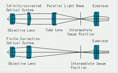
What is an Infinity-corrected Optical System? | Learn about ...


Comments
Post a Comment