41 compound microscope ray diagram
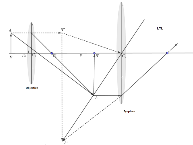
optics - Ray diagram of focussing on a compound microscope ... Here is the ray diagram of a compound microscope. So, when we are focussing, we move the objective lens which tweaks the image distance. My doubt is that, shouldn't the image be seen clearly, wheresoever the first real image forms, if within Fe (Focus of the eyepiece lens). Draw a ray diagram of compound microscope when the class ... Draw a ray diagram of compound microscope, when the final image is formed at the minimum distance of distinct vision. Answer Verified 119.7k + views Hint: A compound microscope is an optical instrument used for observing highly magnified images of tiny objects.
Difference Between Simple and Compound Microscope: Parts ... A compound microscope consists of two convex lenses coaxially separated by some distance. The lens nearer to the object is called the objective. The lens through which the final image is viewed is called the eyepiece. The focal length of objective lens is smaller than eyepiece. Ques. Draw a ray diagram of a compound microscope.
Compound microscope ray diagram
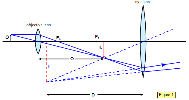
Microscopes - Self Study Point The magnifying power of the compound microscope is defined as the ratio of the angle subtended at the eye by the final image to the angle subtended at the eye by the object when both the final image and the object are situated at the least distance of distinct vision from the eye. Ray diagram for the formation of image by a compound microscope Ray Diagram - Compound Microscope - YouTube How to draw ray diagram of Compound Microscope - Chapter 7 - Lenses - Part 4 Diagram of a Compound Microscope - Biology Discussion ADVERTISEMENTS: In this article we will discuss about:- 1. Essential Parts of Compound Microscope 2. Magnification of the Image of the Object by Compound Microscope 3. Resolution Power 4. Method for Studying Microbes 5. Measurement of the Size of Objects. Essential Parts of Compound Microscope: The essential parts of usually used monocular compound microscope (Fig. […]
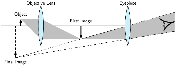
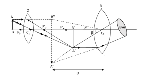
Compound microscope ray diagram. [Term 2] (a) Draw a ray diagram of compound microscope for ... (a) Draw a ray diagram of a compound microscope for the final image formed at least distance of distinct vision? Answer Diagram of Compound Microscope for the final image formed at D (b) An angular magnification of 30X is desired using an objective of focal length 1.25 cm and an eye piece of focal length 5 cm. Describe the compound microscope based on the following ... Puzzles. Describe the compound microscope based on the following headings: 1) Labelled ray diagram of image formation. 2) Magnifying power when final image is formed at least distance of distinct vision. Verified. 140.1k + views. Hint: In this question we have been asked to draw a labelled ray diagram of image formation of a compound microscope. Which ray diagram is correct for a Compound microscope ... Here are two ray diagrams for compound microscope, the first one proposed by the book, and the second one recommended by the teacher: In the first image, the light rays form a real image A'B', which becomes the virtual object for the eyepiece. See, the original rays are carried forward to the eyepiece, which then form a virtual image, A"B". draw a ray diagram to show the image formation by a ... Draw a ray diagram to show the image formation by a compound microscope when the final image is formed at the near point.Define the resolving power of a microscope.Write two factors by which resolving power can be increased?
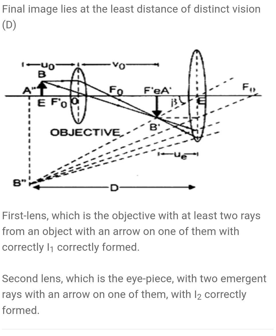
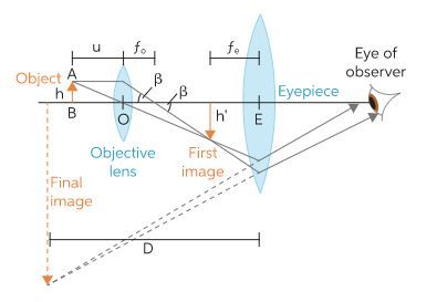
(i) Draw a schematie ray diagram ot a compound microscope ... (i) Draw a schematie ray diagram ot a compound microscope when image is formed at distance of distinct vision. (ii) Write the expression for resolving power of a compound microscope. How can the resolving power ot a microscope be increased ? Compound Microscope Ray Diagram | class 12th physics ... This Fastest drawing Technique to draw Compound Microscope Ray Diagram is for all students of every Board - WBCHSE, HSC, CBSC, ICSE and other state boards fr... Draw a ray diagram of a compound microscope. Write the ... Ray diagram of a compound microscope.When the final image is formed at the least distance of distinct vision,For the image formed at infinity, ue = feand By making focal length of the objective small, the magnifying power can be increased. Draw a Ray Diagram Showing the Image Formation by a ... Draw a ray diagram showing the image formation by a compound microscope. Hence obtained expression for total magnification when the image is formed at infinity. Advertisement Remove all ads Solution A compound microscope consists of two convex lenses co-axially separated by some distance. The lens nearer to the object is called the objective.
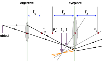
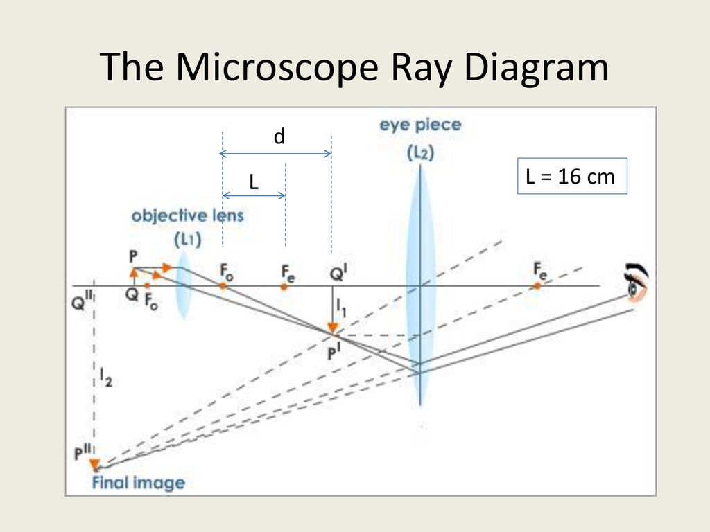
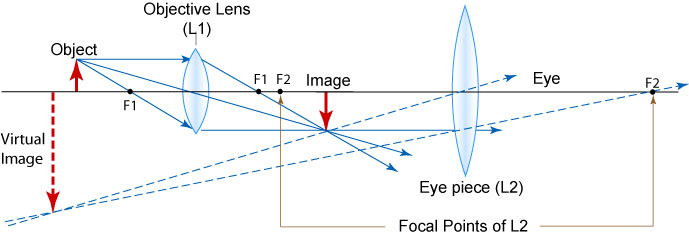
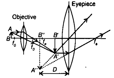
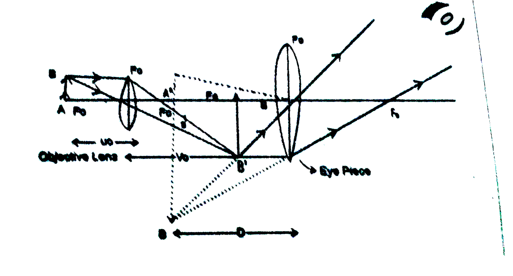
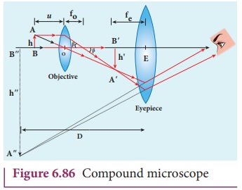
What is a Compound microscope? Applications of Compound ... Ray diagram of Compound microscope. The specimen AB is placed just beyond the principal focus Fo' of the objective lens. A ray of light AO from A goes parallel to the principal axis towards the objective lens and converges. Working Principle and Parts of a Compound Microscope (with ... In a microscope, light is focused on the object as a narrow pencil of light, from where it enters into the objective as a diverging pencil (Figure 4.8). The angle 9 subtended by the optical axis (the line joining the centers of all the lenses) and the outermost ray still covered by the objective is a measure of the aperture called 'half ... Compound Microscope | Class 12 Physics Chapter 9 ... Topic compound microscope explains detail working with ray diagram and magnification of compound microscope. Helpful for cbse class 12 physics chapter 9 ray optics. CBSE 12 Physics 01 Electric Charges and Fields 17 Topics 01.01 Electric Charge. 01.02 Conductors, Semiconductors and Insulators. derivation of compound microscope - Physics ... We now obtain the magnification due to a compound microscope. The ray diagram of figure shows that the (linear) magnification due to the objective, namely h′/h, equals ................ (1) where we have used the result , tanβ = h/fo = h'/L
Draw a neat labelled diagram of a compound microscope and ...
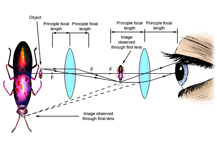
Compound Microscope: Definition, Diagram, Parts, Uses ... Compound microscope is a type of optical microscope that is used for obtaining a high-resolution image. There are more than two lenses in a compound microscope. Learn about the working principle, parts and uses of a compound microscope along with a labeled diagram here.
Electricity - detailed contents
Draw a ray diagram of compound microscope, when final ... Draw a ray diagram of compound microscope, when final image is formed at the minimum distance of distinct vision. Easy Solution Verified by Toppr It consist of two convex lenses, one objective of very small focal length with short aperture. And one Eyepiece with moderate focal length and large aperture.
a) Draw a labelled ray diagram of a compound microscope. (b ...
CBSE NCERT Notes Class 12 Physics Ray Optics Optical ... Simple Microscope. An instrument that gives an enlarged image of a minute object. A simple magnifier or microscope is a converging lens of small focal length. There are 2 types of Microscopes:-. Simple Microscope 2. Compound Microscope. Simple Microscope. The lens is held near the object, one focal length away or less, and the eye is positioned ...
The Compound Microscope - ppt download
(i) Draw a neat labelled ray diagram of a compound ... Draw a neat labeled ray diagram of a compound microscope. Explain briefly its working. asked Aug 1, 2019 in Physics by Rk Roy (63.8k points) jee; jee mains; 0 votes. 1 answer (i) Draw a neat labelled ray diagram of an astronomial telescope in normal adjustment . Explain briefly its working . (ii) An astronomical telescope u
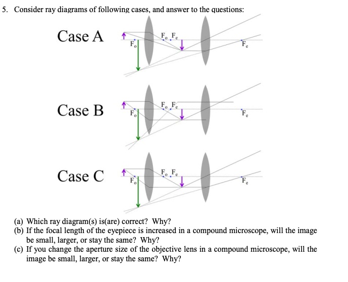
SOLVED:Consider ray diagrams of following cases, and answcr ...
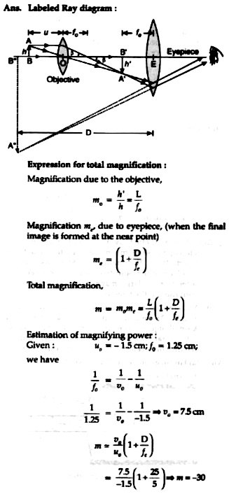
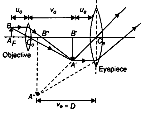
(a) Draw a labelled ray diagram of compound microscope ... (a) Draw a labelled ray diagram of compound microscope, when final image forms at the least distance of distinct vision. (b) Why is its objective of short focal length and of short aperture, compared to its eyepiece? Explain. (c) The focal length of the objective is 4 cm while that of eyepiece is 10 cm. The object is placed at a distance of 6 cm from the objective lens.

Optical microscope - Wikipedia
Derive the formula for angular magnification of a compound ... Draw the required ray diagram. The angular magnification of a compound microscope is the ratio of the angle subtended by the final image at the eye to the angle subtended by the object at the eye, when both are placed at the least distance of distinct vision.

Draw a Ray Diagram Showing Image Formation in a Compound ...
Draw a Labelled Ray Diagram Showing the ... - Shaalaa.com We now obtain the magnification due to a compound microscope. The ray diagram of Figure shows that the (linear) magnification due to the objective, namely h'/h, equals m h' h L m o = h' h = L f o where we have used the result h h' L tan β = ( h f o) = ( h' L)
Draw a ray diagram to show the working of acompound ...
Types of Microscopes: Definition, Working Principle ... Where, D is the least distinct vision; F is the focal length of the convex lens; Simple Microscope Diagram. Principle of Simple Microscope. The working principle of a simple microscope is that when a sample is placed within the focus of the microscope, a virtual, erect and magnified image is obtained at the least distance of distinct vision from the eye that is held at the lens.
MAGNIFICATION IN MICROSCOPE | cell in life
(a) Draw a labelled ray diagram of a compound microscope ... Best answer (a) Labelled diagram of compound microscope. The objective lens form image A' B' near the first focal point ofeyepiece. (b) Angular magnification of objective lens m0 = linear magnification h'/h where L is the distance between second focal point of the objective and first focal point of eyepiece.

Draw a ray diagram to show formation of an image by a ...
Diagram of a Compound Microscope - Biology Discussion ADVERTISEMENTS: In this article we will discuss about:- 1. Essential Parts of Compound Microscope 2. Magnification of the Image of the Object by Compound Microscope 3. Resolution Power 4. Method for Studying Microbes 5. Measurement of the Size of Objects. Essential Parts of Compound Microscope: The essential parts of usually used monocular compound microscope (Fig. […]

Draw the ray diagram of image formation in case of compound ...
Ray Diagram - Compound Microscope - YouTube How to draw ray diagram of Compound Microscope - Chapter 7 - Lenses - Part 4
Exercises, Telescopes and microscopes, By OpenStax (Page 2/2 ...
Microscopes - Self Study Point The magnifying power of the compound microscope is defined as the ratio of the angle subtended at the eye by the final image to the angle subtended at the eye by the object when both the final image and the object are situated at the least distance of distinct vision from the eye. Ray diagram for the formation of image by a compound microscope

Ray Diagram of a Compound Microscope | Diagram, Microscope ...
Draw a ray diagram showing image formation in a compound ...

ABU-SARIM English blog: Ray Diagram for Compound microscope

Draw a labelled ray diagram of an image formed by a compound microscope with final image formed ...

a) Draw a labelled ray diagram of compound microscope, when ...

optics - Can we use a telescope as a microscope and vice ...
a) Draw a ray diagram showing the image formation by a ...

Draw a labelled diagram of an image formed by a compound ...
draw a ray diagram of compound microscope for the final imag ...
Draw a labelled ray diagram of a compound microscope. | Snapsolve
Convex lens use - Microscope

Solved Compound microscope 2 1 2 3 4 In the following ray ...

optics - Ray diagram of focussing on a compound microscope ...

Experts, please give ray diagram of compound microscope ...
Draw a ray diagram of compound microscope for the final image ...

with the help of neat labelled ray diagram ,show formation by ...
Ray Optics And Optical Instruments
Draw a labelled ray diagram of an image formed by a class 12 ...

draw the labelled ray diagram for the formation of image by a ...
a) Draw a ray diagram for final image formed at distance of ...

Solved example: magnifying power of compound microscope

how to draw Compound Microscope ray diagram step wise how to ...

Draw a labelled ray diagram to show image formation by a ...
Draw the ray diagram of Compound microscope when the class 12 ...

Draw the ray diagram of compound microscope in normal ...
schoolphysics ::Welcome::

optics - How to draw ray diagrams for a compound microscope ...

How to draw Compound Microscope Diagram - Final Image at D
Compound microscope - Optical Instruments


Comments
Post a Comment