43 fungi diagram labeled
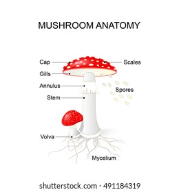
Plant Label Me! Printouts - EnchantedLearning.com Read the definitions then label the leaf margins (including entire, serrate, crenate, lobed, and parted). Read the definitions then label the mushroom diagram, including the cap (pileus), cup (volva), gills (lamellae), mycelial threads, ring (annulus), scales, and stem (stape). Plant Anatomy Printout: Simple Label Me! Label the microbes (fungi, virus, bacteria) | Teaching ... Label the microbes (fungi, virus, bacteria) A cut and stick sheet for pupils to label the parts of the fungi, virus and bacteria. I used it with my low ability year 7 group (to minimise time wasted drawing the diagrams) Report this resource to let us know if it violates our terms and conditions.
Labeled Diagram of the Human Lungs - Bodytomy Given below is a labeled diagram of the human lungs followed by a brief account of the different parts of the lungs and their functions. Each lung is enclosed inside a sac called pleura, which is a double-membrane structure formed by a smooth membrane called serous membrane.

Fungi diagram labeled
› parts-of-a-compoundMicroscope Parts and Functions With Labeled Diagram and ... Microscope Parts and Functions With Labeled Diagram and Functions How does a Compound Microscope Work?. Before exploring microscope parts and functions, you should probably understand that the compound light microscope is more complicated than just a microscope with more than one lens. Fungi: Definition and Types of Fungi (explained with diagram) Fungi: Definition and Types of Fungi (explained with diagram)! Fungi (singular: fungus) are a group of plantlike organisms which lack chlorophyll. There are some 70,000 types of fungi—some unicellular and some multicellular. Most fungi are saprophytes, while some are parasites. Yeast, moulds and mushrooms are fungi. Characteristics of Fungi | Boundless Biology Fungi can be unicellular, multicellular, or dimorphic, which is when the fungi is unicellular or multicellular depending on environmental conditions. Fungi in the morphological vegetative stage consist of a tangle of slender, thread-like hyphae, whereas the reproductive stage is usually more obvious.
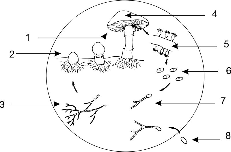
Fungi diagram labeled. Mushroom anatomy labeled biology diagram vector ... 2. Editable Vector .EPS-10 file. 3. High-resolution JPG image. Use for everything except reselling item itself. Description: Mushroom anatomy labeled biology diagram vector illustration. Forest nature exploring and education. Spore bearing fruiting body of a fungus. Structural drawing scheme as learning information. Question Video: Labeling the Structures of a Fungus | Nagwa Question Video: Labeling the Structures of a Fungus. The diagram shows a simplified outline of the structure of a mushroom growing in soil. Which of the following options correctly identifies the structures labeled X and Y on the diagram? [A] X: hyphae, Y: mycelium [B] X: mycelium, Y: xylem [C] X: xylem, Y: endospores [D] X: phloem, Y: lamellae. › fungi › classificationClassification of Fungi (With Diagram) - Biology Discussion The perfect stage of fungus is Gibberella fujikuroi. Gibberellins are natural plant growth hormones. 7. Trichoderma (Fig. 2.53). It is a soil fungus used in biological control of other fungi as it produces allelochemics against them. If the fungus happens to pass into human alimentary canal it produces leucopenia called alimentary canal aleukia. 34 Label The Features Of A General Fungal Life Cycle With ... Label b in the generalized fungal life cycle. General fungi life cycle cycle diagram use createlys easy online diagram editor to edit this diagram collaborate with others and export results to multiple image formats. Creately diagrams can be exported and added to word ppt powerpoint excel visio or any other document.
Fungal cell structure and organization - Oxford Medicine Human pathogenic fungi produce three basic 'cell' types: hyphae, yeast cells, and spores. The organization and subcellular structure of these different cell types and their modes of growth and formation are reviewed. Growth and form is the consequence of how new cell surface is formed. This is generated by the delivery of vesicles to the surface which provides new membrane and the enzymes ... Kyra is making a diagram to compare animal-like protists ... Kyra is making a diagram to compare animal-like protists and fungus-like protists. Which belongs in the area labeled X? are multicellular are heterotrophs - 15116441 Chytrid Fungi Online Batrachochytrium - Examine the prepared slides of frog skin for thalli of this fungus. These are slides of a tadpole's jaw sheath from Crocker Pond in western Maine. Diagram and label the thalli in the hexagon-shaped frog skin cells. 3. Spizellomycetales. Spizellomyces - What is its thallus type? Do you see rhizoids and/or zoospores? Fungi | Diagram of a small section of a hypha. Biology ... Hypha. Fungi are formed from microscopic filaments called hyphae. This is a greatly enlarged diagram of a small section of a hypha. © Copyright D G Mackean. < Back to Fungi.
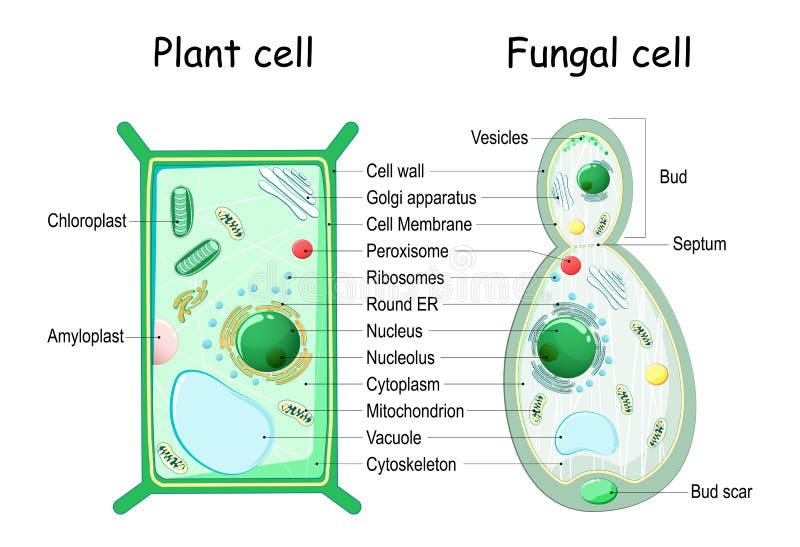
courses.lumenlearning.com › boundless-biologyComponents of the Blood | Boundless Biology - Lumen Learning T cells attack viruses, fungi, some bacteria, transplanted cells, and cancer cells. Natural killer cells attack a variety of infectious microbes and certain tumor cells. One reason that HIV poses significant management challenges is because the virus directly targets T cells by gaining entry through a receptor. Eukaryotic Cell: Structure, Characteristics & Diagram - Embibe 13. Lysosomes are the single membranous structure filled with digestive enzymes which helps to digest worn-out cells and foreign bacteria and viruses. 14. Vacuoles are the membrane-bound structure present in the eukaryotic cell.In animal cells, there are numerous small vacuoles while in plant cells large vacuoles are present. Large vacuoles help in maintaining water balance and keep the cell ... PDF Lab 3 Fungi - WCJC eukaryotic organisms called fungi. Most members of this group are multicellular and macroscopic, whereas some are single-celled and microscopic. Most fungal cells are enclosed by a cell wall containing the polysaccharide chitin. Fungi begin their life cycle as spores that divide rapidly to form threadlike structures called hyphae. These spores are single cells and Fungi - Variety of living organisms - GCSE Biology (Single ... Yeast is an example of a single-celled fungus. Fungal cell structure Fungal cells have a cell wall made of chitin (remember that plant cell walls are made of cellulose).
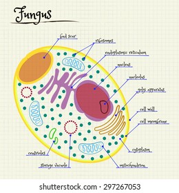
Cross section of a yeast cell. Structure of fungal cell ...
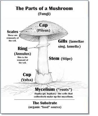
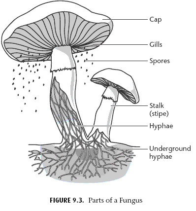
30 Diagram Of Mushroom With Label - Labels For Your Ideas This is a thumbnail of the label the mushroom anatomy diagram. Label the cap stipe and gills. Cap pileus the top part of the mushroom. Rhizopus and mucor are the common saprotrophic fungi that attack a variety of food stuffs. Mucor pusillus causes infection of internal organs in human beings.

Draw a well labelled diagram of fungi. - Brainly.in
Label Mushroom Diagram Printout - EnchantedLearning.com Answers. EnchantedLearning.com. Label the Mushroom Anatomy Diagram. Plants. Read the definitions below, then label the mushroom diagram. This is a thumbnail of the Label the Mushroom Anatomy Diagram. The full-size printout is available only to site members. To subscribe to Enchanted Learning, click here. If you are already a site member, click here.

Mushroom and Fungi Life Cycle Diagram - Label and Describe | TpT
Fungal Cell Diagram Illustrations, Royalty-Free Vector ... Browse 81 fungal cell diagram stock illustrations and vector graphics available royalty-free, or start a new search to explore more great stock images and vector art. Cross section of a yeast cell. Structure of fungus cell. fungal cell diagram stock illustrations. Cross section of a yeast cell.

Cilia 5. Starter – Label the Diagram. - ppt download
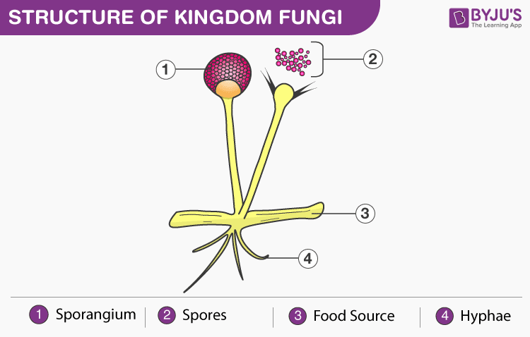
PDF 51 Morphology and General Properties of Fungi Fungus is a member of a large group of eukaryotic organisms that includes microorganisms such as yeasts and molds (British English: moulds), as well as the more familiar mushrooms. These organisms are classified as a kingdom, Fungi, which is separate from plants, animals, protists and bacteria. One major
Fungi: Basidiomycota: The Club Fungi | SparkNotes
› fungi › structure-ofStructure of Fungal Cell (With Diagram) | Fungi The suggested formula for fungus chitin is (C 22 H 54 N 21) n. Electron microscope studies reveal that chitin occurs as elongated variously oriented microfibrillar units. These are laid down in layers and form the basis of the structural rigidity of fungal cell walls. The microfibril layers generally run parallel to the surface.

labelled diagram of fungi - Brainly.in
collegedunia.com › exams › chloroplasts-definitionChloroplasts: Definition, Diagram, Structure and Function Dec 21, 2021 · Diagram of Chloroplast. The diagram of the chloroplast given below represents the chloroplast structure including the different parts of the chloroplast. The parts of a chloroplast such as the inner membrane, outer membrane, intermembrane space, thylakoid membrane, stroma, and lamella are all mentioned.

BACTERIA, VIRUS, FUNGI-- DIAGRAM/LABEL WITH ANSWER SHEET! by ...
Kingdom Fungi- Structure, Characteristics & Classification ... On the basis of nutrition, kingdom fungi can be classified into 3 groups. Saprophytic - The fungi obtain their nutrition by feeding on dead organic substances. Examples: Rhizopus, Penicillium and Aspergillus. Parasitic - The fungi obtain their nutrition by living on other living organisms (plants or animals) and absorb nutrients from their host. Examples: Taphrina and Puccinia.
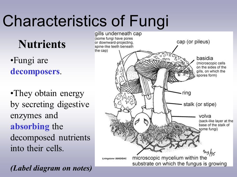
Characteristics of Fungi - ppt video online download
hyphal - fungionline.org.uk Isolation of Fungi using the hyphal tip method. Incubate at 30-34 C for 24 hour at dark condition for first growing fungi or more than two days for slow growing fungi. Observe under microscope and cut your fungal hyphal tip after dichotomous branching or simply you can cut most extended part. On Generative Algorithms: Hyphae inconvergent.

Wild edible fungi a global overview of their use and ...
Rhizopus: Structure with Diagram, Reproduction and Life ... Rhizopus Structure with Diagram. They are fast-growing fungi and have a cottony appearance; The body of rhizopus consists of branched mycelium. The mycelium is coenocytic and composed of three types of hyphae; stolon, rhizoids and sporangiophores; Stolon is the internodal region, it is aerial, forms an arch and touches the substratum forming nodal region ...

File:Structure of fungus.jpg - Wikimedia Commons
microbenotes.com › parts-of-a-microscopeParts of a microscope with functions and labeled diagram Dec 24, 2021 · Parts of a microscope with functions and labeled diagram February 9, 2022 December 24, 2021 by Faith Mokobi Having been constructed in the 16th Century, Microscopes have revolutionalized science with their ability to magnify small objects such as microbial cells, producing images with definitive structures that are identifiable and characterizable.
Kingdom Fungi- Structure, Characteristics & Classification Of ...
Fungi Diagram Illustrations, Royalty-Free Vector Graphics ... Fungi, Mushrooms, Algae and Non-Flowering Plants. Engraved Antique, Representatives of the Algae, Fungi, Bryophyta, Polypodiophyta and other Nonflowering plants Engraving Antique Illustration, Published 1851 fungi diagram stock illustrations.

Identify the following diagram, label it and write detail ...
Hyphae: Definition, Function & Types - Video & Lesson ... Hyphae are the tiny filaments that make up the structure of multicellular fungi. Explore the definition and function of hyphae, with examples of the two major types.

Identify the following diagram, label it and write detail ...
researchtweet.com › microscope-parts-labeledMicroscope, Microscope Parts, Labeled Diagram, and Functions Jan 19, 2022 · Eyepiece Lens: the lens at the top that you look through, usually 10x or 15x power. Tube: Connects the eyepiece to the objective lenses. Illuminator: Illuminator is the most important microscope parts and it serve as light source for a microscope during slide specimen visualization.

Diagram illustrating the ultrastructure of hypha. | Download ...
Animal Plant Fungi Cell Venn Diagram - Studying Diagrams Animal plant fungi cell venn diagram. The animal and plant cell diagram above shows you the table of comparison between the two cells. Venn diagram of the Kingdoms of Fungi Protista Plants. Services of language translation the. Ability to sketch and label a simple plant and animal cell including the following organelles.

Ringworm vector illustration. Labeled fungal skin infection ...
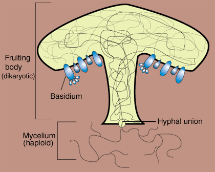
Jared drew a diagram to compare zygote and club fungi ... from the diagram described in the question, area occupy by X simply depicts what zygote is only made of which are multicellular. Club fungi contains both loosely arranged hyphae and production of spores while the similarities between zygote and club fungi is that, they both contains numerous flagella

Fungi
Characteristics of Fungi | Boundless Biology Fungi can be unicellular, multicellular, or dimorphic, which is when the fungi is unicellular or multicellular depending on environmental conditions. Fungi in the morphological vegetative stage consist of a tangle of slender, thread-like hyphae, whereas the reproductive stage is usually more obvious.
template
Fungi: Definition and Types of Fungi (explained with diagram) Fungi: Definition and Types of Fungi (explained with diagram)! Fungi (singular: fungus) are a group of plantlike organisms which lack chlorophyll. There are some 70,000 types of fungi—some unicellular and some multicellular. Most fungi are saprophytes, while some are parasites. Yeast, moulds and mushrooms are fungi.
The Fungi Kingdom
› parts-of-a-compoundMicroscope Parts and Functions With Labeled Diagram and ... Microscope Parts and Functions With Labeled Diagram and Functions How does a Compound Microscope Work?. Before exploring microscope parts and functions, you should probably understand that the compound light microscope is more complicated than just a microscope with more than one lens.

Spore Formation | Definition, Examples, Diagrams
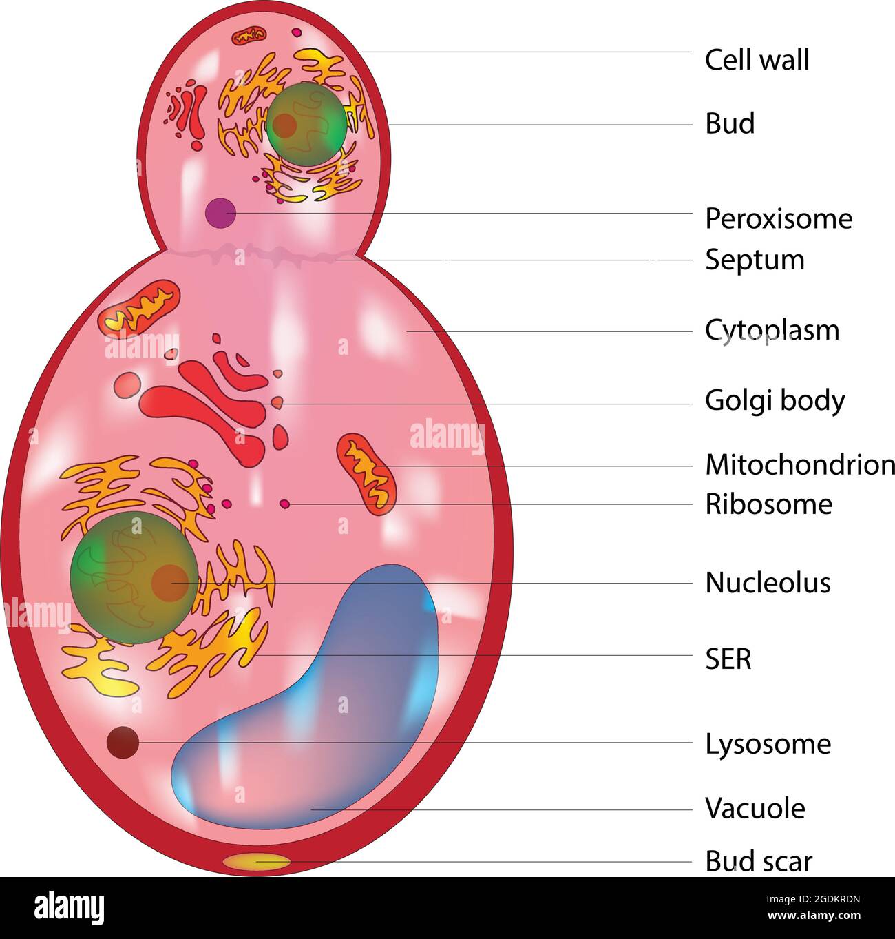
Budding fungus cell structure, Anatomy of fungal cell ...

Label the parts of a mushroom | Parts of a mushroom, Stuffed ...

Fungi
a) What is spore formation? (b) Draw a diagram showing spore ...
MycoTechnology raises $39m in Series D to expand functional ...

Fungi | Microbiology

FUNGAL TAXONOMY (DIVISION) - Microbiology Class

Diagrams of Fungi
Yeast cell structure Images, Stock Photos & Vectors ...
Fungal Cell Structure Stock Illustrations – 158 Fungal Cell ...

Fungi Diagram | Quizlet

81 Fungal Cell Diagram Illustrations & Clip Art - iStock

Culture, Enumeration and Identification of Filamentous Fungi ...

template

Diagram showing infection of barley by the fungal pathogen ...

Structure of Fungal Cell (With Diagram) | Fungi

Classification of Fungi into 5 Phyla flow chart with Examples
Fungi structure Images, Stock Photos & Vectors | Shutterstock

Fungi, Antibiotics, Yeasts, Penicillium.
Fungus - MR. BLACKS ARMY
PPT - Fungi PowerPoint Presentation, free download - ID:2000404

Club Fungi Diagram | Quizlet

81 Fungal Cell Diagram Illustrations & Clip Art - iStock

Budding fungus cell structure, Anatomy of fungal cell ...


Comments
Post a Comment